Implications
Our study provides evidence that, compared to single housing, confinement of domestic pigs does not impose additional chronic stress on the animals. With respect to the stress hormone, cortisol and heart reactions, the confined pigs did not differ from non-confined roommates serving as controls. Heart rate and cortisol levels also did not differ from published values of group-housed pigs. We assume that domestication has adapted domestic pigs to narrow housing conditions without impairing physiological homeostasis. However, this does not exclude mental suffering.
Introduction
Restraint is used as one of the standard approaches for inducing experimental stress in laboratory rodents (for review, see Buynitsky and Mostofsky, Reference Buynitsky and Mostofsky2009). The degree of restraint is mostly severe and the procedure usually lasts for several minutes up to some hours or even weeks. In commercial animal keeping, restraint procedures (confinements) are also not unusual. A characteristic example includes keeping sows in crates during the farrowing and suckling period. Typically, these movement restrictions do not go as far as in the rodent experiments, so some residual freedom to move is given. On the other hand, the duration of movement restriction is often long, depending on the local management practices. However, not much objective information on the degree of stress that pigs experience during chronic restraint exists. As domestic pigs have undergone a long period of domestication that sometimes included poor housing conditions (Albarella, Reference Albarella2007), their susceptibility to restraint, in particular, might have been reduced during the selection process. Consequently, if at all, only slightly increased cortisol concentrations were found in confined pigs (Siard and Stuhec, Reference Siard and Stuhec2003; Siard et al., Reference Siard, Kovac, Ladewig and Stuhec2003; Sorrells et al., Reference Sorrells, Eicher, Harris, Pajor and Richert2007).
Although cortisol is a useful and well-established indicator of stress in farm animals, its use as the sole indicator of stress bears some risks (Mormède et al., Reference Mormède, Andanson, Aupérin, Beerda, Guémené, Malmkvist, Manteca, Manteuffel, Prunet, van Reenen, Richard and Veissier2007). Cortisol concentrations do not only depend on physical or psychological stress but also on conditions of the ambient physical environment, as well as temporal and metabolic factors. Hence, a supplemental approach to judge the severity of stress and, possibly, suffering in confined pigs may be to record their heart reactions (von Borell et al., Reference von Borell, Langbein, Despres, Hansen, Leterrier, Marchant-Forde, Minero, Mohr, Prunier, Valance and Veissier2007). This non-invasive technique can be particularly suitable in situations of movement restraint in which artefacts by physical activities are largely prevented. By separately analysing heart rate (HR) and HR variability (HRV), it is possible to record sympathetic and parasympathetic tones that may reflect stress, emotions and mood (Porges, Reference Porges1995a; Friedman and Thayer, Reference Friedman and Thayer1998; von Borell et al., Reference von Borell, Langbein, Despres, Hansen, Leterrier, Marchant-Forde, Minero, Mohr, Prunier, Valance and Veissier2007).
Using a combined endocrine – the cardiac analysis approach – we tested the hypothesis that the chronic restraint in pigs results in chronic stress. Therefore, we kept growing pigs in metabolic cages and recorded their behaviour, cardiac activity and salivary cortisol.
Material and methods
All procedures involving animal handling and treatment were approved by the Committee for Animal Use and Care of the Agricultural Department of Mecklenburg-West Pomerania, Germany.
Animals and housing
Eighteen male German Landrace barrows at the age of 10 weeks were used at the beginning of the experiment. The animals were born and reared at the Leibniz Institute for Farm Animal Biology under conventional group housing conditions. The experiment was conducted in six consecutive replications with three pigs each. The pigs were randomly divided into two groups. In each replication, two pigs were used in the experimental group and one pig served as a control. Three days before the beginning of the experiment (day −5 with reference to crating day 0), all three pigs were single-housed (pen size = 1.45 m × 0.90 m, 69% slatted floor) without straw in the same room. The pen sides allowed for limited visual and physical contact through bars. The room (4.00 m × 3.40 m) was maintained at 20 ± 2°C and a 12 h : 12 h light–dark cycle (light from 0600 to 1800 h).
Water and food (Ferkelstarter Plus, Trede and von Pein, Itzehoe, Germany) were available ad libitum, with the feeding time set at 0710 h. The feed was refilled when the trough was empty. Measurements were taken for 10 days. The animals were weighed before the start of the experiment (day 5) and on the day after its termination.
On the first 2 days of the study (days −2 and −1), the test and control pigs were in the same housing situation. During these days, the animals were habituated to the flexible belt of the heart beat measurement system as well as to the saliva collection. On the third day (day 0), the two test pigs were transferred to single metabolic cages (1.0 m long × 0.4 m wide × 0.6 m high) at 0730 h and stayed there for the next 7 days (days 1 to 7). These cages were located in the same room as the pen of the control animal. The animals were able to stand up and move 25 cm along the long axis of the cage, but not to turn around. Laterally, a 5 cm space was given. The animals were able to lie down on the side by sticking their legs through the rods. The trough and nipple drinker were located in front of the metabolic cages. The control pig remained in the single pen. The animals had no previous experience with the two housing conditions. As they were housed in single metabolic cages or pens without further equipment, they were brushed daily by a caregiver in order to avoid itches and dandruff development after the observation period.
Behavioural measurements
The pigs were observed with a live camera for 3 h every day between 0800 to 0900 h, 1100 to 1200 h and 1400 to 1500 h. The behaviour was observed by continuous sampling of one focal animal in 10-min periods (i.e. two times in an hour per animal), so that 1 h of behavioural data was recorded per pig distributed over the day. The order of observations was randomized. Observations (duration scanning) included two non-active behaviours: resting on the belly or on the side; and five active behaviours: exploration (examining environment with nose), standing, sitting, locomotion (movement of two legs at least) and escape attempts (including violent biting attacks on the bars of the cage).
Saliva collection and cortisol analysis
The method has been described in detail by Ernst et al. (Reference Ernst, Tuchscherer, Kanitz, Puppe and Manteuffel2006). In short, the sampling of saliva was carried out at 0700 h ± 5 min. It was collected before feeding by allowing the pigs to chew on veterinary cotton buds (Weisweiler GmbH & Co. KG, Münster, Germany) until these were thoroughly moistened (∼30 to 60 s per sample). The buds were placed in tubes and centrifuged at 3500 × g for 10 min at 4°C. Saliva samples were stored at −20°C until analysis. After thawing, the cortisol concentration was measured by an enzyme immunoassay (DSL-10-67100 ACTIVE™, DSL Deutschland GmbH, Germany).
Heart beat measurements
The Polar S810i (Polar Elektro Oy®, Kempele, Finland) device was used for the non-invasive telemetric measurement of heart beat activity (R-R intervals). For more technical details and information on the application of this system in farm animals, see Mohr et al. (Reference Mohr, Langbein and Nürnberg2002). The pigs were adapted to the measuring equipment for 3 days before the experiment started. Electric heart activity was recorded every day in parallel to the behavioural observations (i.e. the total data of one pig comprised 60 min/day). After transferring the data to the PC, an automatic correction for artefacts (Polar Precision Performance Software 4.00.023) was carried out. The procedure had been validated by Marchant-Forde et al. (Reference Marchant-Forde, Marlin and Marchant-Forde2004). In addition, subsequent manual editing (visual control) of the corrected data was carried out to eliminate the still existing artefacts by interpolation. The time segments for the HRV analysis was based on 1 min tachogram intervals, as the small piglets possessed high HRs; hence, comparatively many R-R intervals were included. In the first restriction days, when the animals sometimes behaved vigorously, longer periods, as suggested by von Borell et al. (Reference von Borell, Langbein, Despres, Hansen, Leterrier, Marchant-Forde, Minero, Mohr, Prunier, Valance and Veissier2007), were not suitable for this study. Only segments with error rates of <10% were included in the analysis (Camm et al., Reference Camm, Malik, Bigger, Breithardt, Cerutti, Cohen, Coumel, Fallen, Kennedy, Kleiger, Lombardi, Malliani, Moss, Rottman, Schmidt, Schwartz and Singer1996). Intervals with more than two adjacent dropouts were excluded from data evaluation. The following parameters in the time domain were computed: SDNN (square root of variance of all R-R intervals), RMSSD (root mean square of successive differences of R-R intervals) and RMSSD/SDNN (ratio between RMSSD and SDNN, a global indicator for general changes of the vago-sympathetic balance).
Statistical analyses
In order to compare the behaviour of the control and the experimental group, we used an analysis of variance within the GLIMMIX procedure (SAS Institute Inc., Cary, NC, USA; version 9.2) with the fixed parameters: trial, day, experimental time and housing condition, and interactions between day and experimental time, day and housing condition, experimental time and housing condition. Repeated measurements of the 10-min periods over the observation days (days −2 to 7) per animal were taken into account.
The salivary cortisol concentrations were then compared between the control and the experimental animals. The GLIMMIX procedure was used within the fixed parameters: trial, day and housing condition, and interactions between day and housing condition.
The HR and HRV of the control animals were then compared with the data of the experimental animals using the GLIMMIX procedure. Individual averages from the 10-min measurements were calculated in order to compensate for the large amount of momentary data of active animals. The data of inactive animals were used only if at least three consecutive 1-min intervals existed. The fixed parameters included trial, day experimental hour, activity state and housing condition, as well as interactions between day and experimental hour, day and housing condition, experimental hour and housing condition and also between day, experimental hour and housing condition.
In addition to the main effects, paired comparisons were conducted with the Bonferroni correction for multiple testing in order to compare the control and the experimental groups.
Results
Behavioural measurements
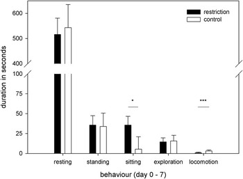
Independent of housing, the animals displayed locomotion, standing, sitting, exploration and resting on the belly and on the side. Resting was the predominant behaviour and accounted for approximately 85% of the animals’ total behaviour during the observation periods in test and control animals. On the restriction days, the sitting behaviour was significantly higher and the locomotion behaviour was significantly lower in the restricted animals (Figure 1).
Total durations (LSM ± s.e.) of behaviour recorded during seven observation days (3 h/day) of control (n = 6; white columns) and experimental animals (n = 12; black columns; *P ⩽ 0.05; ***P ⩽ 0.001).

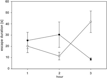
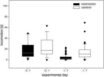
Escape behaviour also differed between the groups. Only the pigs confined in the metabolic cages showed some escape behaviour. However, this rarely happened (Figure 2). The keeping condition thus revealed a significant influence on the locomotion data. Movement-restricted pigs showed significantly less locomotion than the control animals (F1,11 = 21.59, P ⩽ 0.001) over the days 0 to 7 (Figure 3). The daily difference between the control and experimental animals was significant on the second restraint day (restriction = 0.57 ± 0.8 s; control = 5.15 ± 1.2 s: LSM ± s.e.; t = 3.24, P ⩽ 0.05).
Means (± s.e.) of the duration of escape attempts of the confined animals (n = 12) on day 0 (dots) and on day 1 (circles) in the 3 h of observation (the control animals displayed no escape behaviour).

Total durations of observed locomotion behaviour of experimental (n = 12) and control animals (n = 8) during the days before restriction (days −2 and 1) and the restriction days (days −1 to 7; median and first/third quartile; whiskers show 10th and 90th percentiles, dots indicate outliers).

Salivary cortisol
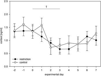
The cortisol concentrations of the two groups are presented in Figure 4. Neither a significant overall effect between the cortisol levels of the experimental and the control group nor a daily effect was found (F1,11 = 0.41, P = 0.6386).
Cortisol levels (LSM ± s.e.) in ng/ml of the experimental (n = 12) and the control animals (n = 8) on the experimental days (†P ⩽ 0.1, significance level between the groups).

HR and HRV
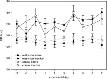
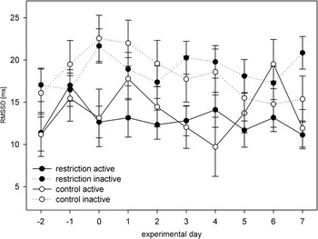
The HR throughout all measuring days is depicted in Figure 5. The data analysis revealed no significant influence of the housing condition (F1,11 = 0.05, P > 0.1). The activity status of an animal had a significant influence (F1,689 = 276.87, P ⩽ 0.001). The calculated parameter of the HRV RMSSD (Figure 6) was also not significantly influenced by the keeping condition. The same result was found in SDNN and RMSSD/SDNN (F1,11 = 0.00, P > 0.1). As in the HR data, the status of activity had a significant influence on RMSSD (F1,689 = 76.29, P ⩽ 0.001), SDNN (F1,689 = 32.27, P ⩽ 0.001) and RMSSD/SDNN (F1,670 = 61.94, P ⩽ 0.001).
Heart rate (LSM ± s.e.) in beats per minute during ten experimental days depending on the state of activity of the experimental (n = 12) and the control animals (n = 8).

RMSSD (LSM ± s.e.) in m.s. during ten experimental days depending on the state of activity of the experimental (n = 12) and the control animals (n = 8; RMSSD = root mean square of successive differences of R-R intervals).

Weight development
There was no difference in weight gain between the confined animals and the controls (LSM ± s.d. = 13.32 ± 2.87 kg and 13.12 ± 3.28 kg, respectively).
Discussion
In this study, we analysed the chronic movement restriction of pigs and its effects on heart beat parameters and cortisol. The results revealed no change of these measures compared to non-restricted single housing.
As we expected, the results show that housing design influences animal behaviour (Arellano et al., Reference Arellano, Pijoan, Jacobson and Algers1992). The pigs in metabolic cages showed a decrease in locomotion and an increase in sitting compared to the control pigs. The decrease in locomotion was obviously a result of less space in the metabolic cages. The increase in the pigs’ sitting behaviour in the cages apparently depended on the design of the cages, as the nipple drinkers were easier to use for the experimental pigs in the sitting position. In addition, the animals occasionally played with this equipment. Escape attempts occurred only in the experimental group, but they were rare, which might indicate a rapid adaptation to the condition. Overall, the predominant resting behaviour occurred throughout the observation periods of all pigs, which is in accordance with the normal behaviour of pigs (Signoret, Reference Signoret1969).
Surprisingly, no significant differences in the resting saliva–cortisol concentrations between both housing groups were detected. In both groups, saliva was collected beginning on the day after the start of movement restriction. On this first day after being transferred, the animals seemed to have completely adapted. Their basal cortisol levels nicely fitted the values of salivary cortisol (∼1 ng/ml), reported for pigs of similar ages as our animals (Ekkel et al., Reference Ekkel, Savenije, Schouten, Wiegant and Tielen1997; Ernst et al., Reference Ernst, Tuchscherer, Kanitz, Puppe and Manteuffel2006; Otten et al., Reference Otten, Kanitz, Couret, Veissier, Prunier and Merlot2010).
It is well known that the basal cortisol alone is not always a reliable marker of chronic stress (Friend et al., Reference Friend, Dellmeier and Gbur1985; Jensen et al., Reference Jensen, Pedersen, Nielsen, Heller, Ladewig and Jorgensen1996; Mormède et al., Reference Mormède, Andanson, Aupérin, Beerda, Guémené, Malmkvist, Manteca, Manteuffel, Prunet, van Reenen, Richard and Veissier2007). On the basis of the cortisol results, it can be concluded that movement restriction is not an additional stress factor for the experimental animals, compared to the control animals. As all animals were adapted to single housing, the additional stress potential of the metabolic cages therefore lay merely in the restriction of mobility. A similar low effect of immobilization was revealed in the study of Siard et al. (Reference Siard, Kovac, Ladewig and Stuhec2003). On the basis of their figures, two of the three repetitions of their experiment, where pigs were housed in metabolic cages and compared to controls in single-housing pens did not show an increase in cortisol neither in plasma nor in saliva (Siard and Stuhec, Reference Siard and Stuhec2003; Siard et al., Reference Siard, Kovac, Ladewig and Stuhec2003).
A slight non-significant drop of salivary cortisol on the second experimental day in both the groups could be the result of reduced social contact after two animals had been brought to the metabolic cages. The pigs then lost the ability to contact each other with the nose through the lattice between adjacent pens. Such a loss of stimulation has already been attributed to reduced cortisol concentrations (Beattie et al., Reference Beattie, O’Conell, Kilpatrick and Moss2000) and, accordingly, has also been revealed in crated sows compared to sows living in groups (Sorrells et al., Reference Sorrells, Eicher, Harris, Pajor and Richert2007).
Apart from the observation of the behaviour and the saliva cortisol, we examined the HR and HRV. Time segments for the HRV analysis were suggested to be 5 min (von Borell et al., Reference von Borell, Langbein, Despres, Hansen, Leterrier, Marchant-Forde, Minero, Mohr, Prunier, Valance and Veissier2007). This general rule bases largely on the argument that this is a suitable interval for most species, so that the values become comparable. However, the HR can differ considerably depending on the species and the ages of the animals, as well as on the experimental design, so that various authors have used adapted lengths of time segments for HRV analyses (Després et al., Reference Després, Veissier and Boissy2002; Schmidt et al., Reference Schmidt, Biau, Möstl, Becker-Birck, Morillon, Aurich, Faure and Aurich2010). We have chosen tachograms that were 1-min long, as piglets display comparatively high HRs and, thus, a high amount of R-R intervals even in this short period. Moreover, it was difficult in the first restriction days to find even enough 1-min periods when the animals were not displaying very vigorous behaviour, so that the electrodes lost contact and, consequently, the error rate of the recording was too high for evaluation. In order to be able to compare the data, the 1-min intervals were used for the whole study, even in later phases when the animals were calmer.
The results did not differ significantly between housing groups. The HRs of both housing groups showed approximately the same trend during the experimental days. The HR depended on the behaviour so that a significant difference resulted between the states of activity, which is a well-known fact (Mohr et al., Reference Mohr, Langbein and Nürnberg2002; Langbein et al., Reference Langbein, Nurnberg and Manteuffel2004; von Borell et al., Reference von Borell, Langbein, Despres, Hansen, Leterrier, Marchant-Forde, Minero, Mohr, Prunier, Valance and Veissier2007). Our RMSSD results showed a considerable decrease during active behaviours. This indicates a deactivation of the parasympathetic branch of the autonomic nervous system (Porges, Reference Porges1995b; Desire et al., Reference Desire, Veissier, Despres and Boissy2004) during activity, independent of the keeping condition. More importantly, the data of resting animals were not different. While resting, possible artefacts of sometimes hard to define active behavioural states are excluded so that resting is the state of choice to demonstrate sustained effects of HRV caused by changed mood.
Throughout the experimental days, we did not measure significant differences between the experimental hours. These results indicate an adaptation of the experimental animals to the housing conditions in the metabolic cages, which is also corroborated by equal weight gains of the confined and single-housed animals. Indices for chronic stress, like a general decrease of RMSSD in the resting state (Langbein et al., Reference Langbein, Nurnberg and Manteuffel2003) were not detectable in the HR and HRV data. In addition, the HR measured in our study coincide with the results of Zebunke et al. (Reference Zebunke, Puppe, Langbein and Manteuffel2008), in which the animals had been the same age and were housed in groups.
Although no classical physiological symptoms of stress due to movement restriction were found in our experiments, a careful conclusion of the results is crucial. Cortisol and HR parameters may not be completely sufficient to indicate chronic psychic stress, although the combination of both is surely a better indicator than each one alone. Altogether, the two parameters indicated virtually no additional response of the hypothalamic-pituitary-adrenal axis and the sympathetic system to movement restriction compared to keeping animals isolated in single pens. This indicates that an adaptation to confinement occurred quickly. In addition, daily quite intense caregiving, such as brushing the skin of the animals, might have been a positive factor. However, the absence of stress, defined as a disturbance of homeostasis (Selye, Reference Selye1973) does not exclude other, subtler welfare impairments (Veissier and Boissy, Reference Veissier and Boissy2007), as, for example, being hindered in locomotion activities or boredom, that is, the lack of stimulation and sensation.
Acknowledgements
We thank Armin Tuchscherer for supporting us with the statistics as well as Ellen Kanitz and Petra Müntzel for helping with the cortisol analysis. We are also grateful to Jan Langbein, Manuela Zebunke and Sandra Düpjan for their advice in the heart rate analysis.