Introduction
The Late Cretaceous rudists of the New World have received significant attention, particularly those of the Caribbean Province. The pioneer and classic works are those of Woodward (Reference Woodward1862), Whitfield (Reference Whitfield1897a, Reference Whitfieldb), Stephenson (Reference Stephenson1922), Trechmann (Reference Trechmann1924), Douvillé (Reference Douvillé1926, Reference Douvillé1927), Sánchez-Roig (Reference Sánchez-Roig1929), Müllerried (Reference Müllerried1931), Boissevain and Mac Gillavry (Reference Boissevain and MacGillavry1932), Palmer (Reference Palmer1933), and Mac Gillavry (Reference Mac Gillavry1937). Subsequent and exhaustive taxonomic data were provided by Chubb (Reference Chubb1955, Reference Chubb1956a, Reference Chubbb, 1967, 1971), Jung (Reference Jung1970), Alencáster (Reference Alencáster1971), and van Dommelen (Reference Dommelen1971). More recent taxonomic papers are those of Mitchell and Gunter (Reference Mitchell and Gunter2002, Reference Mitchell and Gunter2004, Reference Mitchell and Gunter2006), Mitchell (Reference Mitchell2003, Reference Mitchell2010a, Reference Mitchellb, 2013), Grubić (Reference Grubić2004), Oviedo (Reference Oviedo2005), Oviedo et al. (Reference Oviedo, Pons, Pichardo-Barrón, Aguilar-Pérez and Alencáster2007), Mitchell et al. (Reference Mitchell, Gunter and Ramsook2007), Mitchell and Ramsook (Reference Mitchell and Ramsook2009), Mitchell and Pons (Reference Mitchell and Pons2010), and Pons et al. (Reference Pons, Vicens, Pichardo, Aguilar, Oviedo, Alencáster and García-Barrera2010, Reference Pons, Vicens, Oviedo, Aguilar, García-Barrera and Alencáster2013).
Rudist limestones quarried at Santa Elena Peninsula, Costa Rica, were used as ornamental building stone for several official premises in San José and other towns in Costa Rica during first half of the last century, the white rudist shells contrasting with the intense reddish rock when polished. Nevertheless, at that time, these fossils did not attract scientific attention. Geological research and mapping of northwestern Costa Rica, particularly of the Nicoya Peninsula, by the last author together with his doctoral thesis students (Hans-Jürgen Gursky, Monika Gursky-Strebin, Heiko Wildberg, and Carlos Rodríguez) and students from the Escuela Centroamericana de Geología de la Universidad de Costa Rica during the 1970s and 1980s (Schmidt-Effing, Reference Schmidt-Effing1975, Reference Schmidt-Effing1979, Reference Schmidt-Effing1980; Galli and Schmidt-Effing, Reference Galli and Schmidt-Effing1977; Stibane et al., Reference Stibane, Schmidt-Effing and Madrigal1977; Gursky et al., Reference Gursky, Gursky, Schmidt-Effing and Wildberg1984) revealed the presence of shallow-water carbonates with abundant rudists in several localities, corresponding to the lower part of El Viejo Formation. This unit, exposed on the Pacific side of Costa Rica, overlies discordantly the Nicoya Complex, which is built of thick sequences of mainly oceanic basalts in the Nicoya Peninsula, or of serpentinized peridotites in the Santa Elena Peninsula, and that was formed between 88 and 90 Ma (Alvarado et al., Reference Alvarado, Kussmaul, Chiesa, Gillot, Appel, Wörner and Rundle1992). Strata with rudists are also known from Colorado de Abangares, overlying a reworked basaltic complex, and from the ‘Caliza de Colorado’ olistholiths of rudist limestone intercalated in the Las Palmas Formation (pelagic sediments and volcaniclastics) of early Paleocene to early Eocene age in the San Buenaventura area nearby (Schmidt-Effing, Reference Schmidt-Effing1975). The samples collected at that time (1973–1981), housed at the Philipps Universität in Marburg (Germany), constitute the nucleus of our study. Subsequent field work seasons of the first and last authors in 1998, 2002, and 2003 completed the data and sampling.
The aim of this paper is to describe the Upper Cretaceous rudist fauna from Costa Rica. Because of differences in preservation and abundance of specimens, some taxa are investigated in more depth than others.
Localities and stratigraphy
The investigated rudist outcrops are exposed on the western side (Pacific coast) of Costa Rica, in Guanacaste Province (Fig. 1) and correspond to the El Viejo Formation (Schmidt-Effing, Reference Schmidt-Effing1975; Stibane et al., Reference Stibane, Schmidt-Effing and Madrigal1977).
Location of studied outcrops (marked by rudist symbol). Upper left: overview of Costa Rica, with locations of maps 1.1, 1.2, and 1.3 indicated. (1) Ancient quarry, Cuajiniquil, Arroyo Nisperal, and Playa Panamá. (2) Bolsón. (3) Colorado de Abangares.

Playa Panamá (Sheet 1:50 000, Carrillo Norte)
The fossil locality is situated at the northern end of Panama Bay, on the southern edge of the Culebra Bay coast, Papagayo Gulf, north of the Nicoya Peninsula. The outcrop, originally mapped by Gursky et al. (Reference Gursky, Gursky, Schmidt-Effing and Wildberg1984) on top of a high cliff above basaltic layers and corresponding to the basal part of El Viejo Formation, is nowadays reduced to a series of blocks resting on the beach. Although information provided by the blocks is evidently patchy, a reconstruction of both the processes generating the sediments and the patterns of rudist colonization on them, were attempted.
Sedimentary rocks are mainly basalt-derived conglomerates, sandstones, and gravelly sandstones with abundant bioclasts, interpreted as being deposited as a series of flood-related sediment gravity flows, in the area of a fan delta below the action of waves and tides.
All the blocks document the pattern of colonization of a subtidal rocky-pebble bottom by successive rudist larvae invasions. Alencasterites n. gen. mooretownensis (Trechmann, Reference Trechmann1924) occurs in monospecific close-cluster/frame organic reefs (in the terminology of Riding, Reference Riding2002), developed on a coquina substratum, starting with sprawling growth and forming thickets up to 50 cm thick of upright growing specimens. Free space is filled with basalt-derived sandstone. These thickets are capped by coarse sedimentation or by colonization of other species. Angular, decametric-sized blocks of these thickets are also common in between conglomerates. Plagioptychus trechmanni Chubb, Reference Chubb1956a occurs in one-specimen-high close-cluster organic reef (in the terminology of Riding, Reference Riding2002), developed above sandy matrix conglomerates and capped by similar sediments. It also occurs, in sandstones, in a more open fabric of several nonaligned upright individuals with both valves articulated, that are often developed on top of an A. mooretownensis thicket. Mitrocaprina costaricaensis n. sp. occurs, as for P. trechmanni, in sandstones, and most specimens are also articulated. Potosites? aff. P. tristantorresi Alencaster and Pons in Pons et al., Reference Pons, Vicens, Pichardo, Aguilar, Oviedo, Alencáster and García-Barrera2010 appears in one-specimen-high horizons of aligned individuals developed on top of other species thickets. Specimens of both cf. Potosites sp. aff. P. tristantorresi and Chiapasella sp. have been identified in rudstones, the last sometimes forming bouquets of three to four individuals. Barrettia monilifera Woodward, Reference Woodward1862 and Antillocaprinidae such as Antillocaprina sp. aff A. suboccidentalis Chubb, Reference Chubb1967 and Antillocaprinidae indet. mainly appear as major elements in rudstones. Corals, red algae, and nerineid gastropods also occur.
Santa Rosa National Park (Sheets 1:50 000, Murciélago and Ahogados)
Three localities of the El Viejo Formation have been sampled at Santa Rosa National Park, on Santa Elena Peninsula. The first corresponds to outcrops of breccia limestone above the peridotites, south of Cuajiniquil, yielding big fragments of rudist shells and corals; among them, cf. Whitfieldiella gigas (Chubb, Reference Chubb1955) could be identified. The second, Arroyo Nisperal, a streamlet draining to Papagayo Gulf, where peridotite-derived conglomerates with abundant sandy matrix contain nerineid gastropods and rudist shell fragments: Barrettia monilifera, Guanacastea costaricaensis n. gen. n. sp., cf. Potosites aff. P. tristantorresi, and Antillocaprinidae indet. The third is the Ancient Quarry. The limestone bed, massive and ~3 m thick, follows a 12–15 m section of coarse sandstone with peridotite rounded lithoclasts and nerineid gastropod shells, deposited above the peridotite. At the base, there are rudstones with nerineids and diverse rudist shell fragments (Barrettia monilifera and cf. Potosites aff. P. tristantorresi), and the rest is composed of densely spaced, vertically growing radiolitids with a foraminifer packstone matrix. Identified radiolitids are Radiolites sp. aff. R. multicostata (Adkins, Reference Adkins1930), Alencasterites mooretownensis, Guanacastea costaricaensis n. gen. n. sp., and aff. Thyrastylon sp.
Colorado de Abangares (Sheet 1:50 000, Abangares)
North of Colorado de Abangares, at the base of Cerro Cerco de Piedra, rudists appear in two conglomeratic layers intercalated in sandstones immediately above the reworked basaltic conglomerates of the Nicoya Complex. Most recovered specimens are shell fragments of large hippuritids, i.e., Barrettia monilifera and Parastroma trechmanni Chubb, Reference Chubb1967. Additionally, isolated valves of plagioptychids, i.e., Mitrocaprina multicanaliculata Chubb, Reference Chubb1956a, and smaller radiolitids, i.e., Guanacastea costaricaensis n. gen. n. sp. and Bournonia? tetrahedron Chubb, Reference Chubb1967, were identified. Corals, gastropods, scaphopods, and echinoids also occur. A unit named ‘calizas arrecifales’ (reefal limestones) has been mapped nearby (Calvo, Reference Calvo1998), but no rudists were found in it; only a few coral fragments included in breccia limestone were recognized.
Bolsón (Sheet 1:50 000, Tempisque)
In Cerro Cebollín, close to Hacienda El Viejo, north of Bolsón, the El Viejo Formation starts with 3 m of rudist rudstone limestone above the basaltic basement. Rudists appear disarticulate, biocorroded, filled with macroforaminifera, and randomly oriented, together with massive coral fragments and nerineid gastropods. Antillocaprina sp., Barrettia monilifera, Parastroma trechmanni, Plagioptychus cf. zansi Chubb, Reference Chubb1956a, Mitrocaprina costaricaensis n. sp., Plagioptychidae indet., Chiapasella sp., and cf. Potosites aff. P. tristantorresi have been identified. The outcrop was mapped, interpreted as mass flow deposits considered thicker than we observed, and followed up-section by calcareous and volcaniclastic turbidites by Jaccard et al. (Reference Jaccard, Münster, Baumgartner, Baumgartner-Mora and Denyer2001).
Isolated stone slabs of rudist limestone are common in the whole area far from the limestone outcrops. They were used to cover graves by pre-Hispanic inhabitants of the region. Transverse sections of Chiapasella sp. have been identified in one of them.
Methods
Specimens used for this study are maintained in the Paleontological Collections of the Universitat Autònoma de Barcelona (PUAB) and the former Institut für Geologie und Paläontologie, Philipps-Universität, Marburg. In the future, it is planned to keep the main part of the collection in a single institution and depositing a small but representative collection at the Escuela Centroamericana de Geología in San José, Costa Rica.
Specimens were sectioned and polished in the two laboratories of the authors’ institutions; thin sections and acetate peels were prepared at the UAB. All figured specimens are registered and maintained in the PUAB.
Cited repositories include: NHMUK=The Natural History Museum [formerly British Museum (Natural History), London, UK; NMNH=National Museum of Natural History, Smithsonian Institution, Washington, DC, USA; TMM=Bureau of Economical Geology Collection, Texas Memorial Museum, Austin, Texas, USA.
Anatomical conventions are: AM=anterior myophore, am=cavity of anterior myophore, AT=anterior tooth, at=socket of the anterior tooth, C=canals, CT=central tooth, ct=socket of the central tooth, DC=dorsal cavity, iOSL=inner part of the outer shell layer, IMG=inner marginal groove, ISL=inner shell layer, L=ligamental ridge, LV=left valve, MC=main body cavity, OSL=outer shell layer, oOSL=outer part of outer shell layer, P1=first pillar, P2=second pillar, PB=posterior radial band, PC=posterior cavity, PM=posterior myophore, pm=cavity of the posterior myophore, PT=posterior tooth, pt=socket of the posterior tooth, RV=right valve, T=tube, VB=ventral radial band. All transverse sections are figured in standard orientation, i.e., in adapical view and with the dorsal side up, unless indicated. The source of figured sections is specified: AP=acetate peel; PS=polished section; and TS=thin section. The morphoconstructional approach and terminology proposed by Pons and Vicens (Reference Pons and Vicens2008) are used in the descriptions of the outer shell layer structure in radiolitids. Synonymy lists are not exhaustive; only those references relevant to the discussion presented herein are included; the reader can consult Steuber (Reference Steuber2002) for more complete lists.
Systematic paleontology
Family Antillocaprinidae Mac Gillavry, Reference Mac Gillavry1937
Remarks
Mitchell (Reference Mitchell2013, p. 424, 423) split the rudist family Antillocaprinidae into three subfamilies and a large number of new genera using, as the most critical feature, the myocardinal arrangement. He pointed out that this “can only be determinate with well-preserved isolated valves or in transverse sections cut close to the commissure” and also that “their shells were composed of a thick aragonitic inner shell layer that is more than often recrystallized, destroying internal structures.” Unfortunately, the state of preservation of the herein discussed material does not allow adopting Mitchell’s refinement of the taxonomy of the Antillocaprinidae.
Genus Antillocaprina Trechmann, Reference Trechmann1924
Type species
Antillocaprina occidentalis Trechmann, Reference Trechmann1924, p. 407, pl. 25, figs. 1–3 [=Antillocaprina suboccidentalis Chubb, Reference Chubb1967, non Caprinella occidentalis Whitfield, Reference Whitfield1897a] after Chubb (Reference Chubb1971), from the Guinea Corn Formation (upper Maastrichtian), Logie Green, northern Clarendon, Jamaica.
Remarks
Mitchell (Reference Mitchell2013) provided a full discussion of the Antillocaprina type species problem, proposed Caprinella occidentalis as the type species of a new genus (Rotacaprina Mitchell, Reference Mitchell2013), and identified the specimen figured by Trechmann (Reference Trechmann1924, pl. 25, fig. 1) as belonging to Parasarcolites Mitchell and Gunter, Reference Mitchell and Gunter2006.
Antillocaprina sp. aff. A. suboccidentalis Chubb, Reference Chubb1967
(1–3) Antillocaprina sp. aff. A. suboccidentalis Trechmann, Reference Trechmann1924 from Playa Panamá; (1, 2) PUAB-81446, external and internal view, respectively, of LV; (3), PUAB-45202, transverse section of RV. (4) ?Antillocaprina sp. from Bolsón; PUAB-81489, transverse section of RV. (5, 6) Antillocaprinidae gen. and sp. indet. from Playa Panamá; (5) PUAB-81467, transverse section of RV; (6) PUAB-81465, transverse section of unidentified valve. (7–9) Antillocaprinidae gen. and sp. indet. from Arroyo Calera, Santa Rosa National Park; (7) PUAB-74324, transverse section of RV; (8–9) PUAB-74322 and PUAB-74321, respectively, transverse sections of unidentified valves. AM=anterior myophore, AT=anterior tooth, at=socket of the anterior tooth, CT=central tooth, ct=socket of the central tooth, L=ligamental ridge, MC=main body cavity, PM=posterior myophore, PT=posterior tooth, pt=socket of the posterior tooth, T=tube. All sections (PS); scale bar=10 mm.
Type specimens
The syntypes of A. suboccidentalis are: LV, BMNH L63211, from the Maastrichtian, lower part of Logie Green section (Trechmann, Reference Trechmann1924, pl. 25, fig. 3; Chubb, Reference Chubb1971, pl. 28, fig. 3); RV, BMNH L63210, from the Maastrichtian, Great River Valley below Catadupa, St. James (Trechmann, Reference Trechmann1924, pl. 25, fig. 2; Chubb, Reference Chubb1971, pl. 28, fig. 4).
Occurrence
Playa Panamá, Costa Rica. The different species of the genus are widespread in the Caribbean Province.
Description
The incomplete RV is conical in shape, truncated at the apical end, and weathered at the commissure. Its length is 120 mm with a maximum dorsoventral diameter of 80 mm and slightly curved anteriorly. The outer surface is weathered. The transverse section shows the main body cavity is very small, 30 mm by 20 mm. The inner shell layer is thus very thick and composed, also in the rectangular central tooth, of fine but thick walled canals, polygonal toward center and oval toward the outer margin. The crescent-shaped ligamental pillar is located between the central tooth and the posterior socket. Both anterior and posterior sockets are separated from the body cavity by two septa extending from the inner corners of the central tooth.
The LV is convex, with a marginal umbo curved toward the dorsal margin. Its outer surface bears ~ 16 prominent round radial ribs separated by broad grooves. The commissural face, although weathered, shows the rectangular central socked flanked by the anterior and posterior teeth and these by the anterior and posterior myophores, all them forming a horseshoe-shaped structure surrounding the small body cavity except at its ventral side.
Materials
One RV, PUAB-45202, and three LVs, PUAB-81443, PUAB-81445, and PUAB-81446, all more or less weathered, from Playa Panamá.
Remarks
These specimens from Costa Rica resemble Antillocaprina suboccidentalis from Jamaica in the myocardinal structure arrangement, but are much more robust. The LV, although also curved, is much lower and strongly ribbed.
?Antillocaprina sp.
Occurrence
Playa Panamá and Bolsón, Costa Rica. The different species of the genus are widespread in the Caribbean Province.
Description
Weathered shell fragments of one RV, PUAB-81489, allowed recognition, in its transverse section, of the myocardinal structures. The crescent-shaped ligamental pillar is located on the concave side of the long, curved posterior socket. The convex side of the socket is very close to the body cavity margin. The anterior socket, separated from the posterior one by the central tooth, is round in section and is more distant from the body cavity margin. The central tooth is not penetrated by the canals occupying the rest of the inner shell layer. The shell fragment is 80 mm long and has a maximum diameter of 80 mm; the body cavity measures 40 mm dorsoventrally and 30 mm anteroposteriorly. The LV fragments indicate a conical, strongly curved form.
Materials
Two fragments of RVs, PUAB-81489 and PUAB-81490, from Bolsón. Three fragments of LVs, PUAB-45199, PUAB-45204, and PUAB-74309, from Playa Panamá, probably corresponding to the same species.
Remarks
The myocardinal structure arrangement resembles the one reproduced by Chubb (Reference Chubb1971, text-fig. 3) for Antillocaprina lowenstami Chubb, Reference Chubb1967 from Jamaica, although Costa Rican specimens are much larger. There are no more arguments to tentatively attribute our specimens to this genus. The figure by Chubb (Reference Chubb1971, text-fig. 3) is a line drawing. Mitchell (Reference Mitchell2013, fig. 7) presented a photograph of the same specimen and proposed A. lowenstami as the type species of his new genus Clinocaprina.
Antillocaprinidae gen. and sp. indet.
Occurrence
Playa Panamá and Santa Rosa National Park, Costa Rica.
Description
Specimen PUAB-81467 is a conical, strongly ribbed fragment of a RV. It measures 65 mm in length and 55 mm in maximum diameter; the body cavity is 33 mm anteroposteriorly and 18 mm dorsoventrally. The transverse section shows that the inner shell layer is traversed by polygonal canals, also invading the central tooth in which such canals have a smaller diameter. A marginal row of different canals was not observed¸ because it was not developed or disappeared due to weathering. The two sockets for the LV teeth are close to the margin of the body cavity.
Specimen PUAB-81465 is a much-weathered shell fragment in which a transverse section cuts the body cavity and several tubes. The tube arrangement and size resembles those described for the genus Parasarcolites by Mitchell and Gunter (Reference Mitchell and Gunter2006). Transverse sections of the three specimens from Arroyo Nisperal show not much more than the body cavity and the canaliculated inner shell layer with strong costae at the outer surface. A marginal row of smaller canals was observed in one of them.
Materials
Five shell fragments, PUAB-81465 and PUAB-81467, from Playa Panamá, and PUAB-74321, PUAB-74322, and PUAB-74324 from Arroyo Nisperal in Santa Rosa National Park.
Remarks
The poor preservation of the specimens makes it impossible to go further in taxonomy.
Family Hippuritidae Gray, Reference Gray1848
Genus Barrettia Woodward, Reference Woodward1862
Type species
Barrettia monilifera Woodward, Reference Woodward1862 from the lower limestone member of the Back Rio Grande Limestones, Blue Mountain Inlier, Jamaica.
Barrettia monilifera Woodward, Reference Woodward1862
(1–6) Barrettia monilifera Woodward, Reference Woodward1862; (1) PUAB-81475; (2) PUAB-81485; (3) PUAB-45179; (4) PUAB-81481; (5) PUAB-45181; (6) PUAB-81484. (7, 8) Parastroma trechmanni Chubb, Reference Chubb1967 from Bolsón; (7) PUAB-81482; (8) detail (AP) of shell structure at marginal part indicated in 3.7. am=cavity of anterior myophore, AT=anterior tooth, at=socket of the anterior tooth, CT=central tooth, MC=main body cavity, P1=first pillar, P2=second pillar, PM=posterior myophore, pm=cavity of the posterior myophore, PT=posterior tooth. Parentheses indicate that location of structure is deduced because they are filled with inner shell. Transverse sections (PS) of RVs, scale bars=10 mm.
1862 Barrettia monilifera Reference WoodwardWoodward, p. 372, pl. 20, figs. 1–4, pl. 21, fig. 5 (all figures are drawings; figs. 2, 5=type specimen).
1971 Barrettia monilifera; Reference ChubbChubb, p. 208, text-fig. 7, pl. 51, figs. 1–3, pl. 52, figs. 2, 3 (text-fig. 7 and pl. 52, fig. 2=drawing and photograph, respectively, of Woodward’s pl. 20, fig. 2 specimen counterpart).
2010b Barrettia monilifera; Reference MitchellMitchell, p. 10, text-figs. 3D, 4, 6A–D, 8A, 9 (4=photograph of Woodward’s specimen in pl. 20, fig. 2, pl. 21, fig. 5).
Holotype
NHMUK PI OR 42861, cut into several parts, from the lower limestone member of the Back Rio Grande Limestones, Blue Mountain Inlier, Jamaica [Woodward, Reference Woodward1862, pl. 20, fig. 4 (longitudinal section), pl. 21, fig. 5 (transverse section)]. Douvillé (Reference Douvillé1894, pl. 17, fig. 6) provided a photograph of a different transverse section of Woodward’s type specimen. Mitchell (Reference Mitchell2010b) provided a full account on the complex history of the several fragments of the type specimen and of their illustration by different authors.
Occurrence
Playa Panamá, Santa Rosa National Park, and Bolsón, Costa Rica. The species is abundant in Jamaica. Mitchell (Reference Mitchell2010b) considered that the material identified as Barrettia monilifera from Cuba, Puerto Rico, St. Croix, and elsewhere from Jamaica belongs to several other species of Barrettia.
Description
Larger specimens measure up to 140 mm in diameter. All specimens are more or less weathered and lack the external outer shell layer, and some of them show evidence of bioerosion. Bouquets of few individuals attached to each other are not rare. Transverse sections show the presence of ~60 well-developed moniliform infolds (rays). The beads have diameters ranging from 1.1 to 1.8 mm in width, and are separated by distances of 3.0–4.9 mm. The ligamental ridge is no longer than the longest infolds. The P1 knob is circular whereas that of P2 is elongated with parallel sides. The myocardinal structures are cut in some of the sections, showing the posterior myophore aligned (slightly below) with the anterior and posterior teeth, situated between this last and the first pillar. Data gathered from the few specimens allowing measurement show angles a (P1-PM-PT), b (P2-P1-PM), and c (PM-PT-At) values of ~123º, 126º, and 161º, respectively.
Materials
Twelve RVs PUAB-81479–PUAB-81481, PUAB-81483–PUAB-81487, PUAB-45179–PUAB-45182 from Bolsón; two RVs, PUAB-74314 and PUAB-74319, from the ancient quarry and Arroyo Nisperal, respectively, at Santa Rosa National Park; and one RV, PUAB-81429, from Playa Panamá.
Remarks
Measurements of size and spacing of the beads in the rays, as well as the few data available on angles a, b, and c, are within the range indicated by Mitchell (Reference Mitchell2010b) for the species.
Genus Parastroma Douvillé, Reference Douvillé1926
Type species
Parastroma sanchezi Douvillé, Reference Douvillé1926 from the beds with Barrettia in Sánchez Agramonte Domain, Arroyo Hondo, Camaguey Province, Cuba.
Parastroma trechmanni Chubb, Reference Chubb1967
1922 Barrettia cf. monilifera; Reference TrechmannTrechmann, p. 510, pl. 19, fig. 2a, b. [non pl. 20, figs. 1, 2=Whitfieldiella gigas (Chubb, Reference Chubb1955)].
1967 Parastroma trechmanni Reference ChubbChubb, p. 30.
1971 Parastroma trechmanni; Reference ChubbChubb, p. 214, pl. 56, figs. 1–3.
Holotype
RV (NHMUK PI L 44200), from the middle Campanian ‘Barrettia Limestone,’ Haughton Hall, Green Island Inlier, Hannover, Jamaica (Trechmann, Reference Trechmann1922, pl. 19, fig. 2a, b; Chubb, Reference Chubb1971, pl. 56, fig. 1).
Occurrence
Bolsón and Colorado de Abangares, Costa Rica. Haughton Hall Limestone, Green Island Inlier, and Glenbrook Limestone, Grange Inlier, Jamaica.
Description
The better preserved incomplete RV (170 mm in height, 140 mm in diameter) is weathered, bioeroded, and slightly compacted. The specimen was cut into several transverse sections, the upper one showing the main cavity and some elements of the myocardinal arrangement: the socket of the anterior tooth is very wide and opens to the main cavity; the posterior tooth is large, subrectangular, within the corresponding socket, which also opens to the main cavity, and is separated from the anterior socket by a large central tooth; a concentric filling at the right of the posterior tooth indicates the position of the posterior myophore cavity; and a curved filling close to the anterior part of the main cavity indicates the position of the anterior myophore. The structure of the shell shows very thin rays with a single row of transverse curved partitions between them, only visible on its most external part, where low ridges are separated by broad grooves; concave tabulae, which form concentric partitions in transverse section, are seen inwardly. The knobs of the two pillars are not discernible, probably because of collapsing.
Materials
Three incomplete RVs, PUAB-81479, PUAB-81482, and PUAB-81488, from Bolsón, and three RV shell fragments, PUAB-45177, PUAB-45178, and PUAB-81508) from Colorado de Abangares.
Remarks
The arrangement of the myocardinal elements, the concentric tabulae, and particularly the low ridges separated by broad grooves in cross section, point to attributing the Costa Rica specimens to Parastroma trechmanni, and exclude both P. sanchezi Douvillé, Reference Douvillé1926 and P. guitarti Palmer, Reference Palmer1933.
Genus Whitfieldiella Mitchell, Reference Mitchell2010b
Type species
Barrettia gigas Chubb, Reference Chubb1955 (= Barrettia cf. monilifera Whitfield, Reference Whitfield1897b) from the middle Campanian, Haughton Hall Formation, Green Island Inlier, Hannover, Jamaica.
cf. Whitfieldiella gigas (Chubb, Reference Chubb1955)
cf. Whitfieldiella gigas (Chubb, Reference Chubb1955) from south of Cuajiniquil, Santa Rosa National Park, PUAB-74318, commissural face (above) and transverse cross section (PS, below) of RV fragment. Scale bar=10 mm.
Holotype
Designated by Chubb (Reference Chubb1955), the holotype of Whitfieldiella gigas is AMNH 9665/1, from the middle Campanian, Haughton Hall Formation, Green Island Inlier, Hannover, Jamaica (Whitfield, Reference Whitfield1897b, pl. 27, fig. 1, pl. 28, fig. 1).
Occurrence
Santa Rosa National Park, Costa Rica. The species is known from Haughton Hall and Stapleton Limestones of the Green Island and Sunderland Inliers, respectively, in Jamaica (Mitchell, Reference Mitchell2010b) and Chiapas, México (Alencáster, Reference Alencáster1971).
Description
The fragment preserves the commissural face and indicates a conical shell form. At the commissural surface, radial crests corresponding to the location of the rays are limited by depressions corresponding to the inter-rays. The fragment was transversely cut 20 mm below the commissural surface. The minimum diameter of the original specimen, estimated by circumscribing the fragment in a circle, is greater than 260 mm. Eighteen moniliform infolds were observed in the fragment, the longest one 80 mm in length; a total number of ~72 infolds in the original specimen is deduced. Beads are elliptical (length between 2.85 and 4.4 mm, width between 1.92 and 2.85 mm) and the distance between adjacent beads is longer than their length (between 6.21 and 10.91 mm).
Materials
RV fragment, PUAB-74318, from South of Cuajiniquil in Santa Rosa National Park.
Remarks
Although additional diagnostic characters have not been observed, the inferred shell size and form, and the pattern of the infolds, in particular, the measurements of the beads and their spacing, allow a tentative assignment of this specimen to Whitfieldiella gigas.
Family Plagioptychidae, Douvillé, Reference Douvillé1888
Genus Plagioptychus Matheron, Reference Matheron1842
Type species
Plagioptychus paradoxus Matheron, Reference Matheron1842 (=Caprina aguilloni d’Orbigny, Reference Orbigny1840) after Douvillé (Reference Douvillé1888), from the Santonian? of Le Beausset, Bouches du Rhône, France.
Plagioptychus trechmanni Chubb, Reference Chubb1956a
Plagioptychus trechmanni Chubb, Reference Chubb1956a from Playa Panamá. (1) PUAB-45208a, stereo pair, inner view of LV. (2, 3) PUAB-45808d, stereo pair, lateral view of a bouquet of specimens with both valves. (4) PUAB-45808c, stereo pair, group of specimens. (5) PUAB-45203, natural transverse section of LV. (6) PUAB-45207, natural tangential section of two specimens, each with articulated valves. (7–9) PUAB-45205; (7) longitudinal section (PS) through a group of specimens embedded in volcanoclastic matrix, and (8, 9) details of two specimens as indicated in 5.7. AM=anterior myophore, AT=anterior tooth, ct=socket of the central tooth, MC=main body cavity, PC=posterior cavity, PM=posterior myophore, PT=posterior tooth. Scale bars=10 mm.
1924 Plagioptychus jamaicensis; Reference TrechmannTrechmann, p. 407, pl. 25, fig. 4. (non Caprina jamaicensis Whitfield, Reference Whitfield1897a, p. 192, pl. 13, figs. 1, 2, pl. 15, figs. 1, 2).
1956a Plagioptychus trechmanni Reference ChubbChubb, p. 8, text-figs. 1, 2.
1971 Plagioptychus trechmanni; Reference ChubbChubb, p. 181, pl. 32, figs. 6, 7 (new photographs of Trechmann’s type).
Holotype
RV, NHMUK PI L 63220, from the Maastrichtian of Logie Green, northern Clarendon, Jamaica [Trechmann, Reference Trechmann1924, pl. 25, fig. 4 (drawing); Chubb, Reference Chubb1971, pl. 32, figs. 6, 7 (photographs)].
Occurrence
Playa Panamá and Bolsón, Costa Rica. Logie Green, northern Clarendon, and Cambridge-Catadupa railway line, Jamaica (Chubb, Reference Chubb1971).
Description
The RV is straight and cylindrical, higher at the ventral side and shorter at the dorsal one. Longitudinal sections of specimens embedded in sediment reveal the presence of coarse growth lines. The LV is highly arched, particularly approaching the umbo that is dorsally centered and overhangs the commissural plane. Anteroposterior diameter is somewhat larger than dorsoventral one. Pallial canals are present around the anterior, ventral, and posterior valve margins. Canal walls are very close each other and bifurcate up to two times. The anterior myophore is a long inclined platform and reaches the anterior margin. The anterior tooth is radially elongate and its base is prolonged ventralward in a medial septum. The socket of the central tooth is wide and not separated from the posterior cavity. The posterior tooth has a rounded triangular section and is the most prominent element of the myocardinal structures. The ligamental furrow reaches the base of the posterior tooth at its dorsal margin. The posterior myophore is a long ridge parallel to the posterior shell margin.
Materials
A few block samples with several specimens each in sandy matrix. Some specimens could be isolated and are labeled with letters following the sample’s register number. PUAB-81459, PUAB-45203, PUAB-45205–PUAB-45208, and PUAB-74310 from Playa Panamá, and PUAB-45188 and PUAB-45189 from Bolsón.
Remarks
The pattern of the pallial canals was not described by Chubb (Reference Chubb1971) nor indicated on the new photographs of Trechmann’s types, but the arrangement of the myocardinal elements and the external features of the Costa Rica specimens look very similar.
Plagioptychus sp. cf. P. zansi Chubb, Reference Chubb1956a
(1, 2) Plagioptychus sp. cf. P. zansi Chubb, Reference Chubb1956a from Bolsón, PUAB-45191; (2) detail as indicated in 6.1. (3–6) Mitrocaprina multicanaliculata Chubb, Reference Chubb1956a from Colorado de Abangares; (3, 4) PUAB-81499; (4) detail as indicated in 6.3; (5, 6) PUAB-81504; (6) detail as indicated in 6.5. (7, 8) Plagioptychidae gen. and sp. indet. from Bolsón; PUAB-81491; (8) detail as indicated in 6.7. ct=socket of the central tooth, MC=main body cavity, PC=posterior cavity. Transverse sections (AP) (6.1–6.6) or (PS) (6.7, 6.8) of LVs; scale bars=10 mm).
Holotype
NMNH PAL348055, from Logie Green, northern Clarendon, Jamaica [Whitfield, Reference Whitfield1897a, pl. 13, figs. 1, 2, as Caprina jamaicensis (=Plagioptychus zansi)].
Occurrence
Bolsón, Costa Rica. The species is known from the Maastrichtian of Logie Green, northern Clarendon, and also from the Maldon inlier, Marchmont, Westmoreland and St. James, Jamaica (Chubb, Reference Chubb1971).
Description
Only the portion near the commissure is preserved; the umbo was not observed. Diameter is 45 mm anteroposteriorly and 35 mm dorsoventrally. Pallial canals were observed around the anterior, ventral, and posterior valve margins. Canal walls are very close to each other and bifurcate once near the inner margin and often twice near the outer margin. The anterior myophore is very wide. The socket of the central tooth is separated from the posterior cavity.
Materials
One incomplete LV, PUAB-45191, from Bolsón.
Remarks
The pattern of the pallial canals, and of what can be observed concerning the teeth and myophores in the transverse sections, coincide with Chubb’s (Reference Chubb1971) description and, particularly, with his pl. 33, fig. 3. Thus we consider our specimen probably related to Chubb’s species.
Genus Mitrocaprina Boehm, Reference Boehm1895
Type species
Coralliochama bayani Douvillé, Reference Douvillé1888 from the Hippurites limestone of Rennes-les-Bains, Aude, France.
Mitrocaprina costaricaensis new species
Mitrocaprina costaricaensis n. sp. from Playa Panamá, paratypes, stereo pairs, external views of articulated RVs and LVs. (1–3) PUAB-81448, posterior, anterior, and dorsal views, respectively. (4, 5) PUAB-81447, anterior and lower views, respectively. (6, 7) PUAB-45197, dorsal and ventral views, respectively. Scale bars=10 mm.
Mitrocaprina costaricaensis n. sp. from Playa Panamá, transverse cross sections, all (AP), except TS for 8.7–8.9. (1–3) PUAB-81456; (1) holotype, LV; (2) detail of ventral side as indicated in 8.1; (3) RV. (4–6) PUAB-81451; (4) paratype, LV; (5) detail of ventral side as indicated in 8.4; (6) RV. (7–9) PUAB-74308; (7) LV; (8) detail of posterior side as indicated in 8.7; (9) detail of ventral side as indicated in 8.7. AM=anterior myophore, AT=anterior tooth, CT=central tooth, MC=main body cavity, PC=posterior cavity, PM=posterior myophore, pm=cavity of the posterior myophore, PT=posterior tooth. Scale bars=10 mm.
?1971 Plagioptychus toucasianus; Reference ChubbChubb, p. 180, pl. 34, figs. 2–4 (non Plagioptychus toucasianus Matheron, Reference Matheron1842).
2001 Antillocaprina sp. B; Reference ValerioValerio, p. 152, text-fig. 2. CFM-905, a, b.
2001 Caprínido; Reference ValerioValerio, p. 152, text-fig. 2. CFM-919, c, d.
Type specimens
Holotype: PUAB-81456. Paratypes: PUAB-45197, PUAB-81447, PUAB-81448, and PUAB-81451. All are specimens with both valves articulated from Playa Panamá. The holotype and one of the paratypes have been transversely cut at both valves. The other three paratypes are preserved complete and show the ontogenetic variability of the species.
Diagnosis
The RV is gyropleuriform, flat in juveniles to nearly cylindrical in adults. A long narrow cavity is developed in correspondence with the posterior myophore of the LV. LV highly arched with the umbo largely overhanging the commissural plane. Pallial canals are round or oval in section, with an outer row of short pyriform canals with very close walls bifurcating twice.
Occurrence
Playa Panamá and Bolsón, Costa Rica, and (?) the Barrettia Limestone of Haughton Hall, Green Island Inlier, Jamaica (Chubb, Reference Chubb1971, under P. toucasianus).
Description
The RV is gyropleuriform (twisted and exogyrate), flat in young specimens can continue to grow, becoming nearly cylindrical in adults. There are no pallial canals in the inner shell layer. In transverse sections very close to the commissural plane, a long narrow cavity in correspondence with the insertion of the protruding posterior myophore of the left valve can be observed. In some specimens, the intense bioperforation of the shell can give the false impression of accessory cavities. LV is highly arched and the umbo, which is dorsally centered, largely overhangs the commissural plane. Pallial canals are developed around the anterior, ventral, and posterior valve margins. They are arranged in an outer row of pyriform canals with very close walls bifurcating twice, and an inner row with numerous round or oval canals. The anterior myophore is wide and short. The anterior tooth is subquadrangular, occupying a central position, and the medial septum at its base runs obliquely, allowing only a very small posterior cavity. The socket for the central tooth of the RV is very large; the tooth shows a crescent-shaped section, adapting its concavity to the anterior tooth profile. The posterior tooth is subquadrangular, elongated, and located parallel and close to the posterior shell margin. The posterior myophore is long and narrow. The innermost part of the inner shell layer is highly recrystallized and we were not able to decide whether the structure observed in some specimens corresponds to a microcanaliculated structure or is merely a preservation artifact. The outer shell layer is very thin, smooth, and brown, contrasting with the white color of the inner shell layer, and has been only observed in very few specimens, preserved between the coiled umbo of both valves.
Etymology
From Costa Rica.
Materials
Twenty-seven specimens, most of them with both valves, PUAB-81427, PUAB-81447–PUAB-81458, PUAB-81460–PUAB-81466, PUAB-45193–PUAB-45198, PUAB-74308, and PUAB-74313, from Playa Panamá, and a weathered LV, PUAB-81492, from Bolsón.
Remarks
Both the arrangement of the myocardinal elements and the pattern of the pallial canals are completely different from other American Mitrocaprina species, e.g., M. multicanaliculata. If the microcanaliculated structure of the innermost part of the inner shell layer is confirmed, the generic attribution should be reconsidered. Valerio (Reference Valerio2001) figured two specimens from Playa Panamá under different determinations identified by Alencáster. The specimen from the Barrettia Limestone of Haughton Hall, Green Island Inlier, Jamaica, figured by Chubb (Reference Chubb1971) under Plagioptychus toucasianus, seems to correspond to the same species.
Mitrocaprina multicanaliculata Chubb, Reference Chubb1956a
1956a Mitrocaprina multicanaliculata Reference ChubbChubb, p. 11, pl. 1, fig. 14.
1971 Mitrocaprina multicanaliculata Reference ChubbChubb; Chubb, p. 183, pl. 34, fig. 9.
Holotype
A considerably weathered LV, NMNH PAL348073, from Logie Green, northern Clarendon, Jamaica (Chubb, Reference Chubb1956a, pl. 1, fig. 14; 1971, pl. 34, fig. 9).
Occurrence
Colorado de Abangares, Costa Rica. Logie Green, Clarendon, Jamaica (Chubb, Reference Chubb1971).
Description
Most complete specimens show that the valves are considerably arched. The outer shell layer is very thin and only preserved in specimens partly included in matrix. The inner layer is thick, with pallial canals developed around the anterior, ventral, and posterior valve margins. Canals are arranged in six to eight rows, being round and larger at the inner margin and becoming oval and thinner approaching to the outer margin. The posterior cavity is very small and communicates with the large socket corresponding to the central tooth of the RV.
Materials
Seven incomplete LVs, PUAB-81498–PUAB-81500, PUAB-81502–PUAB-81504, and PUAB-81510, from Colorado de Abangares.
Remarks
The species was established by Chubb (1856a) based on a single, considerably weathered LV from Jamaica. Nevertheless, size, external morphology, pallial canal pattern, and myocardinal structures were described and figured. The size of the specimens from Costa Rica is similar, although some specimens are slightly larger. In those specimens with the umbo preserved, it is also considerably arched, with even curvature. The pallial canal pattern is the same and it was confirmed that canals are also present around the anterior side, not preserved in Chubb’s type. As far as can be observed in transverse section, there is no disagreement concerning the myocardinal elements.
Plagioptychidae gen. and sp. indet.
Occurrence
Bolsón, Costa Rica.
Description
The LV is long and flatly arched; the umbo surpasses the commissural plane, but is distant from the dorsal shell margin. As seen in transverse section, the outer part of the inner shell layer is formed by very thin canals, <0.5 mm in diameter, regularly arranged in six to eight festooned rows. Canals are developed at the anterior, ventral, and posterior sides, and are circular at the inner margin, becoming slightly oval outwardly. Larger and broadly spaced oval canals occur inwardly surrounding the outer side of the main body cavity and are particularly well-developed at the base of the septum, separating it from the posterior cavity and at the anterior side. These canals are well developed in some specimens. The central tooth socket is quadrangular with irregular sides. Near the commissure, the shell cavity appears undivided but, adapically, the main cavity is separated from the posterior cavity, and this last from the central tooth socket, by thick septa. Although recrystallization prevents the clear identification of teeth and myophores, the general pattern fits with the arrangement in the Plagioptychidae.
Materials
Six LV, in different stages of completeness and/or weathering, PUAB-45183–PUAB-45185, PUAB-81474, PUAB-81491, and PUAB-81492, from Bolsón.
Remarks
The exclusive canal pattern of the inner shell layer, both the marginal rows of canals and the development of the inner ones, points to a new genus of Plagioptychidae to be described when better-preserved specimens, and particularly RVs, become available.
Family Radiolitidae d’Orbigny, Reference Orbigny1847
Remarks
Originally misspelled Radiolidae, the name was emended to Radiolitidae by Gray (Reference Gray1848).
Genus Radiolites Lamarck, Reference Lamarck1801
Type species
Radiolites angeiodes Lamarck, Reference Lamarck1801 from Rennes-les-Bains, Aude, France.
Radiolites sp. aff. R. acutocostata (Adkins, Reference Adkins1930)
Radiolites sp. aff. R. acutocostata (Adkins, Reference Adkins1930) from the ancient quarry in Santa Rosa National Park. (1) PUAB-81468b, radial section (TS) of RV. (2) PUAB-81468a, transverse sections (TS) of several RVs embedded in an orbitoidid foraminifera matrix. (3) PUAB-81468d, transverse section (TS) of RV. (4) PUAB-81468c, transverse section (TS) of two RVs. L=ligamental ridge, PB=posterior radial band, VB=ventral radial band. Scale bars=10 mm.
Holotype
The holotype of Radiolites acutocostata is a LV, TMM BEG12222, from the Campanian upper Austin chalk on Little Walnut Creek, Travis County, Texas (Adkins, Reference Adkins1930, pl. 7, figs. 3, 4).
Occurrence
Santa Rosa National Park, Costa Rica.
Description
RVs are small, the largest measuring 25 mm in diameter. Growth lamellae of the outer shell layer, with normal cellular structure, are slightly inclined inward and radially folded. Narrow downward folds produce 10–12 very acute ribs at the shell’s outer surface; upward folds between them produce wider sinuses, and reduce considerably the shell thickness when observed in transverse section. Radial structures are upward folds but are subdivided by a wide downward fold. At the outer shell surface, they each produce a sinus with a round short rib in the middle. The ventral radial structure is wider than the posterior one. Both structures are adjacent to each other, only limited by a nonsubdivided sinus. The ligamental ridge is well developed and widens at its distal end. Neither the myocardinal structures nor the LV have been observed in any of the specimens.
Materials
A large block sample, PUAB-81468, with numerous RVs in a red foraminifer wackestone/packstone matrix, from the ancient quarry in Santa Rosa National Park. Cut into several pieces, they are labeled with letters following the register number.
Remarks
Similarities with Radiolites acutocostata, as described by Pons et al. (Reference Pons, Vicens, Pichardo, Aguilar, Oviedo, Alencáster and García-Barrera2010, p. 978, figs. 8.1–8.3, 9.1–9.3), are evident, although the ribs are much more acute and fewer in number in the Costa Rica specimens, because the radial folds are much narrower. The shell is also thinner. Both radial structures are upward folds subdivided by a wide downward fold, each producing a wide sinus with a round short rib in the middle at the outer shell surface; only the posterior radial structure is similar to that in R. acutocostata. The lack of knowledge about both the LV and the myocardinal structures recommends open taxonomy identification for our specimens.
Genus Alencasterites new genus
Type species
Biradiolites mooretownensis Trechmann, Reference Trechmann1924 from the Rio Grande Limestone, upper Campanian, Jamaica.
Other species
No other species are currently known.
Diagnosis
Long, slender, cylindrical radiolitid, bearing 10–15 narrow ribs separated by wide and shallow furrows at the outer shell surface. The outer shell layer of the RV is very thin and the growth lamellae are deeply inclined inward and radially folded. Down-and-outward folds produce the narrow ribs and up-and-inward folds produce the wide furrows at the outer shell surface. A radial-ridge structure is developed at its inner part and a compact structure develops outwardly. Compact structure is dominant at up-and-inward folds, or can be nearly exclusive elsewhere in very thin-shelled individuals. Radial structures are two flat-ending, up-and-inward folds, producing two depressed radial bands at the outer shell surface separated by two ribs and one furrow as in those of the rest of the shell. In very young specimens growing attached one to the other, the outer shell surface can be nearly smooth with radial bands slightly prominent. More or less spaced, concave tabulae occupy the inner shell cavity. The LV has a flat center and a concave margin. A ligamental ridge is absent.
Etymology
In honor of Gloria Alencáster, Mexican paleontologist.
Occurrence
Campanian of Costa Rica and Campanian-Maastrichtian of Jamaica.
Remarks
Characteristics of the type and only species differ from those of the two genera to which it was previously attributed. Radial structures in Biradiolites d’Orbigny, 1850 are two flat-ending, down-and-outward folds separated by one or more down-and-outward folds producing two flat radial bands at the outer shell surface separated by an interband formed by one or more prominent ribs. In Distefanella Parona, Reference Parona1901, the outer shell layer, always with compact structure, is very thin and finely radially folded all around the shell perimeter. The outer shell surface is ornamented with numerous acute ribs and wider intercalated furrows, and the two radial bands are depressed zones with round ribs thinner than those of the rest of the shell perimeter, including the interband. The LV is dome-like.
Alencasterites mooretownensis (Trechmann, Reference Trechmann1924)
Figures 10.1–10.8, 11.1–11.9
Alencasterites mooretownensis (Trechmann, Reference Trechmann1924) from the ancient quarry in Santa Rosa National Park (1–6) and Playa Panamá (7, 8), with indication of details in Figure 11. (1) PUAB-81469b, transverse section (TS) of several RVs. (2) PUAB-81469a, transverse section (TS) of several RVs. (3) PUAB-81469c, longitudinal section (TS) of several RVs attached to each other. (4) PUAB-81470, transverse section (TS) of several RVs. (5) PUAB-74315a, transverse (slightly oblique) section (TS) of several RVs. (6) PUAB-74315b, longitudinal section (TS) of several RVs attached to each other. (7) PUAB-81434, transverse section (TS) of several RVs. (8) PUAB-81433a, longitudinal section (TS) of several RVs attached to each other. PB=posterior radial band, VB=ventral radial band. Scale bars=10 mm.
Alencasterites mooretownensis (Trechmann, Reference Trechmann1924) from the ancient quarry in Santa Rosa National Park (1–8) and Playa Panamá (9), details (TS) of specimens in Figure 10 as indicated. (1, 2) PUAB-81469b, details as indicated in Figure 10.1. (3) PUAB-81469a, detail as indicated in Figure 10.2. (4, 5) PUAB-81469c; (4) details as indicated in Figure 10.3. (6, 7) PUAB-81470; (7) details as indicated in Figure 10.4. (8) PUAB-74315a, details as indicated in Figure 10.5. (9) PUAB-81434a, details as indicated in Figure 10.7. PB=posterior radial band, VB=ventral radial band. Scale bars=10 mm.
1924 Biradiolites mooretownensis Reference TrechmannTrechmann, p. 404, pl. 25, fig. 8.
1926 Biradiolites lumbricoides Reference DouvilléDouvillé, p. 135, text-figs. 3, 4.
1929 Biradiolites mooretownensis; Reference Sánchez-RoigSánchez-Roig, p. 90, pl. 1, fig. 2.
1971 Distefanella mooretownensis; Reference ChubbChubb, p. 186, pl. 35, fig. 5.
2007 Biradiolites mooretownensis; Reference Mitchell, Gunter and RamsookMitchell et al., p. 84, fig. 4K–O.
Holotype
Specimen in NHMUK PI L 63216, from the Rio Grande Limestone, near Mooretown, upper Campanian, Jamaica (Trechmann, Reference Trechmann1924, pl. 25, fig. 8).
Occurrence
Playa Panama and Santa Rosa National Park, Costa Rica, and widespread in the Campanian and Maastrichtian of Jamaica.
Description
Besides the diagnostic characters indicated for the genus, some details can be added. All samples correspond to aggregations of numerous individuals. Shell diameter is currently~10 mm and maximum shell thickness 3 mm, although aggregations of young individuals 3 mm in diameter also occur. Shell lengths of 400 mm, with numerous tabulae filling the inner cavity, are not uncommon. Different models of aggregation ranging from close cluster reef to frame reef (in the terminology of Riding, Reference Riding2002) have been observed: specimens with sediment in between, specimens in contact, ribs of one specimen matching the furrows of the neighbor, very thin shells where in contact with another specimen, and shells of neighbors cemented to each other. Most specimens within a cluster are similarly oriented, i.e., their radial structures face approximately toward the same direction. Clusters are often capped; upper valves and cardinal structures have only been observed in aggregations of young individuals attached on other shells. The deeply inwardly inclined growth lamellae of the RV’s outer shell layer are arranged in sets that, in longitudinal section, appear similar to very thick homogeneous growth lamellae when the structure is compact. Details of the noncompact structure within become evident when this last is developed. This noncompact structure is formed by radial ridges; in radial section, the part of the growth lamellae with continuous growth appears as inclined dark lines and some of the vertical radial ridges can be cut obliquely. In transverse section, because the growth lamellae are cut at a high angle, their continuous and discontinuous growth parts produce a quadrangular mesh pattern, or folding makes the radial ridges of the discontinuous growth part more evident when the growth lamellae are cut at a low angle. A conspicuous inner marginal groove, observable both in radial and transverse sections, is developed.
Materials
Eight blocks, PUAB-45206, PUAB-74311, PUAB-81426, PUAB-81428, and PUAB-81432–PUAB-81435, with numerous individuals each, from Playa Panamá; four more blocks, PUAB-45211, PUAB-74315, PUAB-81469, and PUAB-81470, from Santa Rosa National Park.
Remarks
No other species of the genus are known, and differences with other genera are discussed above in ‘Remarks’ under the genus Alencasterites.
Genus Bournonia Fischer, 1887
Type species
Sphaerulites bournoni des Moulins, Reference Moulins1826 from the Maastrichtian of Dordogne, France.
Bournonia? tetrahedron (Chubb, Reference Chubb1967)
Bournonia? tetrahedron Chubb, Reference Chubb1967 from Colorado de Abangares, PUAB-81497. (1) Transverse section (PS) of the RV. (2) Detail of the anterior part as indicated in 12.1. Reconstruction in the section of the missing shell was made according to what was observed in the valve. PB=posterior radial band, VB=ventral radial band. Scale bars=10 mm.
1967 Bournonia tetrahedron Reference ChubbChubb, p. 28.
1971 Bournonia tetrahedron ;Reference ChubbChubb, p. 195, pl. 40, figs. 11, 12.
Holotype
Bivalve specimen, NHMUK PI LL 30287, from the Maastrichtian, Logie Green, northern Clarendon, Jamaica (Chubb, Reference Chubb1971, pl. 40, figs. 11, 12).
Occurrence
North of Colorado de Abangares, Cerro Cerco de Piedra, Costa Rica.
Description
Small radiolitid RV attached to another shell by its flat anterior side and also by the two long costae developed at its ventral and dorsal margin respectively. The shell is flat conical with a trapezoidal transverse section; the attachment side, being the longest, measures 30 mm and the maximum dimension perpendicular to it is 12 mm. Two prominent, wide, round costae are interpreted as the ventral and posterior radial bands are separated by a wider, slightly concave interband. The outer shell layer is only 1 mm thick but is thicker at the costae. Its structure is compact although noncompact structure develops in coincidence with the anterior and dorsal costae. The LV is not preserved nor are the cardinal structures.
Materials
One specimen, PUAB-81497, from Colorado de Abangares.
Remarks
Characters observed on the specimen coincide with those indicated by Chubb (Reference Chubb1971) for the species. Nevertheless, in our opinion, not only does its generic adscription need a revision, but the genus needs redescription and reconsideration of all included species. The tetrahedral form of the shell indicated in Chubb’s (Reference Chubb1971) description, and origin of its specific name, is a consequence of disregarding the (fifth) flat face formed by the two radial ribs and the interband.
Genus Chiapasella Müllerried, Reference Müllerried1931
Type species
Coralliochama radiolitiformis Trechmann, Reference Trechmann1924 from the Titanosarcolites beds at Logie Green, northern Clarendon, Jamaica.
Chiapasella sp.
Chiapasella sp. from Playa Panamá (1–4) and Bolsón (5–7), transverse sections (TS) except (AP) for 13.5 and 13.6, of RVs with details of some as indicated. (1, 2) PUAB-45200; (2) detail as indicated in 13.1. (3, 4) PUAB-81441; (4) detail as indicated in 13.3. (5, 6) PUAB-81472; (6) detail as indicated in 13.5. (7) PUAB-81473. PB=posterior radial band, VB=ventral radial band. Arrows indicate locations of invaginations in the outer shell layer. Scale bars=10 mm.
2001 Chiapasella sp.; Reference ValerioValerio, p. 152, text-fig. 2. CFM-909, e, f.
Occurrence
Playa Panamá and Bolsón, Costa Rica. The genus is widespread in the Greater Antilles and México.
Description
The specimens from Playa Panamá are small; anteroposterior diameters measure between 40 and 60 mm. RVs are low conical, and LVs are depressed conical with marginal apex. Those from Bolsón measure ~110 mm in anteroposterior diameter, and the specimens of the isolated bouquet are long conical, 40 mm in diameter and >120 mm in length. The outer shell surface of the RV has wide, round costae and narrow furrows at the anterior, ventral, and posterior sides; it is smooth at the dorsal side. The two radial bands are flat and bear fine costae; the ventral one is wider than the posterior. The interband is wider than the bands and has costae and furrows as on the rest of the shell surface. The growth lamellae of the outer shell layer are nearly horizontal and radially folded; down-and-outward folds are very gentle, and up-and-inward folds are acute and produce invaginations, shortly penetrating the outer layer, in correspondence with the outer shell surface furrows. The outer shell layer has discontinuous cellular structure. In the few available specimens with conjoined valves, the LV does not preserve the outer shell layer and the walls of the canals in the inner shell layer become evident. The umbo is located close to the dorsal margin. Myocardinal structures have not been observed.
Materials
Seven specimens from Playa Panamá: a single bouquet of four individuals, one of them with both valves, PUAB-74307; two bouquets of two RVs, PUAB-45200 and PUAB-81442; two individuals with both valves, PUAB-81436 and PUAB-81437; an isolated RV, PUAB-81441; and the transverse section of one individual in a rock block together with other radiolitids and plagioptychids, PUAB-45207. Two RVs from Bolsón: PUAB-81472 and PUAB-81473, and a fragment of a bouquet with three incomplete RVs from an isolated stone slab found near Bolsón, PUAB-81494.
Remarks
Shell features of the Costa Rica specimens are fairly coincident with those originally described by Rutten (Reference Rutten1936) for C. cubensis: size, form, and ribbing of the RV, radial structures, development of the invaginations in the outer shell layer, and a convex LV with a low apex. Mitchell and Ramsook (Reference Mitchell and Ramsook2009) reported this species from the Campanian of the Back Rio Grande Formation in Jamaica, but described it with a strongly coiled LV, and included a long discussion about the unsolved taxonomic questions concerning distinctions among the different species of Chiapasella. In our opinion, the taxonomy of Chiapasella species is far from being clearly established and, despite its similarity with Rutten’s species, we use open nomenclature for the Costa Rica specimens.
Genus Guanacastea new genus
Type species
Biradiolites jamaicensis Trechmann, Reference Trechmann1924 from the Titanosarcolites limestones of Jamaica.
Other species
Guanacastea costaricaensis n. sp.
Diagnosis
Radiolitid with conical, elongated RV presenting seven, or a few more, well-developed acute or swollen costae limited by deep furrows. Growth lamellae are deeply inclined inward. The outer shell layer has normal cellular structure and a variably developed compact marginal part. Radial structures are two down-and-outward folds of the growth lamellae with a subdivided distal end; the ventral one is shorter than the posterior. At the outer shell surface, they appear as two very close, short costae and a long costa with a concave distal end, respectively. The interband is limited to a deep furrow. The LV has a concave margin, fitting with the RV folds, and a more or less convex center. There is no ligamental ridge.
Etymology
From Guanacaste, a province of Costa Rica.
Occurrence
Campanian of Costa Rica and Maastrichtian of Jamaica.
Remarks
Guanacastea differs from Biradiolites mainly in its radial structures. Radial structures in Biradiolites are two flat-ending, down-and-outward folds separated by one or more down-and-outward folds producing, at the outer shell surface, two flat radial bands separated by an interband formed by one or more prominent ribs. Radial structures in radiolitids are “areas of the outer shell layer where inclination, width, and folding, taken one by one or together, of the growth lamellae, are different than those at the rest of the valve perimeter. Because these characters are responsible for the shell surface ornamentation, this last is different at these areas. There are two radial structures one ventral and the other posterior. … Quite frequently, the location of the radial structures is also indicated at the inner margin of the outer shell layer by a disturbance of its normal concave curvature” (Pons and Vicens, Reference Pons and Vicens2008, p. 229, 230). Attempts to identify the radial structures regarding the outer shell surface, without considering the transverse section, too often lead to misinterpretation. Referring to ‘bands’ for the radial structures, when bands are only one among the different types (sinuses, costae, bands, and scars) of external surface morphologies due to different types of radial structures, can lead to misinterpretation; some authors simply look for flat surfaces and, if any rib between them is developed, they ‘identify’ a Biradiolites. Chubb (Reference Chubb1971, pl. 35, fig. 8) and Mitchell (Reference Mitchell2003, pl. 5, fig. 1) identified the two radial structures of the type species as the flat surfaces at one side of each of the two acute costae. Radial structures in Guanacastea are as described in the diagnosis above: two folds of the growth lamellae different from all others in the shell, in the correct position (ventral and posterior), and are indicated at the inner margin of the outer shell layer by a disturbance of its normal concave curvature.
Guanacastea costaricaensis new species
Guanacastea costaricaensis n. gen. n. sp. from the ancient quarry in Santa Rosa National Park (1–3) and Colorado de Abangares (4, 5). (1) PUAB-45209c, transverse section (TS) of several RVs, with holotype specimen indicated (H). (2) PUAB-45209b, radial section (TS) of RV. (3) PUAB-81470, transverse section (TS) of several RVs. (4, 5) PUAB-45175, transverse sections (TS) of several RVs. IMG=inner marginal groove, PB=posterior radial band, VB=ventral radial band. Scale bars=10 mm.
Holotype
Specimen identified with (H) on the slice PUAB-45209c, sectioned in rock block PUAB-45209 from the ancient quarry in Santa Rosa National Park.
Diagnosis
Species of Guanacastea characterized by swollen (wide and outwardly expanded), down-and-outward radial folds of the deeply inwardly inclined growth lamellae. Compact structure is mainly developed on the most marginal part of the outer shell layer, which is commonly microfolded, superposed on the main folds. Thus, ribbed swollen costae, limited by deep furrows, are formed at the outer shell surface.
Occurrence
Santa Rosa National Park and Colorado de Abangares, Costa Rica.
Description
Only specimens included in matrix could be studied from differently oriented sections. Shell diameter is currently less than 30 mm, although specimens of 45 mm have also been observed. The inner contour of the shell is subcircular or, frequently, dorsoventrally elongate; the dorsal side narrower than the ventral side. The outer contour is fluted; approximately seven wide, swollen, ribbed ribs are limited by very deep narrow furrows. Due to the deep inward inclination of the growth lamellae, the normal cellular structure of the outer shell layer was observed only in the center of the folds, where the transverse section of the RV cuts the growth lamellae at a lower angle; otherwise only the characteristic quadrangular pattern of a tangential section can be seen. A narrow inner marginal groove is observed. Compact structure forms a narrow band all around the outer shell perimeter. The ventral radial band is wide and short and its subdivided distal end can be deep or even form an invagination. The posterior radial band is long and nearly parallel-sided, and the subdivision of its distal end is shallow. Myocardinal structures have been observed in some individuals. There is no ligamental ridge. LV teeth are quite close to each other, leaving only a small dorsal cavity, and gliding along two crenelated grooves formed on a thickening of the RV’s inner shell layer. The posterior myophore surpasses the posterior radial structure, and the anterior myophore nearly reaches the ventral radial structure.
Etymology
From Costa Rica.
Materials
Two rock blocks with numerous specimens in foraminifer packstone limestones from Colorado de Abangares, PUAB-45175 and PUAB-45176, and two more from the ancient quarry in Santa Rosa National Park, PUAB-45209 and PUAB-81470.
Remarks
Guanacastea costaricaensis n. sp. differs from G. jamaicensis (Fig. 15.1–15.3) in having wide, swollen, and ribbed costae, instead of being acute, long, and smooth, and by a minor development of compact structure in the marginal part of the growth lamellae.
Guanacastea jamaicensis (Trechmann, Reference Trechmann1924) from the Guinea Corn Formation, Jamaica, transverse cross sections (TS) of RVs. (1) PUAB-81271a. (2) PUAB-81245. (3) PUAB-81245a. PB=posterior radial band, VB=ventral radial band. Scale bars=10 mm.
Genus Potosites Alencáster and Pons in Pons et al., Reference Pons, Vicens, Pichardo, Aguilar, Oviedo, Alencáster and García-Barrera2010
Type species
Potosites tristantorresi Alencáster and Pons in Pons et al., Reference Pons, Vicens, Pichardo, Aguilar, Oviedo, Alencáster and García-Barrera2010 from the Campanian Temazcal limestones, Temazcal, San Luis Potosí, México.
cf. Potosites sp. aff. P. tristantorresi Alencáster and Pons in Pons et al., Reference Pons, Vicens, Pichardo, Aguilar, Oviedo, Alencáster and García-Barrera2010
cf. Potosites sp. aff. P. tristantorresi Alencáster and Pons in Pons et al., Reference Pons, Vicens, Pichardo, Aguilar, Oviedo, Alencáster and García-Barrera2010 from Playa Panamá. (1) PUAB-81439, transverse section (AP) of RV close below the commissure. (2–4) PUAB-45207, transverse section (TS) of RV close below the commissure, and details as indicated; (3) detail of anterior side as indicated in 16.2; (4) detail of posterior side as indicated in 16.2. (5–7) PUAB-81444, RV; (5) detail of radial section (TS); (6) details of transverse sections (PS). AT=anterior tooth, DC=dorsal cavity, IMG=inner marginal groove, PB=posterior radial band, PM=posterior myophore, PT=posterior tooth, VB=ventral radial band. Scale bars=10 mm.
Occurrence
Playa Panamá and Bolsón, Costa Rica. Potosites tristantorresi is known from the Campanian Temazcal limestones of San Luis Potosí, México.
Description
The RV is conical with numerous round costae, wider than the furrows between them. The specimen from Bolsón is the largest, measuring 90 mm in diameter, whereas Playa Panamá specimen diameters are between 60 and 80 mm. The outer shell layer is thick (up to 20–30 mm), with horizontal, extended, and strongly radially folded growth lamellae, producing a radially undulated limb; downward folds are wider than upward ones. Growth lamellae are very thin, closely stacked, and have noncompact structure, cellular with small, irregular, elongated, discontinuous cells, or compact structure. The inner marginal groove is well-developed and enhanced by the inwardly inclined inner marginal part of the growth lamellae. Radial structures are two wide, gently upward-directed folds with a finely folded marginal part, producing two unequal, finely ribbed, and slightly depressed radial bands. The LV fragment seems to bear a single row of pseudocanals in the inner shell layer, but it is too weathered to confirm its presence. A ligamental ridge is absent. The myocardinal structures are robust. The two LV teeth are subquadrate, the anterior one larger than the posterior, and present a fluted outer margin that glides along two furrows in the inner margin of the thin inner shell layer of the RV. Myophores extend considerably toward the ventral side of the shell and display a fluted outer margin.
Materials
Six specimens from Playa Panamá: a LV fragment, PUAB-45201, preserving weathered teeth and myophores; a block rock with several specimens together with other rudists, PUAB-45207; three RVs, PUAB-81438–PUAB-81440; and a RV fragment, PUAB-81444. One RV, PUAB-45186, from Bolsón.
Remarks
Characteristics of the RV are mostly coincident with those described for Potosites tristantorresi, differing mainly in size. Our specimens from Playa Panamá are small and conical instead of large and very high cylindrical. Costa Rica specimens could be related to the Mexican species, perhaps being young specimens, or as a new (and smaller) species of the same genus.
Genus Thyrastylon Chubb, Reference Chubb1956b
Type species
Radiolites adhaerens Whitfield, Reference Whitfield1897a from Logie Green, Jamaica.
aff. Thyrastylon sp.
aff. Thyrastylon sp. from Santa Rosa. (1– 3) PUAB-81468h, RV; (1– 2) two parallel transverse cross sections (TS); (3) detail as indicated in 17.2. (4, 5) PUAB-81468e, specimen with both valves (left) and transverse section (TS) of another specimen attached to a young Radiolites sp. aff. R. acutocostata (Adkins, Reference Adkins1930) (right); (4) longitudinal section (TS); (5) detail as indicated in 17.4. (6) PUAB-81468g, longitudinal section (TS) of both valves. (7–9) PUAB-81468f, longitudinal section (TS) of both valves; (8–9) details as indicated in 17.7. iOSL=inner part of the outer shell layer, ISL=inner shell layer, OSL=outer shell layer, oOSL=outer part of outer shell layer, PB=posterior radial band, VB=ventral radial band. Scale bars=10 mm.
Occurrence
Santa Rosa National Park. The genus Thyrastylon is widely distributed in the Caribbean Province.
Description
Only sections of specimens are available for study, including transverse sections of RVs and more or less oblique, longitudinal sections of articulated RVs and LVs. RVs measure <20 mm in diameter. The growth lamellae are slightly inwardly inclined near the inner margin and develop a conspicuous inner marginal groove; they flatten and are gently radially folded outwardly. Down and upward-directed radial folds of the growth lamellae produce wide round ribs and narrower sinuses, respectively, at the outer shell surface. The outer shell layer structure is normal cellular with, locally, somewhat irregular cells or even discontinuous elongate cells, particularly when normal growth of the shell is disturbed due to contact with other individuals. Radial structures are two up-and-inward folds. The ventral one is deeply sunken and very acute, becoming an invagination in the outer shell layer; its outermost part, with compact structure, is surrounded by the extended neighboring down-and-outward folds, similar to those in Lapeirousia Bayle, Reference Bayle1878 but without developing a pseudopillar. The posterior one is gentle and wide, with a flat apical end. At the outer shell surface, radial structures appear, respectively, as a scar across the shell and as a smooth wide depressed band.
The LV is gently convex. Its outer shell layer has compact structure and bears a row of thin canals opening to the inner shell margin. Canals have only been observed in radial sections and, consequently, their number cannot be estimated. In correspondence with the ventral radial structure, the outer shell layer of the LV is much thickened; although only observed in radial section of a specimen embedded in matrix, it appears as a protruding upward structure. Myocardinal structures have not been observed.
Materials
A large block rock, PUAB-81468, with several specimens together with other radiolitids, included in a red foraminifer wackestone/packstone matrix, from the ancient quarry in Santa Rosa National Park. Cut into several pieces, they are labeled with letters following the register number.
Remarks
The pattern of radial structures, together with the row of canals in the LV, differ from that observed in other radiolitid genera having normal cellular outer shell layer structure in the RV, compact in the LV, and lacking a ligamental ridge. Lapeirousia has both radial structures with invaginations and developing inner pseudopillars. Radial bands in Thyrastylon, although deeply sunken, are not closed, and the ventral (and largest) one is marked by several narrow longitudinal ribs. Probably due to the very deep and acute fold of the ventral radial structure, a protruding upward structure is developed in our specimens, instead of oscules in the LV as in Lapeirousia and Thyrastylon. Mitchell (Reference Mitchell2010a) described concentric slits developed on the LV of Thyrastylon. Nevertheless, the lack of abundant and well-preserved material recommends open nomenclature instead of the proposal of a new taxon.
Discussion
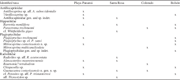
The identified rudist taxa at each of the four studied localities deserve some biostratigraphic discussion. Most taxa are common to all localities (Table 1).
Rudist taxa occurrences at the four studied localities from Costa Rica.
All identified Antillocaprinidae remain in open nomenclature and thus, they are uninformative for biostratigraphic purposes. One has been related (‘aff.’) with Antillocaprina suboccidentalis, known as are all Antillocaprina species, from the Maastrichtian (Mitchell, Reference Mitchell2013). Another, attributed as questionable to Antillocaprina, has been considered related to A. lowenstami, taken by Mitchell (Reference Mitchell2013) as the type species of the new genus Clinocaprina, considered middle Campanian in age. In one of the Antillocaprinidae indet. specimens, the tube arrangement is similar to that in Parasarcolites, considered to be upper Maastrichtian (Mitchell and Gunter, Reference Mitchell and Gunter2006).
The multiple-fold Hippuritidae have developed an array of shell characters, the study of which allowed the distinction of different phylogenetic lineages and recognition of the species sequence within each. Consequently, they are useful for biostratigraphy. The three species identified, Barrettia monilifera, Parastroma trechmanni, and cf. Whitfieldiella gigas indicate a middle Campanian age.
The stratigraphic range of many Caribbean Plagioptychidae is not accurately known, because several species have only been reported from a single locality, with incomplete descriptions, based on very few and/or incomplete specimens. Thus, their identification at other localities is difficult. Among the identified species, Plagioptychus trechmanni, P. zansi, and Mitrocaprina multicanaliculata have only been reported from the Maastrichtian of Jamaica. A specimen misidentified by Chubb (Reference Chubb1971) as P. toucasianus from the middle Campanian is included in the synonymy of Mitrocaprina costaricaensis n. sp. The still unnamed, and probably new taxon, Plagioptychidae indet., is unknown outside Costa Rica. In summary, the Plagioptychidae do not provide much biostratigraphic information.
Among the Radiolitidae, Radiolites acutocostata and Potosites tristantorresi, herein tentatively identified, are known from the early Campanian of México. Alencasterites mooretownensis is known from the Campanian and Maastrichtian. Bournonia? tetrahedron is known from the Maastrichtian. Chiapasella sp. is one of the Campanian species of Chiapasella. Guanacastea costaricaensis n. gen. n. sp. is not known outside Costa Rica; the other species of the genus, G. jamaicensis, is known from the Campanian and Maastrichtian. Although Thyrastylon is considered a Maastrichtian genus, aff. Thyrastylon sp. is unknown outside Costa Rica.
Conclusions
The Campanian rudist fauna of Costa Rica is fairly diverse, including 18 different taxa in four rudist families. All of these rudist bivalves colonized shallow waters above the raised oceanic crust west of the Caribbean, and constitute its westernmost record in the Caribbean Province.
Antillocaprinidae and Hippuritidae specimens mainly occur in basalt-derived conglomerates and gravelly sandstones or in rudstone limestones, thus, the material is fragmented and eroded, limiting its detailed study. All identified Antillocaprinidae taxa remain in open nomenclature. Nevertheless, Barrettia monilifera, Parastroma trechmanni, and also cf. Whitfieldiella gigas, were identified among the Hippuritidae, indicating a middle Campanian age for the Costa Rica rudist biohorizon.
Plagioptychidae specimens have been mainly recovered in conglomerates and rudstones but are quite well-preserved individuals, often with both valves articulated, and occur in more suitable settings. Thus, they could be reasonably investigated in detail. The two identified species from genus Plagioptychus, P. trechmanni and P. zansi, were only previously reported from their type localities in the Titanosarcolites Series of Jamaica. Knowledge of the former has been improved by the description of pallial canal patterns. Mitrocaprina multicanaliculata was previously only known through the type specimen, a considerably weathered LV from the Titanosarcolites Series of Jamaica. The description of its characters has been improved. A new species of Mitrocaprina, M. costaricaensis n. sp., has been described based on abundant, well-preserved specimens with conjoined valves, thus becoming one of the best known American Mitrocaprina species. It seems that some misidentified material from the Barrettia Limestones of Jamaica could correspond to this species. Although described in open nomenclature due to the incompleteness of the available specimens, a likely new genus of Plagioptychidae has been described herein. In summary, the Plagioptychidae of the Caribbean Province, although insufficiently known, appear to be highly diversified, with several genera not known in the Old World (Mediterranean Tethys).
Radiolitidae specimens, although mostly embedded in limestone matrix, are well preserved and could be acceptably investigated through differently oriented sections. A new genus, Alencasterites n. gen., has been proposed for the species A. mooretownensis, previously assigned either to Biradiolites or Distefanella, known from the Campanian-Maastrichtian of Jamaica, and reported herein from the Campanian. A second new genus, Guanacastea n. gen., has been proposed for Biradiolites jamaicaensis from the Campanian-Maastrichtian of Jamaica and includes G. costaricaensis n. sp. from the Campanian of Costa Rica. A likely new genus of Radiolitidae, with canals in the LV, has been also reported although left in open nomenclature because of the lack of sufficiently well-preserved and complete material; it has been related to the Caribbean genus Thyrastylon. One species from Jamaica, Bournonia tetrahedron, known from only three individuals (of which two are immature, attached to another rudist shell) has been recognized in Costa Rica and its original generic attribution has been questioned. A Chiapasella with primitive characters, similar to those originally described for C. cubensis, remains under open nomenclature. Two other species left in open nomenclature, Radiolites sp. aff. R. acutocostata and cf. Potosites aff. P. tristantorresi, have been related to taxa reported in the Campanian of México. In summary, following main points become evident from this study: (1) the attribution of several American Radiolitidae species to Old World genera (Biradiolites, Bournonia, Distefanella) are erroneous; in fact, these taxa correspond to endemic American genera; (2) the stratigraphic range of some taxa appears to be wider than previously considered; (3) Radiolitidae taxa (as well as the other rudist families) from the outcrops exposed in the western side (Pacific coast) of Costa Rica are common to the Caribbean Province (Antilles and México).
Most identified taxa were common at the four localities studied. A middle Campanian age is attributed to them, based mainly on the multiple-fold Hippuritidae occurrences. The four localities recorded slightly different depositional settings. There are no sufficient data supporting a single episode of rudist colonization occurring in different settings, or instead, close successive episodes related to the geodynamic evolution of the Central America fore arc (Pacific margin of the Caribbean).
Acknowledgments
Field work of RSE in Costa Rica from 1974 to 1984 was supported by Deutsche Forschungsgemeinschaft Bonn and Escuela Centroamericana de Geología de la Universidad de Costa Rica. Final paleontological research has been financed by project CGL2011-25581 of the Spanish Ministerio de Economía y Competitividad. T. Aguilar and P. Denyer, Escuela Centroamericana de Geología, San José, Costa Rica, gave us useful indications both in the field and at San José. A.L. Valerio, Museo Nacional de Costa Rica, San José allowed examination of the Playa Panamá material that she collected. M.M. Chavarría, Santa Rosa National Park, Guanacaste, Costa Rica, provided all needed facilities to work in the park, guided us in the field, contributed to fossil collecting, and found the location of the ancient quarry. J. Gallemí, Museu de Geologia de Barcelona (MCNB), helped with the first English version of the text. Concerns and suggestions of S. Mitchell, another anonymous reviewer, and the editors helped to improve the manuscript. All of these persons and institutions are gratefully acknowledged.