Introduction
Chondrosarcomas are primary bone neoplasms which may arise spontaneously in normal bone and secondarily in diseased bone.Reference Hug and Slater1 Although they account for up to one-third of primary malignant bone tumours, intracranial chondrosarcomas are considerably rarer, making up less than 1 per cent of all intracranial tumours.Reference Berkmen and Blatt2 The embryology of these tumours explains their sites of origin.Reference Jaffe3 Within the skull, they are believed to arise anywhere that endochondral bone ossifies intramembranously, leading to a predilection within the clivus and petrous bone and near the sella turcica and frontoethmoid region.Reference Rosenberg, Nielsen, Keel, Renard, Fitzek and Munzenrider4–Reference Lau, Wharton, Antoun, Bottrill and Moffat9 It is hypothesised that the tumours arise within rests of fetal cartilage and notochord in these regions, where the petroclival, spheno-petrosal, petro-occipital and spheno-occipital synchondroses converge.Reference Lau, Wharton, Antoun, Bottrill and Moffat9, Reference Seidman, Nichols, Raju, Mehta and Levy10
Although they more commonly occur spontaneously, chondrosarcomas may also arise as a result of malignant degeneration in endochondromas, seen both in Maffucci's syndrome (a mesodermal dysplasia causing enchondromas and haemangiomas) and in Ollier's disease (manifesting as multiple enchondromas).Reference Jaffe3, Reference Seidman, Nichols, Raju, Mehta and Levy10 They are also reported to arise in association with Paget's disease, osteocartilaginous exostoses and bone trauma.Reference Burkey, Hoffman, Baker, Thornton and McClatchey11, Reference Brown, Hug and Weber12
As a rule, chondrosarcomas are locally invasive and rarely metastasise, causing injury through compression and invasion of critical structures within the skull base, including the cranial nerves and the carotid artery.Reference Weber, Brown, Hug and Liebsch13 Because of the complex anatomy of the skull base, margin-free tumour extirpation in this region is often not possible without significant morbidity to the patient. As a result, treatment has focused on local control, using a combination of surgery and radiation or proton beam radiotherapy.Reference Hug and Slater1, Reference Korten, ter Berg, Spincemaille, van, Laan, Van and Wel14, Reference Munzenrider, Liebsch, Efird, Johnson and Didolkar15
In an effort to further characterise these uncommon neoplasms, we performed a retrospective review of patients with chondrosarcomas involving the temporal bone and skull base, treated at our institution over the past 30 years.
Patients and methods
We undertook a retrospective review of all patients with chondrosarcomas involving the skull base and temporal bone, treated at our institution over the past 30 years. Patients were identified through a review of clinical and operative records and of a pathology database. Patients were excluded from the study if there was insufficient clinical data, including lack of current clinical information (within the past five years) or clear documentation in the medical records of the final outcome (e.g. death). Patients were also excluded if there was not a clear histopathologic diagnosis of chondrosarcoma.
From the clinical records, data were tabulated on each patient, including gender, age at presentation, presenting symptoms and signs, and treatment (both surgical and radiotherapeutic). Data tabulated also included length of survival from initial presentation, and whether the patient was currently either: free of disease (e.g. gross total surgical resection had been undertaken and there had been a normal post-operative magnetic resonance imaging (MRI) screening); alive with disease (e.g. subtotal resection of tumour, with or without radiation therapy and persistence of tumour on MRI screening, or initial gross total resection with recurrence on MRI); deceased from disease; or deceased from an unrelated cause.
This study comprised a review of existing clinical data with patient identifiers removed. It qualified for exemption from an institutional review board protocol based upon Department of Health and Human Services criteria 45 CFR 46.101(b)(4). The determination that this study was exempt from a protocol requirement was made by the joint committee on clinical investigation of the Johns Hopkins University School of Medicine.
Results
Patient demographics
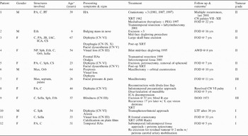
A total of 12 patients were identified who met all the criteria for inclusion in this retrospective review (Table I). There were eight females and four males. The average age at presentation was 42 years, with a range of six to 67 years.
Clinical Information

* At first presentation. †Length of time is from first operation to time to writing. Yrs=years; M=male; PA=petrous apex; C=clivus; PF=posterior fossa; HA=headache; XRT=radiotherapy; CN=cranial nerve; PEG=percutaneous endoscopic gastrostomy; FOD=free of disease; Eth=ethmoid; F=female; JB=jugular bulb; IAC=internal auditory canal; ICA=internal carotid artery; CS=cavernous sinus; NP=nasopharynx; Sph=sphenoid; Orb=orbit; Sella=sella turcica; bilat=bilateral; AWD=alive with disease; Max=maxilla; R=right; L=left; DOD=died of disease; w/=with; LTF=lost to follow up; mths=months
Anatomical presentation
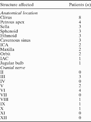
The chondrosarcomas involved a variety of structures throughout the skull base (Table II). The most common site of involvement was the clivus, in eight patients (67 per cent). Four patients (33 per cent) had involvement of the petrous apex, three (25 per cent) had involvement of the sella turcica with suprasellar extension, three (25 per cent) had involvement of the cavernous sinus, three (25 per cent) had involvement of the sphenoid and three (25 per cent) had involvement of the ethmoid. Two patients (17 per cent) had involvement of the orbit, internal carotid artery and maxilla. One patient (8 per cent) had involvement of the jugular bulb.
Structures Responsible for Presenting Clinical Features of Chondrosarcomas
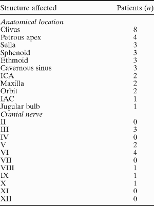
Sella=sella turcica; ICA=internal carotid artery; IAC=internal auditory canal
Presenting symptoms and signs
The most common presenting features were diplopia and facial pain or headache, in association with cranial nerve deficits (Tables I and II). Diplopia was due to a VIth cranial nerve deficit in four patients and proptosis in one patient. Headache or facial pain was noted in three patients (25 per cent). Other presenting symptoms included visual loss or blindness (n=3), facial dysaesthesias (n=2), a bulging mass in the nose (n=1), and otalgia (n=1). When evaluated by cranial nerve deficits at presentation (Table II), the VIth cranial nerve was the most commonly affected (n=4), followed by the IIIrd (n=3), Vth (n=2), VIIIth (n=1), IXth (n=1) and the Xth (n=1).
Patients requiring a single operation
All patients underwent some form of surgical excision of their tumour (Table I). Seven of the 12 patients (58 per cent) only required a single operation to achieve control of the disease. Of these, three also underwent post-operative radiation therapy. Of these seven cases, three had well differentiated (grade one) tumours, three had moderately differentiated (grade two) tumours and one had a poorly differentiated (grade three) tumour. All of these seven patients were free of disease at the time of writing, after a follow up ranging from five to 33 years from their original presentation. There did not appear to be any unique anatomical features of the tumour in these seven (single operation) patients, as compared with those who underwent multiple procedures for persistent disease or recurrence (see below). However, there did appear to be an association between the histologic grade of the tumour and the need for more than one surgical procedure. Patients with well differentiated tumours (i.e. grade one) were more likely to require only a single operation (3/4 patients) as opposed to those with grade two tumours (2/6 needed only a single operation). Both patients with grade three tumours underwent only one operation, although one of these patients had an inoperable recurrence 17 years later, to which she subsequently succumbed.
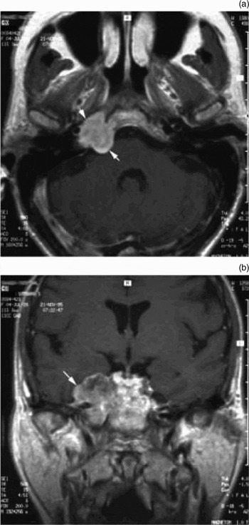
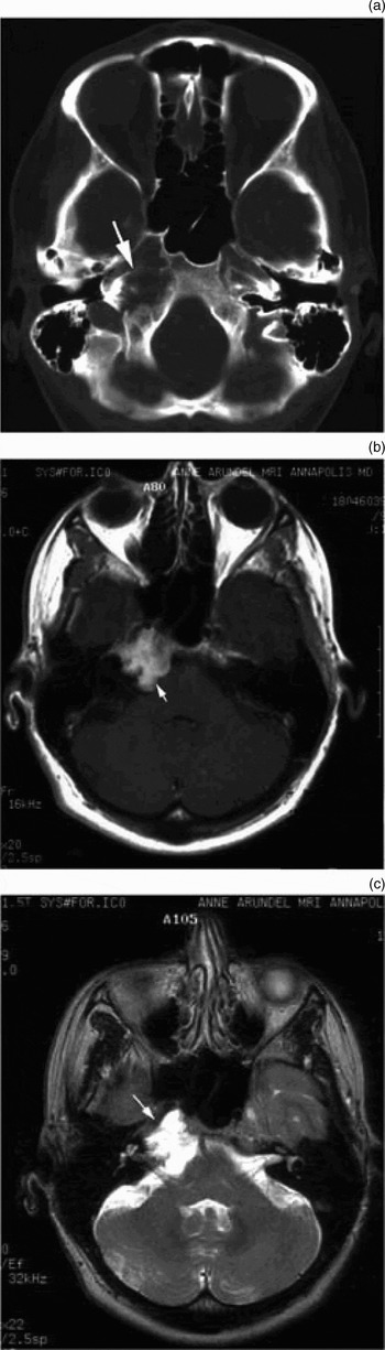
Of these seven patients requiring only one operation, three underwent anterior approaches (Table I). Patient six, with maxillary and orbital involvement, and patient seven, with maxillary, septal and palatal involvement, both underwent a mid-face degloving procedure for tumour removal. Patient 10, with clival and sphenoid involvement, underwent a transsphenoethmoid approach. Three of the seven patients underwent laterally based infratemporal fossa approaches. Patient three underwent an infratemporal fossa approach to the skull base for tumour involving the petroclival region, extending down to the jugular bulb, and involving the internal carotid, internal auditory canal and cavernous sinus (Figure 1). Patient five underwent an infratemporal fossa approach and an anterior petrosectomy for tumour involvement of the petroclival junction, cavernous sinus and sphenoid. Patient five underwent an infratemporal pre-auricular approach for tumour involving the petroclival junction (Figure 2). The last patient who required only a single operation (patient 11) underwent a frontal craniotomy for tumour in the region of the sella, with suprasellar extension.
(a) Axial and (b) coronal magnetic resonance imaging scans for patient three. The tumour (arrows) involves the petroclival region, extends down to the jugular bulb, and involves the internal carotid artery, internal auditory canal and cavernous sinus.

Patient five presented with tumour involvement of the petroclival junction, cavernous sinus and sphenoid. (a) Axial computed tomography scan showing an expansive lesion adjacent to the sphenoid and carotid artery (arrow). (b) T1-weighted magnetic resonance image (MRI) demonstrating the tumour in the petroclival region (arrow). (c) T2-weighted MRI showing the same tumour, just anterior to the internal auditory canal (arrow).

Patients requiring multiple operations
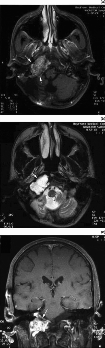
The remaining five patients required multiple operations due to tumour recurrence or persistence. Patient one (Table I) required four operations over a 20 year period. This patient had originally presented in 1981 with a petroclival chondrosarcoma extending into the posterior fossa (Figure 3). He had undergone a large suboccipital, posterior fossa resection, with post-operative radiation therapy, for a medium (grade two) chondrosarcoma. He had two additional recurrences in 1987 and 1997, requiring resection via the same approach. Because of subsequent recurrence within the temporal bone, in 2001 he underwent a complete labyrinthectomy with gross total tumour removal. At the time of writing, he had unilateral deficits of cranial nerves VII to XII and was alive without obvious disease (by MRI) two years from his last procedure and 22 years from his initial presentation.
Patient one. (a) Axial T1-weighted magnetic resonance image (MRI) showing tumour (arrows) in the petroclival region, extending into the posterior fossa. (b) The same tumour (arrow) viewed on a T2-weighted MRI. (c) Coronal T1-weighted MRI of the same tumour (arrow), with contrast enhancement.

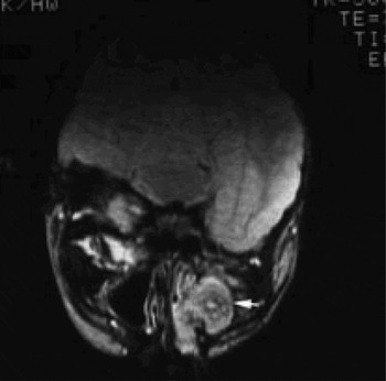
Patient two (Table I) presented with a bulging nose at the age of six years. Investigations revealed a chondrosarcoma involving the ethmoid sinuses (Figure 4). He underwent a mid-face degloving procedure for excision. However, due to tumour persistence, the patient required an additional operation, via the same approach, within a year of the initial surgery. Histopathology demonstrated a moderately differentiated (grade two) tumour. At his most recent follow up, 16 years after the initial surgery, this patient was free of disease.
Patient two; T1-weighted magnetic resonance image showing a chondrosarcoma (arrow) involving the ethmoid sinuses.

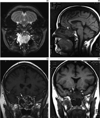
Patient four (Table I) presented at the age of 54 years with progressive visual loss, headaches and otalgia. Investigations demonstrated a large chondrosarcoma involving the nasopharynx, sphenoid, ethmoid, clivus, sella turcica and orbit (Figure 5). The patient originally underwent a mid-face degloving procedure in 1995. Histopathological analysis of the tumour identified a grade two chondrosarcoma. Four years later, because of tumour recurrence, the patient underwent a transantral re-resection. A third recurrence two years later led to resection via an infratemporal fossa approach. Eight years after her initial presentation and two years after her most recent infratemporal fossa excision, the patient was alive with residual disease that appeared stable.
Magnetic resonance imaging (MRI) series from patient four. (a) Axial T2-weighted MRI showing a large chondrosarcoma (arrow) eroding into the sphenoid and ethmoid sinuses and nasopharynx. (b) Saggital T1-weighted MRI demonstrating tumour involvement (arrowhead) of the sella turcica and upper clivus. Coronal T1-weighted MRI images are also shown, (c) without and (d) with contrast enhancement.

Patient nine presented at the age of 37 years with progressive visual loss. Investigation led to the diagnosis of a skull base tumour involving the clivus and sella, with extension to the suprasellar region and to the sphenoid and ethmoid bones. She underwent an anterior approach to the skull base via a mid-face degloving procedure. Histopathological analysis of the tumour identified a high grade (grade three) chondrosarcoma. The patient did well until 17 years later, when she re-presented with visual loss in her only seeing eye. Evaluation demonstrated a large recurrence of the tumour, which was felt to be unresectable. Despite receiving radiation therapy, the patient died secondary to central nervous system compression following radiation treatment.
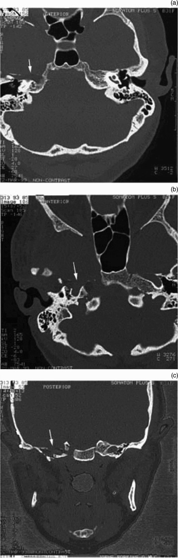
Patient 12 presented at the age of 51 with temporal headaches. Evaluation led to the diagnosis of a tumour involving the petroclival region of the skull base, adjacent to the intrapetrous carotid artery (Figure 6). She underwent a subtemporal-infratemporal fossa approach with an anterior petrous apicectomy. Histopathological analysis of the tumour identified a low grade (grade one) chondrosarcoma. Because of persistent disease, the patient underwent a re-excision of the residual tumour, with mobilisation of the petrous carotid artery, two months later. At her most recent follow-up, five years post-operatively, she was free of disease.
Axial (a & b) and coronal (c) computed tomography images of patient 12; arrows indicate the tumour.

Long-term follow up
The duration of patients' follow up ranged from five to 33 years from their initial presentation. Only one patient died as a result of the tumour or its treatment (patient nine). At the time of writing, the remaining patients were either free of disease (by MRI evaluation; nine patients), or alive with persistent disease (by MRI; one patient). One patient (patient 10) had been noted to be free of disease at their 30 year follow up, but was lost to follow up over the past five years.
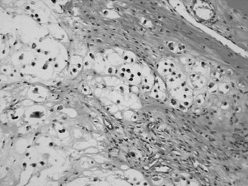
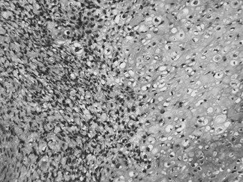
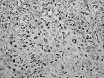
Long-term results were also compared for each histological grade. Of the four patients with low grade (i.e. grade one) chondrosarcoma (Figure 7), all were free of disease at follow up ranging from five to 33 years. Of these four cases, three achieved tumour control after one operation, while one (patient 12) required a second operation within two months of the first for persistent disease. Six patients had grade two chondrosarcoma (Figure 8); five were free of disease after follow up ranging from seven to 22 years, and one was alive with disease at eight years. Of these six individuals with grade two tumours, two had complete tumour removal in a single operation, while four required multiple surgical procedures for tumour control, due to recurrence. Two individuals had poorly differentiated (grade three) chondrosarcoma (Figure 9). One was free of disease 11 years following her operation (patient seven), while the second died as a result of a large tumour recurrence 17 years after her original surgery (patient nine).
Histopathology of a well differentiated (grade one) chondrosarcoma, characterised by spindle cell stroma containing chondrocytes in lacunae with a lobular configuration. There is minimal cellular atypia, with only occasional mitotic figures (H&E; original magnification ×400).

Histopathology of a moderately differentiated (grade two) chondrosarcoma, showing a cellular, sarcomatous stroma at an interface with chondroid material containing dysplastic chondrocytic elements within lacunae. The latter shows vesicular nuclear chromatin, hyperchromatic staining and pleomorphic features. The stroma is highly cellular, being composed of streaming, predominantly spindle-shaped elements (H&E; original magnification ×400).

Histopathology of a poorly differentiated (grade III) chondrosarcoma, showing a central area with marked cellular pleomorphism and nuclear aberrations, with only minimal chondroid differentiation (H&E; original magnification ×400).

Discussion
Chondrosarcomas involving the temporal bone and skull base are uncommon lesions, accounting for less than 1 per cent of tumours within the head and neck, and approximately 6 per cent of skull base lesions in some series.Reference Koch, Karnell, Hoffman, Apostolakis, Robinson and Zhen16, Reference Kveton, Brackmann, Glasscock, House and Hitselberger17 Other series have shown a much smaller incidence in the skull base.Reference Berkmen and Blatt2, Reference Seidman, Nichols, Raju, Mehta and Levy10
As already noted from its embryologic origins, chondrosarcomas tend to arise in areas of synchondrosis. Within the skull base, this occurs in the petroclival, petro-occipital, spheno-occipital and spheno-petrosal synchondroses.Reference Lau, Wharton, Antoun, Bottrill and Moffat9 The findings in this current series of 12 patients support this assertion, as chondrosarcomas were most likely to arise in relation to the petroclival junction, occurring here in two-thirds of patients, while the remainder arose in relation to the sella, sphenoid and ethmoid regions (Table II). Extension of these tumours also caused involvement of additional critical structures, including the cavernous sinus in three patients, the orbit in two, the internal auditory canal in one and the jugular bulb in one patient.
These areas of tumour involvement are similar to findings from previous studies. In a review of 200 cases of chondrosarcoma of the skull base, Rosenberg and colleaguesReference Rosenberg, Nielsen, Keel, Renard, Fitzek and Munzenrider4 also showed that two-thirds of cases involved the petroclival junction, approximately one-fourth of cases arose from the spheno-occipital region and 6 per cent were centred in the spheno-ethmoid complex. In contrast, in a literature review of 48 patients with skull base chondrosarcoma, Seidman et al. Reference Seidman, Nichols, Raju, Mehta and Levy10 showed a 44 per cent involvement of the petrous bone, but only an 8 per cent involvement of the clivus.
Clinically, the patients in our study presented with a variety of symptoms (Table II), although ophthalmologic complaints were most likely, occurring in two-thirds of patients. Eye complaints included diplopia, due to involvement of the abducens nerve (VIth cranial nerve; four patients), proptosis (one patient) or loss of vision due to involvement of the optic nerve (IIIrd cranial nerve; three patients). This is not surprising, since prior reports have shown that approximately 50 per cent of patients with chondrosarcomas involving the skull base will present with ocular symptoms (i.e. diplopia or visual impairment) as the initial manifestation of the disease.Reference Volpe, Liebsch, Munzenrider and Lessell18
Headache or facial pain was another common presenting feature, being reported by three patients. Facial dysaesthesias due to involvement of the trigeminal nerve (Vth cranial nerve) occurred in two patients. In contrast, in the report by Seidman et al.,Reference Seidman, Nichols, Raju, Mehta and Levy10 hoarseness and dysphasia were the most common presenting symptoms in patients with temporal bone and skull base chondrosarcomas.
When evaluating our cases according to the cranial nerves involved, we found that the VIth cranial nerve was the most commonly injured by tumour, followed by the IIIrd cranial nerve. In contrast, Seidman's report showed that the Xth cranial nerve was most commonly involved, followed by the VIth cranial nerve. Another series of temporal bone chondrosarcomas, reported by Coltrera et al.,Reference Coltrera, Googe, Harrist, Hyams, Schiller and Goodman7 showed that 75 per cent of their 12 patients presented with VIth cranial nerve deficits, similar to our study.
Most contemporary studies rely on MRI for characterisation and diagnosis of chondrosarcomas, while earlier studies relied upon computed tomography (CT). While CT is useful for defining the extent of bony destruction (Figures 2 and 6), MRI is superior in defining the tumour extent and the involvement of critical structures within the skull base (Figures 1–5).Reference Brown, Hug and Weber12 The characteristic MRI appearance of chondrosarcoma is a low to medium signal on T1-weighted images, a high signal on T2-weighted images and enhancement with contrast (e.g. gadolinium). These lesions will often have a ‘soap bubble’ appearance, helping differentiate them from other bony lesions such as chordomas. Following treatment (radiation or surgery), serial post-operative MRIs are recommended for screening for recurrent disease. Whereas the MRI appearance of post-operative changes will be fairly stable over several scans spanning one to two years, recurrent disease will manifest as an enlarging lesion.
Histopathologically, chondrosarcomas are divided into four sub-types: conventional (hyaline/myxoid), dedifferentiated, clear cell and mesenchymal.Reference Rosenberg, Nielsen, Keel, Renard, Fitzek and Munzenrider4, Reference Evans, Ayala and Romsdahl19 Within the skull base, the conventional type is overwhelmingly the most common.Reference Rosenberg, Nielsen, Keel, Renard, Fitzek and Munzenrider4 These conventional chondrosarcomas can be further subdivided, based upon cellular features, into low grade or well differentiated (grade one), medium grade or moderately differentiated (grade two), and high grade or poorly differentiated (grade three), based upon the tumour's cellularity and atypia (although some features of grade three tumours overlap with those of the undifferentiated variety).Reference Rosenberg, Nielsen, Keel, Renard, Fitzek and Munzenrider4 In the current study, four patients (33 per cent) had grade one tumours, six patients (50 per cent) had grade two tumours and two patients (17 per cent) had grade three tumours (Figures 7–9). These numbers are in contrast to Rosenberg's pathological review of 200 cases of skull base chondrosarcomas, which found 50 per cent grade one tumours, 21 per cent grade two tumours and no grade three tumours. The reason for the preponderance of grade two tumours in our current series is unclear.
Generally, chondrosarcomas, particularly the lower grade varieties, rarely metastasise but cause morbidity through local invasion of surrounding structures.Reference Sheth, Yasko, Johnson, Ayala, Murray and Romsdahl20 Thus, treatment has focused upon complete surgical excision, with radiation therapy being reserved for high grade tumours or following incomplete tumour resection. Within the skull base, the close proximity of chondrosarcomas to vital structures makes these guiding treatment principles even clearer. However, because these are in fact benign, often low grade lesions, the aim of treatment should be disease control with minimal surgical morbidity.
Because these tumours can arise in a variety of locations within the skull base, it is not surprising that a variety of surgical approaches, both anteriorly and laterally based, have been described. These ingenious approaches include the infratemporal fossa approach (as popularised by Fisch),Reference Fisch21 the petrosal approach and its variations,Reference Al-Mefty, Fox, Rifai and Smith22–Reference Megerian, Chiocca, McKenna, Harsh and Ojemann24 the extended frontal approach,Reference Sekhar, Schramm and Jones25 the supra-infratentorial presigmoid avenue,Reference Samii and Ammirati26 the transsphenoethmoid approach,Reference Lalwani, Kaplan and Gutin27 and the combined intradural presigmoid transversarium transcondylar approach,Reference Ammirati, Ma, Cheatham, Mei, Bloch and Becker28 to name just a few. As can be shown from this and prior series, the variable anatomic locations of these tumours require the surgeon to be familiar with a variety of anterior and lateral approaches to the skull base, depending upon the specifics of the presentation.
The decision to operate, particularly on asymptomatic lesions, can be a challenging one. Morbidity from these lesions results from growth and impingement upon surrounding structures. For asymptomatic lesions, an initial, conservative ‘watch and wait’ approach can be adopted, together with serial scanning. For those lesions showing rapid expansion or ones that become symptomatic during the observation period, intervention can then be planned.
In the current series, four of the patients required some form of lateral approach to reach the tumour; these approaches included variations on the infratemporal fossa approach type B or C.Reference Fisch21 In contrast, six patients required anterior approaches to the tumour. These included mid-face degloving approaches in five patients and a transsphenoethmoidectomy approach in one case. One patient required a standard suboccipital posterior fossa craniotomy, while another required a frontal craniotomy. In each case, the approach was based upon the specifics of the tumour, and was decided upon after consultation between our institution's skull base surgeons and neurosurgeons. As noted by Crockard et al.,Reference Crockard, Steel, Plowman, Singh, Crossman and Revesz29 such a multidisciplinary team approach is critical for effective management of these tumours.
Radiation therapy was used in five of the 12 patients in this series. The literature suggests that radiation therapy for chondrosarcomas is generally ineffective.Reference Hug and Slater1 However, in this series, it was reserved for cases in which there was subtotal tumour resection or recurrence in the setting of inoperable tumour. Radiation treatment was given to one of the patients with a grade one tumour, three patients with grade two tumours and one patient with a grade three tumour. The only death in the series occurred in the patient with a 17-year recurrence of an inoperable grade three tumour, who received radiation in an attempt to control further tumour growth. At the time of writing, tumour growth in the remaining patients had been controlled, after follow up ranging from seven to 33 years. Other large series have shown similar efficacy for radiation therapy, as an adjunct to surgery, for treatment and control of chondrosarcoma.Reference Rosenberg, Nielsen, Keel, Renard, Fitzek and Munzenrider4 Proton radiation has also been advocated as a more useful modality by some,Reference Hug and Slater1 although none of the patients in the current series received it.
• This study evaluated the clinical presentation and treatment outcomes in 12 patients with chondrosarcomas involving the skull base and temporal bone
• Patients with chondrosarcomas involving the skull base and temporal bone may present in a variety of ways
• The decision to operate, particularly on asymptomatic lesions, can be a challenging one
• For asymptomatic lesions, an initial, conservative ‘watch and wait’ approach can be adopted, with serial scanning. For those lesions showing rapid expansion or becoming symptomatic during the observation period, intervention can then be planned
Perhaps the strongest feature of the current series is the extended follow-up data available for all patients, over time periods ranging from six to 67 years from the original presentation and from three to 33 years from the most recent treatment. As some of the patients within this series demonstrate, recurrence can occur many years after treatment for these insidious tumours (as many as 17 years in this report). Thus, even five-year survival rates may not accurately reflect a tumour-free state. Prior studies have conflictingly shown that survival rates may either be related to histologic gradeReference Evans, Ayala and Romsdahl19 or bear no relation to histologic grade.Reference Rosenberg, Nielsen, Keel, Renard, Fitzek and Munzenrider4 In the large series of 200 patients reported by Rosenberg et al.,Reference Rosenberg, Nielsen, Keel, Renard, Fitzek and Munzenrider4 the five- and 10-year local control and disease-specific survival rates were approximately 98 per cent for all tumours, regardless of histological type. In contrast, in the review by Seidman et al.,Reference Seidman, Nichols, Raju, Mehta and Levy10 10-year survival rates were reported to be 83, 64 and 29 per cent for grade one, two and three tumours, respectively. By comparison, in the current study, five-year local control and disease-specific control rates were 92 and 83 per cent, respectively. While those with grade one tumours were more likely to have their disease controlled with a single operation than those with grade two tumours, the data from our study suggest that the long-term control rates did not differ greatly between these histologic grades. However, as only two patients in the current study had grade three tumours, meaningful survival statistics cannot be generated.
These results, combined with data from prior studies, show that patients with chondrosarcomas, despite the anatomically challenging location within the skull base, can be effectively treated with a combination of surgery and radiation therapy. Even with recurrences or persistent disease, long-term control rates of over 10 years are generally possible.