Introduction
Reconstruction of a perforated tympanic membrane in chronic otitis media aims to achieve and maintain morphological closure and functional gain over time.Reference Kazikdas, Onal, Boyraz and Karabulut1 Since the introduction of tympanoplasty by Wullstein in 1952,Reference Wullstein2 and Zollner in 1955,Reference Zoellner3 different types of graft materials have been used for the repair. As a result of ease of accessibility at the surgical site, abundant availability, low basal metabolic rate and similar thickness to the tympanic membrane, temporalis fascia grafts have been extensively used worldwide, with successful closure of tympanic membrane defects in over 90 per cent of normally ventilated middle ears.Reference Dermirpehlivan, Onal, Arslanoglu, Songu, Ciger and Can4, Reference Kulkarni, Kulkarni, Burse, Sancheti and Roy5 However, temporalis fascia has much poorer healing in cases of tubal dysfunction, an adhesive process, tympanic fibrosis and total perforation, and in revision surgical procedures. Consequently, during the last decade, there has been renewed interest in the use of cartilage grafts as an alternative.Reference Mohamad, Khan and Hussain6–Reference Tek, Karaman, Uslu, Habesoglu, Kilicarslan and Durmus10
Cartilage, as a graft material for myringoplasty, was first used by Salen in 1963, who used autologous septal cartilage.Reference Salen11 Heerman was the first to introduce the cartilage palisade technique, in the early 1960s, in which preserved perichondrium cartilage strips were placed parallel to the malleus covering the middle ear.Reference Salen11 Since then, surgeons have adopted the palisade technique with different modifications.Reference Amedee, Mann and Eiechelmann12–Reference Dornhoffer14 The concept of composite cartilage perichondrial grafts as island grafts for tympanic membrane reconstruction was yet another advancement in cartilage tympanoplasty, as popularised by Goodhill in patients with total perforation and a shallow middle-ear cavity.Reference Goodhill15 The greater firmness, high resistance to resorption and retraction, and long-term survival owing to its nourishment by diffusion, makes cartilage a favourite material for tympanoplasties.Reference Aidonis, Robertson and Sismanis16, Reference Arriage17 Nevertheless, there is growing concern regarding using cartilage sheets or palisades in cases of chronic otitis media with atelectasis, adhesions and or total perforation, because of the possibility of hiding a future generation of cholesteatoma and the acoustic transfer loss due to its inherent thickness.Reference Poe and Gadre18–Reference Dornhoffer20
To overcome these shortcomings, and utilising the favourable properties both of temporalis fascia and cartilage grafts, a technique of temporalis fascia reinforcement with cartilage has been put forward.Reference Kulkarni, Kulkarni, Burse, Sancheti and Roy5, Reference Tek, Karaman, Uslu, Habesoglu, Kilicarslan and Durmus10, Reference Mundra, Sinha and Agarwal21 We introduce yet another novel technique of cartilage rim augmented fascia tympanoplasty (‘CRAFT’), which involves underlay placement of temporalis fascia, which acts as reinforcement, achieved by placing a horseshoe-shaped conchal cartilage as an annular graft under the tympanic annulus. We also introduce the placement of ‘rescue bars’. These bars are thin cartilage strips placed in the mesotympanum to reconstruct the shallow middle ear for better ventilation, to resist negative pressure due to tube dysfunction and prevent mediatisation of the graft in selected cases. The morphological and functional outcomes were compared with the traditional temporalis fascia technique and the results statistically analysed; this was accompanied by a literature review of cartilage tympanoplasty.
Materials and methods
This retrospective study was conducted over a period of two years, at a tertiary care teaching hospital in Oman, between 2013 and 2015. All patients who had undergone myringoplasty and/or tympanoplasty with the cartilage rim augmented fascia tympanoplasty technique were included in the study. Ethical approval was obtained from the Sultan Qaboos University Hospital ethical committee. Those patients who underwent temporalis fascia tympanoplasty during this two-year period were included for comparison.
The cartilage rim augmented fascia tympanoplasty technique was performed by a single surgeon, but the temporalis fascia tympanoplasty technique was performed by multiple surgeons. All cases were consecutive and not randomised.
For morphological evaluation, all patients awaiting primary tympanoplasties with large central, subtotal or total perforations, irrespective of ossicular chain status, with a dry ear for a period of three months, with or without hearing loss prior to the surgery, were included. The two groups (cartilage rim augmented fascia tympanoplasty and temporalis fascia tympanoplasty) had perforations that were similar in size and position. For functional assessment of hearing, those patients with an ear drum perforation with pure conductive hearing loss, with or without a normal ossicular chain, were included. Patients were excluded from the study if they had chronic suppurative otitis media with cholesteatoma (unsafe type of disease), any history of previous ear surgery, and/or presented with sensory neural or a mixed type of hearing loss.
Surgical technique
After obtaining informed consent, all operations were performed under general anaesthesia with endotracheal intubation via a routine post-aural approach. The infiltration was carried out locally with 2 per cent xylocaine (2 per cent lidocaine hydrochloride; AstraZeneca, Södertälje, Sweden) with adrenaline (1:1000; Hospira, Lake Forest, Illinois, USA) with a 1 in 200 000 solution.
Temporalis fascia was harvested by a standard approach, and subsequently 1 cm sized rounded conchal cartilage was harvested from the cymba concha, approaching through the same incision. The perichondrium on either side of the cartilage was removed and the cartilage was carved with a number 11 surgical blade, resulting in a horseshoe-shaped cartilage of 1–2 mm width and 0.5 mm thickness that allowed an acceptable compromise between sufficient mechanical stability and acoustic transfer, as described by Murbe et al.Reference Murbe, Zahnert, Bornitz and Huttenbrink9
Cartilage strips of 4 mm length and 0.2 mm width were used to splint the graft, to prevent Eustachian tube blockage and the collapse of middle-ear volume due to negative pressure, thus acting as ‘rescue bars’ in selected cases (Figure 1).
Components of cartilage rim augmented fascia tympanoplasty: (a) harvested conchal cartilage; (b) sculptured conchal cartilage in the form of annular graft and rescue bars; and (c) harvested temporalis fascia graft.

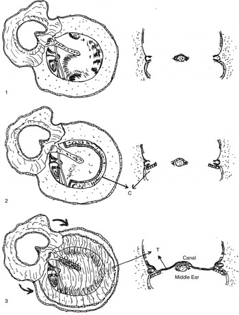
Under an operating microscope, the tympanomeatal flap was elevated up to the tympanic annulus after freshening the edge of the perforation, and the middle ear was entered in the conventional manner. The middle-ear mucosa was inspected and ossicular mobility was checked. Ossicular reconstruction was accomplished using either sculptured homologous incus or cortical bone, depending upon the defects. The cartilage graft was then placed in a meticulous manner under the tympanic annulus, from the level of the supratubal recess, along the anterior, inferior and posterior tympanic annulus, stopping just short of the oval window to avoid contact with the ossicular chain. Temporalis fascia was placed by underlay technique lateral to the cartilage graft, extending to the adjacent bony wall, and then the tympanomeatal flap was repositioned, draping over the bony wall (Figure 2). Rescue bars were placed in a horizontal or vertical manner in the mesotympanum, without affecting the free mobility of the ossicles, if the middle ear was too shallow or there was adhesion or atelectasis.
Diagrammatic representation of cardinal steps of cartilage rim augmented fascia tympanoplasty: (1) elevation of tympanomeatal flap and inspection of middle ear; (2) placement of cartilage annular graft; and (3) placement of temporalis fascia over the annular graft as underlay graft. C = cartilage annular graft; T = temporalis fascia

The post-aural wound was sutured in layers, a medicated gauze pack was inserted and a mastoid bandage was applied for 24 hours. Analgesics and an appropriate antibiotic were given post-operatively. The sutures and the pack were removed after one week. Follow-up examinations were performed at 2, 8, 12 and 24 weeks. Post-operative audiograms were conducted after six months.
All patients were analysed based on the protocol described by the Committee on Hearing and Equilibrium guidelines for the evaluation of results of treatment of conductive hearing loss, to standardise the reporting.22 The tympanic membrane findings were examined and recorded in the out-patient department. All patients subsequently underwent pure tone audiometric assessment.
Pure tone air conduction and bone conduction thresholds were recorded at octave intervals from 0.5 to 8 kHz and at 3 kHz. The pure tone average (PTA) was then calculated as the average of 0.5, 1, 2 and 3 kHz, rounded to the nearest number. The air–bone gap was calculated by subtracting the PTA of bone conduction from the PTA of air conduction. The means, standard deviations and summaries of air–bone gaps in bins (0–10 dB, 11–20 dB, 21–30 dB and more than 30 dB) were calculated. The pre-operative air–bone gap was compared with the post-operative air–bone gap six months after surgery.
Morphological success (anatomical closure) was defined as a healed graft, with no residual perforation, adhesion, mediatisation, lateralisation and/or atelectasis (Figure 3). The functional outcome was considered favourable when there was an air–bone gap closure of within 20 dB after surgery.
Post-operative photographs (a & b) showing healed cartilage temporalis fascia composite graft in cartilage rim augmented fascia tympanoplasty.

Statistical analysis
Descriptive statistics were used to summarise the data. Associations between two categorical variables were assessed using the chi-square test. An independent samples t-test was applied to compare the mean scores between two unrelated groups on the same continuous, dependent variable. A two-tailed level of significance was set at 0.05. All statistical analysis was carried out using SPSS software (version 22.0; IBM, Armonk, New York, USA).
Results
A total of 115 patients (63 males and 52 females) between the ages of 9 and 64 years were enrolled in the study. The mean patient age was 31.8 years. The average follow up after surgery was 11.1 months. In 54.8 per cent of patients, the left side was affected; in 45.2 per cent, the pathology was on the right side. All patients had had a dry ear for no less than three months prior to surgery. Fifty-eight patients underwent temporalis fascia tympanoplasty and 57 underwent cartilage rim augmented fascia tympanoplasty surgery.
Of the 58 patients (50.4 per cent) in the temporalis fascia tympanoplasty group, 60.3 per cent (n = 35) were male and 39.7 per cent (n = 23) were female. The mean patient age was 31.6 years (range, 9–64 years). The average follow-up duration was 14.6 months. In 18 patients, the presenting complaint was ear discharge, with no documented hearing loss, hence these patients were not considered for functional assessment after surgery, which was carried out in the remaining 40 cases.
Of the 57 patients (49.5 per cent) in the cartilage rim augmented fascia tympanoplasty group, 49.1 per cent (n = 28) were male and 50.9 per cent (n = 29) were female. The mean patient age was 32 years (range, 11–61 years). The average follow-up duration was 7.6 months. Forty of the 57 patients were available for functional assessment because of associated conductive hearing loss.
In the temporalis fascia tympanoplasty group, graft take up was achieved in 70 per cent of cases (40 out of 58). In the cartilage rim augmented fascia tympanoplasty group, the drum had healed in 94.7 per cent of cases (54 out of 57). This difference was statistically significant (Table I).
Group comparison of graft take-up rates

* Statistically significant. CRAFT = cartilage rim augmented fascia tympanoplasty
The mean pre- and post-operative PTAs in the temporalis fascia tympanoplasty group were 36.39 ± 9.31 dB and 27.40 ± 9.89 dB respectively; in the cartilage rim augmented fascia tympanoplasty group, pre- and post-operative PTAs were 37.86 ± 11.75 dB and 22.57 ± 7.58 dB respectively. In the temporalis fascia tympanoplasty group, the average pre- and post-operative air–bone gaps were 23.37 ± 8.07 dB and 17.59 ± 9.36 dB respectively; in the cartilage rim augmented fascia tympanoplasty group, these values were 27.52 ± 10.06 dB and 14.41 ± 7.00 dB respectively.
Of the 40 patients in the temporalis fascia tympanoplasty group with pre-operative conductive hearing loss, 33 had a normal intact mobile ossicular chain, whereas the ossicular chain was discontinuous in 7 patients. Of the 40 patients in the cartilage rim augmented fascia tympanoplasty group with pre-operative conductive hearing loss, 27 had a normal intact mobile ossicular chain, whereas the ossicular chain was discontinuous in 13 patients.
In those with a normal ossicular chain, the post-operative air–bone gap was within 20 dB in 69.7 per cent of cases (23 out of 33) in the temporalis fascia tympanoplasty group, and in 92.6 per cent of cases (25 out of 27) in the cartilage rim augmented fascia tympanoplasty group. This difference was statistically significant (p = 0.027) (Table II). Among the defective ossicular chain cases, the post-operative air–bone gap was within 20 dB in 57.1 per cent in the temporalis fascia tympanoplasty group, as against 76.9 per cent of cases in the cartilage rim augmented fascia tympanoplasty group. Although this indicates an advantage for the cartilage rim augmented fascia tympanoplasty group, the difference was not statistically significant (p = 0.613) (Table III).
Group comparison of post-operative air-bone gaps in patients with normal mobile ossicular chain

* Statistically significant. CRAFT = cartilage rim augmented fascia tympanoplasty
Group comparison of post-operative air-bone gaps in patients with defective ossicular chain

CRAFT = cartilage rim augmented fascia tympanoplasty
Discussion
Temporalis fascia has been used as a graft material for tympanic membrane defect reconstruction for years, and is still a preferred choice worldwide, with a success rate of around 80–90 per cent for primary tympanoplasty.Reference Murbe, Zahnert, Bornitz and Huttenbrink9, Reference Altuna, Navarro, Martinez, Lobato and Algaba23–Reference Nichlani, Jagade and Ganeshan25 However, the fascia can undergo atrophic changes, and lack of vascularisation tends to result in more failures, especially in cases of tubal dysfunction, a poorly ventilated middle ear, revision surgery, adhesive otitis media or atelectasis, and defects of the entire tympanic membrane.Reference Onal, Uguz and Kazidas7–Reference Murbe, Zahnert, Bornitz and Huttenbrink9 The last decade has seen a renewed interest among otologists in using cartilage grafts as an alternative to temporalis fascia, because of its unique ability to provide a better outcome in high-risk situations, as described above. The intrinsic firmness of the cartilage, its nourishment by diffusion and imbibition, its ability to resist increased negative middle-ear pressure and to stabilise the absent fibrous annulus, and the comparatively lower rejection rate, have been emphasised as advantages.Reference Kazikdas, Onal, Boyraz and Karabulut1, Reference Aidonis, Robertson and Sismanis16
Many authors have described different cartilage tympanoplasty techniques in the literature. Tos identified 23 different cartilage grafting techniques and, in 2008, proposed a classification for future research.Reference Tos26 Cartilage can be used as parallel full thickness strips (palisade technique), in plates or foils of different sizes, or as modified cartilage (perichondrium composite island grafts).Reference Tos26, Reference Yung27 Heerman introduced the cartilage palisade technique in 1962.Reference Yung27 Later, many authors adopted this technique, with further modifications. In 1989, Amedee et al. reported 52 cases of palisade cartilage tympanoplasty to treat recurrent perforations and atelectasis,Reference Amedee, Mann and Eiechelmann12 and Milewski reported additional cases in 1993.Reference Milewski13 Dornhoffer modified Heerman's technique by using several cartilage plates like pieces of a jigsaw puzzle to reconstruct posterior perforations.Reference Dornhoffer14 Goodhill described the technique of using a composite perichondrium graft with a circumferential cartilage batten as a rescue procedure, to overcome the problems of ventilation failure in shallow middle-ear cavities.Reference Goodhill15 Several authors have described other composite graft techniques.Reference Dornhoffer20, Reference Duckert, Muller, Makielski and Helms28–Reference Eavey31
In general, all previous studies have shown a definite advantage for cartilage grafts in terms of better anatomical closure of tympanic membrane perforations, but cartilage has not convincingly proved to be a better material for hearing gain over other graft materials. A systemic review, published in 2012, revealed conflicting evidence regarding outcomes of graft uptake and hearing gain for temporalis fascia and cartilage graft materials used in tympanoplasties.Reference Mohamad, Khan and Hussain6 To date, only three randomised controlled trials (RCT) have been published,Reference Yung, Vivekanandan and Smith32–Reference Cabra and Moñux34 two of which did not show any significant difference between the two techniques, morphologically or audiologically.Reference Yung, Vivekanandan and Smith32, Reference Mauri, Neto and Fichs33 In contrast, the RCT by Cabra and Moñux showed a better morphological outcome with cartilage tympanoplasty, but with no significant difference in terms of hearing level.Reference Cabra and Moñux34 All other studies were retrospective and showed contradicting outcomes regarding the success of perforation closure between the two graft materials. None of the studies showed any advantage for cartilage graft over temporalis fascia in terms of hearing gain either.Reference Kazikdas, Onal, Boyraz and Karabulut1, Reference Onal, Arslanoglu, Oncel, Songu, Kopar and Demiray35–Reference Couloigner, Baculard, El Bakkouri, Viala, François and Narcy37 Three studies were conducted in paediatric populations;Reference Ozbek, Ciftçi, Tuna, Yazkan and Ozdem36–Reference Albirmawy38 two of these showed better morphological outcomes with the use of cartilage over temporalis fascia, making a strong argument for using cartilage graft in patients with Eustachian tube dysfunction.Reference Ozbek, Ciftçi, Tuna, Yazkan and Ozdem36, Reference Albirmawy38
The main concern is that cartilage can have a negative impact on the hearing mechanism, because of its thicker and harder nature.Reference Tek, Karaman, Uslu, Habesoglu, Kilicarslan and Durmus10, Reference Poe and Gadre18 Cartilage material has also been criticised regarding post-operative middle-ear surveillance in cholesteatoma cases.Reference Murbe, Zahnert, Bornitz and Huttenbrink9, Reference Gerber, Mason and Lampert19, Reference Dornhoffer20 Wen et al. revealed transmission losses at lower frequencies when large tympanic membrane defects were reconstructed with thick pieces of cartilage.Reference Wen, Hsu, Chen and Lee39 Transmission losses can be reduced by thinning the cartilage to 0.1–0.2 mm, which can provide similar acoustic properties to the tympanic membrane. Zehnert et al. carried out an experimental study which concluded that 500-μm thick cartilage has acceptable acoustic transfer, with good mechanical stability.Reference Zahnert, Huttenbrink, Murbe and Bornitz40
In order to maximise the hearing gain of cartilage graft and to avoid the resistance of acoustic transfer, a reinforcement technique in which cartilage is used as an annular graft reinforcing the temporalis fascia placed as an underlay has been suggested, using a combination of cartilage graft and temporal fascia, utilising the benefits of these two grafts. Tek et al. used cartilage graft to reinforce the temporalis fascia anteriorly and medially as an underlay graft, to prevent medialisation of the fascial graft from the anterior part.Reference Tek, Karaman, Uslu, Habesoglu, Kilicarslan and Durmus10 Graft take up was then better than with temporalis fascia, but, contrary to the expectation, there was no significance difference in the hearing gain over traditional temporalis fascia in their series.Reference Tek, Karaman, Uslu, Habesoglu, Kilicarslan and Durmus10 In a similar technique, Mundra et al.Reference Mundra, Sinha and Agarwal21 and Kulkarni et al.Reference Kulkarni, Kulkarni, Burse, Sancheti and Roy5 reported anatomical success (perforation closure) rates of 95.74 per cent and 98.3 per cent respectively. In the first study, a hearing level of up to 20 dB after surgery was achieved in 84.75 per cent of cases.Reference Mundra, Sinha and Agarwal21 In the second study, the post-operative air–bone gap was 13.36 ± 5.22 dB, which was statistically acceptable.Reference Kulkarni, Kulkarni, Burse, Sancheti and Roy5 The main limitation in both studies was the lack of a comparative analysis with a temporalis fascia graft. Hence, we conducted a similar study, but with a new reinforcement technique, and compared the results statistically with temporalis fascia grafting, to validate the outcome (Table IV).Reference Kazikdas, Onal, Boyraz and Karabulut1, Reference Dermirpehlivan, Onal, Arslanoglu, Songu, Ciger and Can4, Reference Tek, Karaman, Uslu, Habesoglu, Kilicarslan and Durmus10, Reference Yung, Vivekanandan and Smith32–Reference Albirmawy38
Summary of studies comparing morphological and functional outcomes between cartilage and temporalis fascia tympanoplasties

Post-op = post-operative; ABG = air–bone gap; RCT = randomised controlled trial; CRAFT = cartilage rim augmented fascia tympanoplasty
Our technique of cartilage rim augmented temporalis fascia underlay grafting, or cartilage rim augmented fascia tympanoplasty, is a modification of cartilage reinforcement of temporalis fascia. It aims to maximise the hearing gain by reducing the surface area of the cartilage matter, and uses a horseshoe-shaped annular graft to reinforce the temporalis fascia by placing it under the fibrous annulus so that graft medialisation can be prevented, especially in cases where there is negative middle-ear pressure (as in chronic Eustachian tube dysfunction). The perichondrium is removed on either side to prevent a possible adhesion with middle-ear mucosa, especially in revision cases. The gel foam placement is not needed practically as the annular cartilage graft will keep the fascia in place without displacement. We believe that placement of an isolated cartilage piece below the anterior annulus, as described in previous studies, may cause migration of cartilage because of the lack of adequate support.Reference Kulkarni, Kulkarni, Burse, Sancheti and Roy5, Reference Tek, Karaman, Uslu, Habesoglu, Kilicarslan and Durmus10, Reference Mundra, Sinha and Agarwal21 Hence, the cartilage strip was carved into the shape of a horseshoe, with a thickness less than 0.5 mm for acceptable acoustic transfer, and with a width of less than 2 mm. This design minimises the resistance of acoustic transfer. The cartilage strip is placed from the supratubal recess to just below the stapes foot plate, bridging the annulus at the anterior, inferior and posterior walls, so that the free ossicular mobility will not be affected. We also designed 4 mm length cartilage strips of less than 2 mm width to place across the mesotympanum if the middle-ear space was too shallow to restore middle-ear volume for adequate ventilation. Such a method has not been reported previously to the best of our knowledge. Goodhill et al. proposed a similar design, but it was a composite cartilage perichondrium graft and was not used as a reinforcement method.Reference Goodhill15
In order to validate the outcome, cartilage rim augmented fascia tympanoplasty was compared with temporalis fascia tympanoplasty, both in terms of anatomical closure and post-operative hearing gain. In the cartilage rim augmented fascia tympanoplasty group, 54 out of 57 patients (94.7 per cent) showed a healed graft, whereas in the temporalis fascia tympanoplasty group, 40 out of 58 patients (70 per cent) had normal perforation closure. Among 40 patients who had pre-operative documented conductive hearing loss in the cartilage rim augmented fascia tympanoplasty group, the post-operative air–bone gap was within 20 dB in 92.6 per cent of cases (25 out of 27); in the temporalis fascia tympanoplasty group, the post-operative air–bone gap was within 20 dB in 69.7 per cent of cases (23 out of 33). This finding was statistically significant, reconfirming the effectiveness of the reinforcement technique in terms of better hearing gain compared to the other method.
The outcome of the temporalis fascia group is at the lower end of what was expected, both morphologically and functionally, possibly because multiple surgeons had performed the temporalis fascia technique. However, as reported in Table IV, international studies have shown temporalis fascia tympanoplasty outcomes ranging from 64.4 to 97.2 per cent.
Among the defective ossicular chain patients, the post-operative air–bone gap was 76.9 per cent in the cartilage rim augmented fascia tympanoplasty group, as against 57.1 per cent in the temporalis fascia tympanoplasty group. Even though this difference was not significant, cartilage rim augmented fascia tympanoplasty was associated with a better outcome over temporalis fascia tympanoplasty. This implies that the annular cartilage graft has no impact on the mobility of the reconstructed ossicular chain, and has outcomes comparable to temporalis fascia tympanoplasty.
The influence of different types of ossicular defects might have influenced the tympanoplasty outcomes and so might have affected the overall hearing assessment outcome, which was not studied separately; hence, this must be reported as a limitation of this study. The role of cartilage rim augmented fascia tympanoplasty with the application of rescue bars in difficult middle-ear tympanoplasties needs to be further studied and validated.
• There has been renewed interest among otologists in using cartilage grafts as an alternative to traditional temporalis fascia
• Different cartilage tympanoplasty techniques have been described, involving palisades, sheets and annular grafts
• Use of cartilage as annular graft to reinforce temporalis fascia placed as an underlay graft was described
• This reinforcement technique can maximise hearing gain and prevent resistance of acoustic transfer
• Cartilage rim augmented fascia tympanoplasty is a new, reliable technique, with superior graft take up and better hearing gain than temporalis fascia tympanoplasty
• The new technique has advantages in revision surgery and in cases of defective middle-ear ventilation due to chronic Eustachian tube dysfunction
Conclusion
Cartilage rim augmented fascia tympanoplasty is a reliable, newly introduced cartilage reinforcement temporalis fascia tympanoplasty technique. It has superior graft take-up rates and a definite advantage over temporalis fascia tympanoplasty in terms of hearing gain. The critical adverse effects of using cartilage reported in earlier studies can be minimised by adopting this modified technique, as exemplified in our study.
Acknowledgements
The authors would like to thank those who were enrolled in this study. We also express our gratitude to the operating theatre staff for helping to perform the surgery.
Competing interests
None declared.