I. INTRODUCTION
Rare earth orthovanadate is an important class of inorganic materials because its interesting properties enable its potential applications in catalysis,Reference Fang, Hong, Zhou, Dai, Weng and Wan1, Reference Martínez-Huerta, Coronado, Fernández-Garcia, Iglesias-Juez, Deo, Fierro and Banares2 polarizers,Reference Terada, Shimamura, Kochurikhin, Barashov, Ivanov and Fukuda3 laser host materials,Reference Fields, Birnbaum and Fincher4 and phosphors.Reference Huignard, Gacoin and Boilot5 In addition, monodisperse LnVO4 nanoparticles have some unique size- and shape-dependent properties, exhibiting great potential as building blocks for bottom-up fabrication of nanodevices. With these promising applications, rare earth orthovanadates have drawn extensive attention and have been widely studied.
In the past several years, several methods were developed for the controlled synthesis of rare earth orthovanadate nanopaticles, such as sol–gel method, complexing agent–assisted method, hydrothermal synthesis, and microwave-assisted synthesis.Reference Fan, Song, Bu, Sun and Zhao6–Reference Qian, Zhu, Chen, Gui, Gong, Yuan, Zai and Qian12 Although there are numerous studies concerning the synthesis and characteristics of rare earth orthovanadate nanomaterials, the synthesis of monodisperse LnVO4 nanoparticles with high quality and controllable morphology has been a great challenge for the chemists. Only a few universal synthetic strategies for the synthesis of monodisperse LnVO4 nanoparticles were reported.Reference Liu and Li13 In most cases, complex organic ligands were used to control the morphology of LnVO4 nanoparticles. Recently, an oleic acid–assisted method has been developed for the synthesis of monodisperse square-like rare earth orthovanadate nanoparticles with hydrophobic surface.Reference Deng, Yang, Xiao, Gong and Wang14, Reference Liu and Li15 To the best of our knowledge, a facile method that does not use complex surfactants and additions for the synthesis of monodisperse LnVO4 nanoparticles has not been reported until now.
Most reports on the synthesis of LnVO4 nanoparticles focused on a single compound, such as YVO4 or LaVO4 nanoparticles. There are few reports about the general approaches of the synthesis of LnVO4 nanoparticles. We believe that the lanthanide contraction effect, which greatly influences the crystal structures and physical properties of lanthanide compounds, is worthy for in-depth investigation. LnVO4 is an ideal model for the systematic study of the influence of lanthanide contraction, which would reveal some general rules underlying the crystal nucleation and growth process and further guide the synthesis of other lanthanide compound nanoparticles.
In this article, we report a facile solvothermal method for the synthesis of hydrophilic monodisperse rare earth orthovanadate nanoparticles in ethylene glycol (EG) without the need for surfactants. EG, as an important solvent, has many unique characteristics, such as high viscosity, water-miscibility, and adequate coordination effect with metal ions. These characteristics endow EG with great advantages in the field of nanoparticle synthesis. Using EG can effectively inhibit aggregation of nanoparticles and obtain a relatively narrow size distribution. EG has been used successfully in the synthesis of various kinds of nanostructures of uniform size and morphology, including metal, transition metal oxides, and so on.Reference Ge, Hu, Biasini, Beyermann and Yin16–Reference Irn, Lee, Wiley and Xia20 However, there are few examples on the synthesis of rare earth nanostructures of uniform size and controllable morphology in EG. On the other hand, rare earth elements are oxyphilic and, thus, have strong interaction with EG. The EG-based solvothermal approach was developed by our group and has proven to be an effective and controllable method for fabrication of rare earth nanostructures. Earlier, we have reported the synthesis of ceria and rare earth nanostructures in EG.Reference Liang, Wang, Zhuang, Xu, Kuang and Li21, Reference Liang, Xu, Kuang and Wang22 Herein, a series of monodisperse LnVO4 nanoparticles have been successfully prepared by using Ln3+, NH4VO3, and NaOH as reactants via a solvothermal approach. The effects of the synthesis parameters such as pH and the reactant concentration have been investigated in detail. The fluorescent properties of lanthanide ions–doped YVO4 nanoparticles have been investigated. The doped YVO4 nanoparticles radiate multi-color fluorescence with different dopants, which is advantageous for further applications.
II. EXPERIMENTAL SECTION
Ln(NO3)3 hydrate (Ln = lanthanides), NH4VO3, NaOH, and EG were purchased from Beijing Chemical Reagent Company. All the chemicals were of analytical grade and used as received. In a typical synthesis procedure, 3.75 mmol of NH4VO3 and 3.75 mmol of NaOH were mixed and dissolved in 15 mL of de-ionized water to form solution A. 1 mL of 0.5 M Ln(NO3)3 was added to 30 mL of EG to form solution B. Then, 1 mL of solution A was added into solution B with vigorous stirring. The mixture was transferred to a 50 mL autoclave, heated at 180 °C for 10 h under solvothermal conditions and cooled to room temperature. The precipitate was separated by centrifugation, extensively washed with water and ethanol, and dried at 60 °C in air.LnVO4 nanoparticles were obtained on following the above procedure.
The phase purity of the products was examined by XRD on a Bruker (Karlsruhe, Germany) D8 Advance x-ray diffractometer using Ni-filtered Cu Ka radiation (λ = 1.5418Å). The size and morphology of the products were observed by using the JEOL JEM-1200EX transmission electron microscope (JEOL Ltd., Tokyo, Japan) with a tungsten filament at an accelerating voltage of 120 kV. High-resolution transmission electron microscopy (HRTEM) images were obtained on a JEOL JEM-2010F transmission electron microscope (JEOL Ltd., Tokyo, Japan). The size and morphology of the nanoparticles were further determined by a JEOL JSM-6700F scanning electron microscope. Luminescence spectra were recorded with a Hitachi (Hitachi, Tokyo, Japan) F-4500 fluorescence spectrophotometer.
III. RESULTS AND DISCUSSION
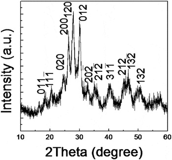
The phase purity of the as-prepared LaVO4 precipitates was characterized by the x-ray diffraction analysis. All the x-ray diffraction peaks of the as-prepared samples shown in Fig. 1 were indexed as arising from the pure monoclinic phase with lattice constant a = 7.043Å, b = 7.279 Å, c = 6.721 Å, β = 104.86° (JCPDS 50-0367).
XRD pattern of LaVO4 nanoparticles.

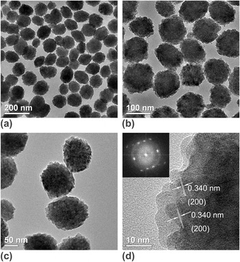
Figures 2(a)–2(c) show TEM images of the as-prepared LaVO4 nanoparticles with different magnifications. The TEM images show that the LaVO4 nanoparticles are slightly elliptical with an aspect ratio about 1.15. LaVO4 nanoparticles have uniform morphology with a narrow size distribution (105 ± 9 nm). The TEM and HRTEM images also show that the larger nanoparticles comprised many smaller plate-like nanoparticles. HRTEM image of a selected nanoparticle [Fig. 2(d)] displayed clear crystal fringes of the nanoparticle, revealing that the prepared LaVO4 nanoparticles are of high crystallinity, which was also confirmed by the typical single crystal diffraction dots in the FFT-pattern. Further calculation on the crystal fringes revealed that the distance of the lattice planes (0.34 nm) extend on the same direction in each part of the nanocrystal, which could be {200} planes of monoclinic phase LaVO4. Therefore, the larger nanoparticles can be proposed as an aggregation of many small nanoparticles that attached with the same orientation.
(a)–(c) TEM images of LaVO4 nanoparticles at different magnifications. (d) HRTEM image of LaVO4 nanocrystal; the inset shows the fast Fourier transform pattern.

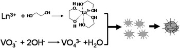
Based on the TEM and HRTEM analysis, the formation process of the LaVO4 nanoparticles is proposed to follow a two-step nucleation and aggregation growth process.Reference Libert, Gorshkov, Goia, Matijevi and Privman23, Reference Privman, Goia, Park and Matijevié24 As shown in Fig. 3, the La3+ ions have a strong coordination effect with the EG molecules. In the first step, when La3+ ions reacted with the VO43− ions, nano-crystalline LaVO4 formed in the supersaturated solution. The EG molecules were attached on the surface of the newly formed LaVO4 nanoparticles. In the second step, the newly formed LaVO4 nanoparticles self-assembled with each other to form a secondary particle because of the surface energy minimization principle. The EG ligands on the surface effectively control the growth rates and the self-assembly modes of the nano-crystalline precursors, which result in nanoparticles with relatively uniform size and morphology.
An illustration of LaVO4 nanoparticle formation process.

This synthetic method is universal for the synthesis of the family of lanthanide orthovanadate nanomaterials. The XRD patterns and SEM images of as-prepared LnVO4 (Ln = Pr to Lu) samples are shown in Fig. S2. For LaVO4, the XRD patterns can be indexed as the pure monoclinic phase of LaVO4 (JCPDS 50-0367). For PrVO4 to YbVO4, the samples were well assigned to the pure tetragonal phase. The peak positions and relative intensities in those samples were almost identical, which indicated that this class of compounds shares a similar crystal structure. A very small difference in 2θ values of XRD peaks can be seen, which gradually increases from PrVO4 to YbVO4, indicating the gradual shrinkage of the crystal structure according to the atomic number. For LuVO4, no obvious peaks were found from the XRD pattern, revealing that the sample is non-crystalline.
This gradual variation in crystal structure of LnVO4 compounds is believed to be caused by the decrease of the ionic radii of Ln3+ ions. As is well known, LnVO4 compounds adopt two kinds of crystal structure: monazite-type and zircon-type. For monazite-type crystal structure, the lanthanide elements adopt a nine oxygen coordination structure, whereas for the zircon-type crystal structure, the lanthanide elements adopt an eight oxygen coordination structure.Reference Fan, Song, Bu, Sun and Zhao6 La3+ ions prefer the higher coordination number with larger ionic radii, which led to the formation of monoclinic crystal structure. For Pr3+ to Yb3+ ions, the lanthanide ions have a tendency to form the zircon-type structure with a relatively low coordination number with decreasing ionic radii. However, for Lu3+ ions, the crystallization process is more difficult because of the larger steric hindrance arising from the smallest ionic radius compared with other lanthanide elements, which leads to the non-crystalline product.
As discussed earlier, the formation process of these LnVO4 nanoparticles followed a nucleation and self-assembly growth mode. TEM observation confirmed that the nanoparticles comprised many small primary nano-sized precursors. Figure S2 shows typical SEM images of LnVO4 (PrVO4 to LuVO4) nanoparticles. It can be seen that LnVO4 nanoparticles have uniform morphology and narrow size distribution. The morphology of the LnVO4 nanoparticles has shown gradual changes in growth modes with decreasing ionic radii. For PrVO4 to EuVO4, the LnVO4 nanoparticles are spherical [Figs. S2(a), S2(c), S2(e), and S2(g)]; for GdVO4 to DyVO4, the products are elliptical [Figs. S2(i), S2(k), and S2(m)]; for HoVO4 to YbVO4 the samples have a roundish-sheet morphology [Figs. S2(o), S2(q), S2(s), and S2(u)]. HoVO4 samples were roundish nanodisks with an average size of about 180 nm. Obvious branched growth modes of HoVO4 nanodisks can be found (Fig. S4), which could be ascribed a transitional morphology from spherical to flake-like morphology. The morphologies of ErVO4 samples are still nanodisks, and few branched morphologies were found by careful observation. TmVO4 and YbVO4 nanoparticles shared the similar roundish-sheet morphologies, but the thickness of the nanoflakes became thinner and the edges were broken. This edge-damage phenomenon is similar to the results reported by Liu et al.Reference Deng, Yang, Xiao, Gong and Wang14 The as-prepared LuVO4 sample were irregular nanoparticles. As LnVO4 compounds have an identical anion, the crystal structures and physical properties are clearly influenced by the cations. This morphologic evolution of LnVO4 nanomaterials according to atomic number is proposed to have close relationship with the transformation of lanthanide elements in the coordination ability and ionic radius.
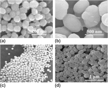
The acidity of the synthetic system proved to greatly influence the morphology of the as-prepared products. Figures 4(a) and 4(b) displays the LaVO4 nanomaterials synthesized at different acidity. Slightly elliptical nanoparticles (aspect ratio 1.15) have been obtained at pH 6, whereas elliptical nanoparticles (aspect ratio 1.57) were formed at pH 3 (1.0 ml, 1 M HCl was added).

SEM images of LaVO4 nanomaterials synthesized at (a) pH 6 and (b) pH 3; SEM images of PrVO4 nanomaterials synthesized at (c) pH 6 and (d) pH 3.

As shown in Eq. (1), the formation state of the V–O ions is influenced by pH. By tuning the acidity in the system, the existence state of V–O ions can be turned, which may influence the morphology of the products. Relatively high acidity in the synthetic system is responsible for the elliptical growth of LnVO4 nanoparticles. Further investigations revealed that the acidity of the synthetic systems have a general influence rules for other lanthanide orthovanadates. Figures 4(c) and 4(d) display the SEM images of PrVO4 nanomaterials derived at different acidity. Spherical and elliptical PrVO4 nanoparticles could be selectively synthesized by changing the acidity of the synthetic system.
The high content of EG and the strong interaction between Ln3+ ions and EG are responsible for the formation of nanomaterials with uniform morphology and narrow size distribution. In the typical synthesis, the molar ratio of Ln3+/VO43− is 2:1. The Ln3+ ions are excessive compared with VO43− ions according to the stoichiometric proportion. Experiments revealed that the higher concentration of lanthanide ions is necessary to achieve a narrow size distribution of the products. If the reactant molar ratio was 1:1 according to the stoichiometric proportion, only irregular nanoparticles were formed, which indicates that the interaction between lanthanide ions and EG is important for the formation of LnVO4 nanoparticles with uniform morphology.
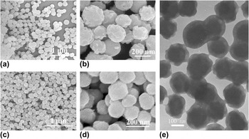
With the reactant molar ratio of 4:1, core-shell-structured nanomaterials can be obtained. Figure 5 shows SEM and TEM images of EuVO4 nanomaterials. At the reactant ratio of 2:1, the as-prepared EuVO4 samples were spherical nanoparticles with smooth surface [Figs. 5(c) and 5(d)]. At the reactant molar ratio of 4:1, the SEM images [Figs. 5(a) and 5(b)] show that the as-prepared nanoparticles are covered by ragged layers and the surface is rough. From TEM [Fig. 5(e)], the core-shell structure of the product can be confirmed. The driving force for the formation of core-shell structure arises from the excessive Eu3+ adsorbed and the epitaxial growth on the surface of EuVO4 nanoparticles. However, experiments found that this epitaxial growth mode is not suitable for all lanthanide elements.
The influence of reactant molar ratio: (a) and (b) SEM images of EuVO4 samples obtained at n(Eu3+):n(VO3−) = 4:1; (c) and (d) SEM images of EuVO4 samples obtained at n(Eu3+):n(VO3−) = 2:1; (e) TEM image of the core-shell structure.

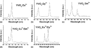
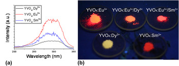
LnVO4 compounds are a family of useful fluorescent host materials. YVO4 was selected as a host materials, and Eu3+, Dy3+, and Sm3+ ions were selected as excited ions to investigate the fluorescent properties of lanthanide orthovanadates. Annealed at high temperature (600 °C) for 2 h, the nanomaterials maintained their morphologies without aggregation. The emission spectra of the annealed Ln3+-doped YVO4 materials under the 270 nm excited UV irradiation are shown in Fig. 6. YVO4:Eu3+ phosphor exhibits typical red fluorescent light under UV irradiation. The characteristic emission peaks located at ≈593 and 618 nm correspond to the typical emissions for 5D0→7FJ (J = 1, 2) transition [Fig. 6(a)]. For YVO4:Dy3+ phosphor, the emission peaks were at ≈483 and 572 nm, corresponding to the 4F9/2→6H15/2 and 4F9/2→6H13/2 energy level transition, respectively [Fig. 6(b)]. For YVO4:Sm3+ phosphor, the emission spectra have typical peaks at ≈564, ≈602, and ≈645 nm, corresponding to the 4G5/2→6H5/2, 4G5/2→6H7/2, and 4G5/2→6H9/2 energy level transition, respectively [Fig. 6(c)]. The excitation spectra were recorded as shown in Fig. 7(a) by controlling the emission peaks of Eu3+-, Dy3+-, and Sm3+-doped YVO4 at 618, 572, and 602 nm, respectively. It can be seen that the absorption spectra present an intense broad band at the region of 260–340 nm, which can be attributed to the charge transfer of VO43− ions, revealing that the VO43− ions play an important role as energy transfer carriers.Reference Wang, Qin, Zhang, Wang, Wei, Zhu and Kim25, Reference Wang, Xue and Liu26 From the investigation of emission spectra, it is clear that the as-prepared YVO4 nanomaterials are good host materials for various luminescent ions.
Fluorescent spectra of Ln3+-doped YVO4 under 270 nm excited UV irradiation.

(a) The excitation spectra of Ln3+ ions–doped YVO4; setting the emission peaks of Eu3+-, Dy3+-, and Sm3+-doped YVO4 at 618, 572, and 602 nm, (b) photograph of Ln3+ ions–doped YVO4 powders under UV irradiation.

To demonstrate the fluorescent diversity of lanthanide-doping lanthanide orthovanadate phosphors, different lanthanide ions were co-doped into the crystal structure of YVO4. Figures 6(d) and 6(e) display the emission spectra of Eu3+/Dy3+ and Eu3+/Sm3+ co-doped YVO4 phosphors, respectively. From the emission spectra, it can be seen that the emission peaks of the doping ions were independent and did not interact. As a result, the multi-color emissions were achieved by co-doping different lanthanide ions into the host materials. Figure 7(b) shows the photograph of as-prepared YVO4 phosphors under the irradiation of UV lamp. By doping with different lanthanide ions, the emission properties of phosphors can be effectively tuned. Lanthanide orthovanadate nanomaterials were demonstrated to be excellent host materials for various lanthanide ions. The doping approach of lanthanide ions has been demonstrated to be an effective method to generate a large number of emission spectra by tuning the kinds and content of the doping ions.
IV. CONCLUSIONS
A general solvothermal approach for the preparation of lanthanide orthovanadate nanomaterials was developed in this article. The obtained LnVO4 nanoparticles have narrow size distribution. The morphologies transform from sphere to ellipse and platelet-shaped particles depending on the ionic size. And the crystal structure of LnVO4 nanoparticles changed from monoclinic-type to zircon-type structure with the decreasing of Ln ionic radius. The LnVO4 nanoparticles follow a nucleation and aggregate formation mechanism. The shapes and compositions of LnVO4 nanoparticles could be controlled by tuning the pH and the reactants ratio. The as-prepared lanthanide orthovanadates were good fluorescent host materials. The emission properties can be monitored by varying the kinds and content of doping ions. This approach should be an effective method to generate a large number of emission spectra and to get phosphors with desirable properties.
ACKNOWLEDGMENTS
This work was supported by NSFC (90606006), the Foundation for the Author of National Excellent Doctoral Dissertation of China, and the State Key Project of Fundamental Research for Nanoscience and Nanotechnology (2006CBON0300).