Introduction
There are currently very few Ce4+-dominant mineral species approved by the International Mineralogical Association's Commission on New Minerals, Nomenclature and Classification (IMA–CNMNC): cerianite-(Ce) (CeO2, Fm $\bar{3}$ m, Graham, Reference Graham1955), dyrnaesite-(La) (Na8Ce4+La2(PO4)6, Pnma, Rønsbo et al., Reference Rønsbo, Balić-Žunić and Petersen2017; Balić-Žunić, Reference Balić-Žunić2017), håleniusite-(La) (La(1–y)[Ce3+(y–x)Cex4+]O(1+x)F(1–x), Im3m, Holtstam et al., Reference Holtstam, Grins and Nysten2004) and stetindite-(Ce) (Ce4+(SiO4), I41/amd, Schlüter et al., Reference Schlüter, Malcherek and Husdal2009). Cerianite-(Ce) and stetindite-(Ce) are isostructural with U4+ and Th4+ oxides and zircon, respectively, and the oxidation state of Ce4+ is therefore crystal-chemically constrained. Håleniusite-(La) is isostructural with cerianite-(Ce), but the amount of Ce4+ depends on the dominance of O over F, which is not demonstrated without doubt because of analytical problems (probably related to interference between Ce and F spectra lines in wavelength-dispersive spectroscopy mode). Armellinoite-(Ce), Ca4Ce4+(AsO4)4⋅H2O, is the fifth mineral containing Ce4+ discovered so far. The name honours Gianluca Armellino (born 1962) of Millesimo, Savona province, Liguria, Italy. Mr. Armellino is an avid and sophisticated mineral hunter and systematic collector specialising in the minerals of Liguria and other specific localities. He is the discoverer of the present new mineral species as well as the co-discoverer of poppiite, cortesognoite, molinelloite and piccoliite. Furthermore, Mr. Armellino also first found more than 40 mineral species new for the Liguria region. Some of them (bariopharmacoalumite, barnesite, brandtite, calderónite, čechite, franciscanite, gageite, hummerite, natrodufrénite, norrishite, roscoelite, schröckingerite and talmessite) represented the first discovery for either Italy or Europe. He is author or co-author of more than 20 articles mainly concerning the above-mentioned new finds, including that on the type locality of the new species studied, which was first briefly described as an unnamed mineral by Kolitsch et al. (Reference Kolitsch, Ciriotti, Cadoni, Armellino, Piccoli, Ambrino, Blass, Odicino and Ciuffardi2011, Reference Kolitsch, Ciriotti and Blaß2013). In keeping with general nomenclature guidelines, its name must include a Levinson suffix. Therefore, the mineral name must be armellinoite-(Ce). Similarly, the originally approved names of cerianite and stetindite were subsequently changed into cerianite-(Ce) (Nickel and Mandarino, Reference Nickel and Mandarino1987) and stetindite-(Ce) (Hålenius et al., Reference Hålenius, Hatert, Pasero and Mills2016). The mineral and its name were approved by the IMA–CNMNC (IMA2018-094, Cámara et al., Reference Cámara, Ciriotti, Kolitsch, Bosi, Bittarello, Brizio, Vignola and Blass2018). The holotype material (consisting of a small chip of rock, a probe mount and the crystal used for determination of crystal structure, optical properties and powder X-ray diffraction data) is deposited in the Mineralogical Collection of the Museum of the Earth Sciences Department of the University of Milano (catalogue number MCMGPG-H2018-003).
Occurrence
The sample containing armellinoite-(Ce) was collected by Gianluca Armellino in March 2008 in the dumps of the Montaldo di Mondovì mine, Frazze Rocconi, Borgata Oberti, Montaldo di Mondovì, Corsaglia Valley, Cuneo Province, Piedmont, Italy (~44°19'28“N, 7°51'8“E). Beginning in 1838 this mine worked an Fe–Mn ore deposit for iron (Sismonda, Reference Sismonda1841a, Reference Sismonda1841b; Conti, Reference Conti1873), though operations had definitely ceased by the mid 1950s. The deposit is hosted in metaquartzites and quartz-bearing marbles of the ‘Klippen of Deviglia’ Unit (Vanossi, Reference Vanossi1980) of the Mesozoic coverage of the innermost Brianzonese Zone (Dalla Giovanna and Vanossi, Reference Dalla Giovanna and Vanossi1991; Vanossi et al., Reference Vanossi, Cortesogno, Galbiati, Messiga, Piccardo and Vannucci1986). The deposit is mentioned in the literature with different names, not all fully correct, such as Ponte dei Gorrazzi (located upstream), Molinetto and Roccon (nearby villages) and Borgata Oberti (very close) (Piccoli et al., Reference Piccoli, Kolitsch, Blaß and Ciriotti2007a; Kolitsch et al., Reference Kolitsch, Ciriotti and Blaß2013). The deposit consists of thin (5 cm) layers, boudins, massive recrystallised braunite (up to 20 cm thick) and, subordinately, mineralised veins in marbles. Secondary hematite-bearing veinlets cross-cut both metaquartzites and marbles (Cabella et al., Reference Cabella, Lucchetti and Marescotti1995). Mastrangelo and Natale (Reference Mastrangelo and Natale1977) described the mineralisation as a layer interposed between Werfenian quartzites and dolomitic limestones of the Middle Trias, which shows a thickness of 5–6 m and is, in substance, a metaquartzite, more or less feldspathic and phyllitic, abundantly impregnated by iron and manganese oxides, with thin intercalations consisting only of oxides. The mineralisation consists of cryptomelane-group minerals ± hematite ± braunite, with variable amounts of quartz, calcite, Mn- and Fe-rich muscovite and aegirine (Cabella et al., Reference Cabella, Lucchetti and Marescotti1999; Kolitsch et al., Reference Kolitsch, Ciriotti, Cadoni, Armellino, Piccoli, Ambrino, Blass, Odicino and Ciuffardi2011; Giai, Reference Giai2020). Pale pink montmorillonite can be considered the indicator mineral for rare species, mainly arsenates.
From a geological perspective, starting from the Cambrian–Ordovician, the deposition of pelitic–arenaceous sediments was followed by the setting of numerous intrusive bodies, both basic and acid. Later, this succession was affected by two metamorphic events: one between the Ordovician and the Silurian; a second one attributable to the Variscan orogeny in the amphibolitic facies, which led to the formation of micaschists and bodies of orthogneiss, eclogites and amphibolites. This polymetamorphic complex was then involved in the post-Variscan extensional tectonics, which gave rise to the formation of different palaeogeographic environments, including the formation of oceanic crust. In these extensional tectonics, datable in two events to the Upper Carboniferous and the Permian, along with the deposition of sediments – mainly of a silico-clastic character –, masses of volcanic rocks are set, both effusive (rhyolites/rhyodacites and andesites) and intrusive (granodiorites). The area was affected by a metamorphism that peaked at blueschist-facies conditions, marked at the Montaldo mine by widespread aegirine, and in some cases up to the development of a jadeite–quartz paragenesis (Vanossi et al., Reference Vanossi, Cortesogno, Galbiati, Messiga, Piccardo and Vannucci1986; Cabella et al., Reference Cabella, Cortesogno, Gaggero and Lucchetti1994; Piana et al., Reference Piana, Musso, Bertok, D'atri, Martire, Perotti, Varrone and Martinotti2009).
The first mineralogical description of the Montaldo mine is provided by Jervis (Reference Jervis1873) who reports ‘manganoan-oligist’ (hematite) and braunite. Cabella et al. (Reference Cabella, Gaggero and Lucchetti1992, Reference Cabella, Lucchetti and Marescotti1995, Reference Cabella, Lucchetti and Marescotti1999) give a description of the Montaldo deposit from the mineralogical–petrological and genetic points of view and report the presence of mineral phases bearing rare earth elements (REE). Up to 32 mineral species have been described for this mine (Giai, Reference Giai2020), which is also the type locality of another arsenate: piccoliite (NaCaMn3+2(AsO4)2O(OH), Kolitsch, Reference Kolitsch2008, Cámara et al., Reference Cámara, Biagioni, Ciriotti, Bosi, Kolitsch, Paar, Blass and Bittarello2017). Other minerals reported from the mine dump are aegirine, anatase, arsenogoyazite, berzeliite, calcite, chernovite-(Y), cinnabar, cryptomelane, dolomite, gasparite-(La), goethite, hollandite, montmorillonite, muscovite (alurgite variety), orthoclase, quartz, rutile, spessartine, svabite, talmessite, tilasite, titanite, todorokite, wallkilldellite (Barelli, Reference Barelli1835; Mariani and Scaini, Reference Mariani and Scaini1978; Cabella et al., Reference Cabella, Lucchetti and Marescotti1999; Piccoli, Reference Piccoli2001; Piccoli et al., Reference Piccoli, Kolitsch, Blaß and Ciriotti2007a,Reference Piccoli, Maletto, Bosio and Lombardo2007b; Kolitsch et al., Reference Kolitsch, Ciriotti, Cadoni, Armellino, Piccoli, Ambrino, Blass, Odicino and Ciuffardi2011, Reference Kolitsch, Ciriotti and Blaß2013; Giai, Reference Giai2020) and wakefieldite-(Y) with an approximate (Y0.40Nd0.15Ce0.15Ca0.15Th0.15)(V0.92As0.08)O4 composition (Cadoni et al., Reference Cadoni, Ciriotti and Ferraris2011).
Some mineral components, such as cinnabar and several primary arsenates, argue in favour of the hypothesis of the formation of large volumes of oxidic Mn and Fe ores on the seafloor of the ‘Piemontese–Ligurian’ oceanic basin, originated by hydrothermal–exhalative processes, which were deposited on top of an ophiolitic sequence. Armellinoite-(Ce) is believed to have formed by reaction between the ore minerals and hydrothermal solutions during subsequent metamorphism.
Appearance and physical properties
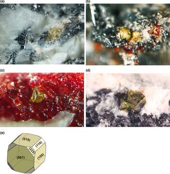
The holotype specimen consists of very small (< 200–100 μm) and rare, pale yellow to brown–yellow pseudo-octahedral translucent crystals embedded in matrix (quartz and hematite, Fig. 1). Streak is white and lustre is resinous to vitreous. The mineral is non-fluorescent. Estimated Mohs hardness is ~3–3.5. It is brittle with irregular fracture and fair cleavage parallel to {110} and {100}. Density could not be measured because of small grain size; calculated density is 4.29 g/cm3, using the empirical formula for armellinoite-(Ce) and the unit-cell volume obtained by single-crystal X-ray diffraction. The same crystal used for the single-crystal X-ray diffraction study was used subsequently to determine the optical properties. Armellinoite-(Ce) is uniaxial (–), ω = 1.795(5), ɛ = 1.765(5) (white light), non-pleochroic. Dispersion was not observed.
(a–d): Four pictures of lemon-yellow to greenish yellow armellinoite-(Ce) crystals in quartz-hematite-cryptomelane/hollandite-tilasite-muscovite-braunite and montmorillonite matrices. Field of view: 6.3, 1.2, 1.6 and 0.4 mm, respectively (collection G. Armellino; photos P. Ambrino / G. Armellino). Pictures (b), (c) and (d) are reproduced with permission from Kolitsch et al. (Reference Kolitsch, Ciriotti, Cadoni, Armellino, Piccoli, Ambrino, Blass, Odicino and Ciuffardi2011). (e): Habit of the crystal shown in photo (d), calculated with KrystalShaper (JCrystalSoft, 2017 v. 1.4.0).

Raman spectrospcopy
The Raman spectrum of armellinoite-(Ce) was obtained using a micro/macro Jobin Yvon LabRam HRVIS, equipped with a motorised x–y stage and an Olympus microscope. The back-scattered Raman signal was collected with a 50 × objective and the spectrum was obtained from a randomly oriented crystal. The 632.8 nm line of a He–Ne laser was used as excitation; laser power was controlled by means of a series of density filters. The minimum lateral and depth resolution was set to a few μm. The system was calibrated using the 520.6 cm–1 Raman band of silicon before each experimental session. Spectra were collected with multiple acquisitions (2 to 6) with single counting times ranging between 20 and 180 s. The spectra were recorded using the Jobin Yvon LabSpec 5 software from 70 to 4000 cm–1.
The Raman spectrum of armellinoite-(Ce) (Fig. 2) is dominated by two strong bands at 838 and 857 cm–1, with a weak shoulder at 820 cm–1 that can be ascribed to the ν 3 symmetric stretching mode of AsO4. A weak band at 945 cm–1 could be ascribable to the antisymmetric stretching mode of AsO4. A band at 507 cm–1 and lower-intensity bands at 468 and 450 cm–1 could be attributed to antisymmetric bending ν 4 of AsO4. Bands at 386 and 360 cm–1 could be attributed to symmetric bending ν 2 of AsO4. Lattice, CeO8 and CaO8 polyhedral modes are present in the range below 360 cm–1 (331, 315, 278, 187, 151, 132 and 174 cm–1). A weak band at 1651 cm–1 in the Raman spectrum (inset A in Fig. 2) is related to the bending vibrations of H2O groups, whereas a series of weak bands observed in the range between 2800 and 3721 cm–1 (inset B in Fig. 2) are related to stretching of O−H bonds of the H2O groups.
Raman spectrum of armellinoite-(Ce) in the 70–1100 cm–1 region with the regions (a) and (b) (insets) at ~1650 cm–1 and between 2800 and 4000 cm–1, respectively.

Chemical composition
Chemical analyses (12) were carried out on a further crystal of the holotype specimen by electron probe microanalysis (EPMA) using a Cameca SX50 instrument at the ‘Istituto di Geologia Ambientale e Geoingegneria, Rome, Italy, CNR’ in wavelength-dispersive spectroscopy (WDS) mode, operating at an accelerating potential of 15 kV, a sample current of 15 nA, a beam diameter of 1 μm and analysing P, Ca, As, Sr, Y, Ce and Th. Analyses were completed at the Earth Science Department of the University of Milano (ESD-UMI) with a Jeol JXA-8200 electron microprobe (WDS mode, 15 kV, 5 nA, beam diameter 3 μm) for elements F, Pr, Nd, Sm, Gd, Dy, Er, Yb, Ho and Lu (15 analyses). The elements Na, Mg, Al, Si, K, Sc, V, Fe, Ba, La, Eu, Tb and U were also analysed but found to be below detection limits. The analyses were subject to ZAF-corrections using the PAP routine (Pouchou and Pichoir, Reference Pouchou, Pichoir, Heinrich and Newbury1991). Cerium was considered as Ce4+ based on bond-valence values (see below). The analysed crystal shows zonation in terms of Ce4+, Nd, Sm, Y and Th4+, with a strong negative correlation between Ca, Y and Ce4+ and a less clear negative correlation between Th4+ and Ce4+. Yttrium, Nd and Sm correlate positively, suggesting the substitution of CaCe4+ by 2REE 3+. The sample showed beam damage with 15 nA and a beam diameter of 1 μm, probably due to evaporation of the H2O content. The damage was largely reduced by the setting at ESD-UMI, however the slightly high total reported in Table 1 may still reflect the contribution due to devolatilisation. The H2O content, whose presence is confirmed by Raman spectroscopy (see above) and the crystal-structure determination, was calculated by stoichiometry: (F + H2O) = 2.00 atoms per formula unit (apfu). The empirical formula, calculated on the basis of 17 anions (O+F), A(Ca3.89Th0.08Sr0.02La0.03)Σ4.02B(Ce4+0.76Nd0.13Y0.08Gd0.03Sm0.02Pr0.01Dy0.01Ho0.01)Σ1.05[(As4.00P0.01)Σ4.01O4]4⋅(H2O0.85F0.15)Σ2.00 leads to the end-member formula Ca4Ce4+(AsO4)4⋅H2O, which requires CaO 25.66, Ce4+O2 19.68, As2O5 52.60, H2O 2.06, total 100 wt.%. The Gladstone–Dale compatibility index is [1 – (K p/K c)] = –0.008 (superior) (Mandarino, Reference Mandarino1979, Reference Mandarino1981).
Analytical data (wt.%) for armellinoite-(Ce).

* H2O was calculated on the basis of (H2O + F) = 1.00 apfu; S.D. – standard deviation.
X-ray crystallography
Powder X-ray diffraction data were obtained from a crystal of the holotype specimen (the same used for the crystal-structure determination and for optical measurements) by the Debye–Scherrer geometry using an Oxford Diffraction Xcalibur single-crystal X-ray diffractometer, operated at 50 kV and 30 mA, with monochromatised MoKα radiation and equipped with a CCD detector at 80 mm from the crystal position. Observed interplanar spacings and intensities of indexed reflections using Highscore (Degen et al., Reference Degen, Sadki, Bron, König and Nénert2014) and d(I) values calculated using Vesta 3.0 (Momma and Izumi, Reference Momma and Izumi2011) are reported in Table 2. The tetragonal (I41/a) unit-cell parameters refined using GSAS (Larson and Von Dreele, Reference Larson and Von Dreele1994) are a = 10.736(2), c = 12.003(4) Å, V = 1383.6(4) Å3 with Z = 4. These values are slightly smaller than those determined from the single-crystal structure determination.
Powder X-ray diffraction data for armellinoite-(Ce).*

* Only calculated reflections with I rel > 3%(I rel) are listed. The strongest lines are shown in bold.
The aforementioned single crystal of armellinoite-(Ce) (0.15 mm × 0.15 mm × 0.18 mm), optically homogeneous and free of inclusions under a transmitted-light polarising microscope, was used to solve the crystal structure of the mineral. A full sphere of X-ray diffraction intensity data up to 2θ = 65.11° was collected at room temperature at the Institut für Mineralogie und Kristallographie, Universität Wien, with a Nonius KappaCCD single-crystal X-ray diffractometer equipped with a CCD area detector and using monochromatised MoKα radiation (50 kV and 30 mA). Crystal data and data collection information are reported in Table 3.
Crystal data and experimental conditions for single-crystal XRD study.

* Number of reflections I>2σ(I) = 1136.
Structure solution and refinement
The crystal structure was solved by direct methods (SIR97, Guagliardi et al., Reference Guagliardi, Moliterni, Polidori and Spagna1999) and refined to R 1 = 0.023 in SHELXTL-2018/3 (Sheldrick, Reference Sheldrick2015). Three cation sites (T, A and B) and four anion sites were detected in the asymmetric unit. Details on the data collection and structure refinement are reported in Table 3. Final fractional atom coordinates, site occupancies, incident bond-valence values (in valence units, vu; bond-valence parameters from Gagné and Hawthorne, Reference Gagné and Hawthorne2015) and isotropic displacement parameters are reported in Table 4. Anisotropic displacement parameters are reported in Table 5. Bond distances are given in Table 6. Neutral atom scattering curves from The International Tables for Crystallography (Wilson, Reference Wilson1992) were used for the refinement of these sites: T = As; A = Ca; B = Ce; and O = O, with observed mean intrapolyhedral bond lengths of 1.689, 2.451 and 2.367 Å, respectively (Table 6). The observed <T−O> value is in good agreement with the grand mean value reported for arsenates in the modern literature (1.687(27) Å, Gagné and Hawthorne, Reference Gagné and Hawthorne2018). Refinement of the cation-site occupancies showed only small deviations from full occupancy of A and B. Therefore, these site occupancies were modelled considering both Ca and Ce scattering curves and constraining their relative fractions to be equal to 1. The unconstrained occupancy of the T site showed a value 31.67 electrons per site (eps), in agreement with some P measured by EPMA. With regard to the A site, the observed mean bond length and the site scattering of 22.3 eps is compatible with a site dominated by Ca with minor amounts of a heavier scatterer such as Th4+ and La according to EPMA data and the polyhedral size, because the measured amounts of Sr (= 0.02 apfu) are too low for Sr2+ to be the sole substituent of Ca2+. The [8]B-centred polyhedron is smaller than the [7]A-centred polyhedron and, following the higher refined B-site scattering value (51.96 eps), a REE is expected to be also present at the B site, but with a lower scattering power than that of Ce4+. As confirmed by the chemical analyses, indicating B(Ce4+0.76Nd0.13Y0.08La0.03Gd0.03Sm0.02Pr0.01Dy0.01Ho0.01)Σ1.08 apfu, the presence of 0.08 Y apfu helps to lower the site scattering and the polyhedron size is more compatible with Ce4+. At convergence of the model with four O sites, the first maximum that was found in the difference-Fourier map showed a low electron density and a bond distance (2.75 Å) compatible with it being an additional oxygen atom (water molecule) bonded to the A site. This maximum had close symmetrically equivalent positions at only 0.73(3) Å (Table 6), clearly indicating a split site. This site was added to the model as an O5 anion site (using the oxygen scattering curve) and its partial occupancy was refined with the constraint of dividing its multiplicity by four. The refinement yielded full occupancy within error limits. The next maximum evident in the difference-Fourier map was at a distance of 0.95 Å from O5 and was added to the model as an H atom and refined with an isotropic displacement parameter constrained to be equal to 1.2 times that obtained for the O5 site. The crystallographic information file has been deposited with the Principal Editor of Mineralogical Magazine and is available as Supplementary material (see below).
Sites, site occupancies, bond-valence sums (vu), fractional atom coordinates (Å) and equivalent isotropic displacement parameters (Å2) for armellinoite-(Ce).

Notes: Bond-valence parameters from Gagné and Hawthorne (Reference Gagné and Hawthorne2015) were used assuming end-member composition. The temperature factor has the form exp(–T) where T = 8π2U(sin(θ)/λ)2 for isotropic atoms. $BVS with H contribution = 2.09 vu.
Anisotropic displacement parameters* for armellinoite-(Ce) (Å2).

* The temperature factor has the form exp(–T) where T = 2π2Σij(h (i)h (j)U (i,j)a*(i)a*(j)).
Selected bond distances (Å) and distortion parameters* for armellinoite-(Ce).

* Mean quadratic elongation (λ) and the angle variance (σ2) (Robinson et al., Reference Robinson, Gibbs and Ribbe1971).
Bond-valence calculations agree with a cation with slightly higher charge than 2+ at the A site, thus confirming Ca dominance at this site with minor amounts of Th4+. Regarding the B site, calculations using bond-valence parameters for either Ce4+ or Ce3+ (from Gagné and Hawthorne, Reference Gagné and Hawthorne2015) both yield bond-valence sums (3.72 vu and 4.12 vu, respectively) consistent with Ce4+. The structure topology and charge arrangement evidently require relatively small tetravalent cations at the B site.
Structure description
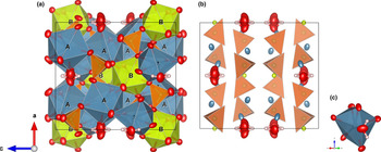
The structure of armellinoite-(Ce) has isolated TO4 (T = As5+) tetrahedra that link Ca2+ or Ce4+ cations via common oxygen ligands, to form 2D blocks or double-layered (DL) structural units parallel to (001) (Fig. 3b). The DL units are formed by pairs of AsO4 tetrahedra arranged opposite to each other and oriented in opposite directions. It corresponds to the type DL7 of DL vanadate of Kovrugin et al. (Reference Kovrugin, Siidra, Zaitsev, Spratt, Shilovskikh, Agakhanov and Turner2016), which was so far represented only by pottsite, ideally (Pb3Bi)Bi(VO4)4⋅H2O (Williams, Reference Williams1988; Kovrugin et al., Reference Kovrugin, Siidra, Zaitsev, Spratt, Shilovskikh, Agakhanov and Turner2016). In fact, this mineral is isostructural with armellinoite-(Ce).
Polyhedral representation of the crystal structure of armellinoite-(Ce). Blue = AO8 polyhedra, pale green = BO8 polyhedra, orange = TO4 tetrahedra, red ellipsoids = oxygen atoms; small grey spheres = hydrogen atoms. Displacement ellipsoids are drawn at the 90% probability level. (a) Polyhedral representation (view along the a axis; the unit cell is outlined); (b) schematic view showing the ‘double layers’ of TO4; (c) detail of the H2O group forming one of the ligands of the A site. Figure drawn with Vesta 3.0 (Momma and Izumi, Reference Momma and Izumi2011).

Both armellinoite-(Ce) and pottsite have a pentavalent cation at the T site. While pottsite has a (2+33+2) charge combination, armellinoite-(Ce) has (2+44+). Different charge arrangements are found in synthetic compounds with the topology of Na4Th(MoO4)4, i.e. (1+44+). However, these have T 6+O4 tetrahedra required for electroneutrality (Sillén and Sundvall, Reference Sillén and Sundvall1943; Klevtsova et al. Reference Klevtsova, Gaponenko, Glinskaya, Zolotova, Podberezskaya and Klevtsov1979; Ovanysian et al., Reference Ovanysian, Ishkakova, Efremov and Trunov1988; Dahale et al., Reference Dahale, Keskar and Singh Mudher2006). In armellinoite-(Ce), Th4+ (<0.29 apfu) and possibly very minor trivalent REE substitute for (Ca+Sr) and (Ce4++Y3+) and are equally distributed in two different sites (A and B), both with rather distorted square antiprismatic coordination. The suggested cation distribution at these sites is A(Ca3.89Th0.08Sr0.02La0.03)Σ4.02 and B(Ce4+0.76Nd0.13Y0.08Gd0.03Sm0.02Pr0.01Dy0.01Ho0.01)Σ1.05 (see above). In the case of pottsite, bond-valence calculations, structure refinement and chemical analyses led Kovrugin et al. (Reference Kovrugin, Siidra, Zaitsev, Spratt, Shilovskikh, Agakhanov and Turner2016) to the following site assignment: (Pb0.500Bi0.380Ca0.030) and (Pb0.068Ca0.032) for two split A(1) and A(2) sites, respectively, corresponding to the single A site of armellinoite-(Ce), having thus a mean charge > +2. The Bi site in pottsite is occupied by 0.84 Bi apfu and has a distorted square antiprismatic coordination environment, showing a rather symmetrical coordination that indicates a low degree of stereochemical activity of the lone electron pair of the Bi3+ ion (Kovrugin et al., Reference Kovrugin, Siidra, Zaitsev, Spratt, Shilovskikh, Agakhanov and Turner2016). The presence of vacancies on the Bi site is confirmed by bond-valence calculations and justified by charge arrangements.
The O5 anion site (H2O) is partially occupied [refined occupancy 0.273(7), therefore fixed to 0.25] and positionally disordered [O5⋅⋅⋅O5' = 0.73(3) Å]. This would result in a refined water content of ~1.3 H2O apfu. However, a limited substitution of H2O by F occurs at the O5 site and brings the actual (H2O + F) content closer to 1.00 apfu. The H2O group at the O5 site is a ligand of the cation at the A site (Fig. 3c). Conversely, an ordered model has been described for pottsite by Kovrugin et al. (Reference Kovrugin, Siidra, Zaitsev, Spratt, Shilovskikh, Agakhanov and Turner2016; see their figure 2d), where the O5 anion (labelled as OW) is at the 4a Wyckoff position with coordinates (0, ¼, ⅛). The disordered model in armellinoite-(Ce) implies that there are different local atomic arrangements in which one cation at the A site is bonded to one H2O at O5 with a bond length of 2.75 Å, whereas another of the other three adjacent A sites would coordinate with the same H2O group at 2.84 Å. The other two cation sites are at 3.49(4) Å and 3.55(2) Å, and provide no bonding. This disordered arrangement explains the multiple bands observed by Raman spectroscopy (Fig. 2, inset b). If we consider the different possible arrangements in terms of donor (O5) and acceptor distances (O3 and O4), and the empirical calibration of Libowitzky (Reference Libowitzky1999), 3592−304×109⋅exp(−d(O⋅⋅⋅O)/0.1321), we obtain 3218, 3561, 3568 and 3574 cm–1 for the O⋅⋅⋅O distances 2.71, 3.04, 3.07 and 3.11 Å, respectively. The stronger Raman shift at 3721 cm–1 is probably related to a more frequent atomic arrangement in which the H bonded to the O at O5 is not bonded locally to any other anion site. Some of those bands are present in the inset (b) of Fig. 2. In pottsite, the water content ranges between 0.8 and 1.0 H2O apfu (Kovrugin et al., Reference Kovrugin, Siidra, Zaitsev, Spratt, Shilovskikh, Agakhanov and Turner2016) and Raman spectra of pottsite from the type locality reported by Frost et al. (Reference Frost, Henry, Weier and Martens2006) show no Raman bands in the water region, yet the infrared (IR) spectrum of the same material (Frost et al., Reference Frost, Henry, Weier and Martens2006) has a band distribution similar to the one observed by Raman in armellinoite-(Ce), with the stronger IR band at 3695 cm–1, similar to the stronger observed Raman band for armellinoite-(Ce). Other IR bands observed by Frost et al. (Reference Frost, Henry, Weier and Martens2006) for pottsite are 3648, 3621, 3604 and 3400 cm–1, very similar to the Raman shifts observed for armellinoite-(Ce). This raises a question about the correct location of the Ow ( ≡ O5) site in the pottsite structure model, making a disordered model plausible. The high scattering power of the cations in pottsite may have made it more difficult to observe split positions for oxygen atoms. In fact, the model reported by Kovrugin et al. (Reference Kovrugin, Siidra, Zaitsev, Spratt, Shilovskikh, Agakhanov and Turner2016) reports a particularly large isotropic displacement parameter for the Ow site (0.105(11) Å2; see their Table 3), although they also report some Cl at this site (0.07 apfu), that may slightly compensate for the higher charge assigned to the A(1) site due to 0.38 apfu of Bi3+. The measured F content of armellinoite-(Ce) probably compensates the presence of high-charge cations at the A site such as Th4+ and La3+ (see above).
Relation to other species
Armellinoite-(Ce), ideally Ca4Ce4+(AsO4)4⋅H2O, is the first natural alkaline REE arsenate. Along with pottsite, ideally (Pb3Bi)Bi(VO4)4⋅H2O (Williams, Reference Williams1988; Kovrugin et al., Reference Kovrugin, Siidra, Zaitsev, Spratt, Shilovskikh, Agakhanov and Turner2016), it can form the pottsite group (Mills et al., Reference Mills, Hatert, Nickel and Ferraris2009, Table 7).
Comparison of armellinoite-(Ce) and pottsite to become members of the pottsite-group minerals.

Notes: n.d. = not determined.
A series of synthetic compounds are known that have the pottsite and armellinoite-(Ce) topology and space-group type I41/a, although they are reported as anhydrous compounds: Na4Zr(MoO4)4 (Klevtsova et al., Reference Klevtsova, Gaponenko, Glinskaya, Zolotova, Podberezskaya and Klevtsov1979), Na4Pu(MoO4)4, Na4U(MoO4)4 and Na4Th(MoO4)4 (Lazoryak and Efremov, Reference Lazoryak and Efremov1987; Dahale et al., Reference Dahale, Keskar and Singh Mudher2006). Other homeotypic compounds, such as Na5La(WO4)4 (Sillén and Sundvall, Reference Sillén and Sundvall1943) and Na3.68Dy1.44(SeO4)4 (Ovanysian et al., Reference Ovanysian, Ishkakova, Efremov and Trunov1988), have an excess of Na in a position corresponding to that of the H2O group in armellinoite-(Ce) and pottsite. This Na atom is in [4]-coordination with a large mean bond length (2.81 Å), that is more suitable for a larger cation like K+ or an (NH4)+ group. The substitution of Ca by Na in these compounds is helped by the larger (WO4) and (MoO4) polyhedra.
Whereas chemical analyses of armellinoite-(Ce) show the presence of some other lanthanides and actinides, along with Y, possible substitutional series with other trivalent REE (including Ce3+) or even Eu2+ would imply substitution by Ca, leading to a charge-arrangement (2+33+2) as in pottsite. Size requirements would favour a combination of heavy REE (HREE) at the B site and light REE (LREE) at the A site. Substitution of Ca by Sr would further influence the HREE–LREE exchange. Again, by comparison with pottsite, a corresponding vanadate analogue of armellinoite-(Ce) may also be discovered in the future within a V-rich geochemical environment.
Acknowledgements
Henrik Friis, Travis Olds and Pete Leverett are thanked for useful suggestions. F.C. acknowledges financial support by the grant Ricerca Locale 2020, Università di Milano, and from the Italian Ministry of Education (MUR) through the project “Dipartimenti di Eccellenza 2018–2022”. M.E.C. and P.B. acknowledges financial support of AMI – Associazione Micromineralogica Italiana.
Supplementary material
To view supplementary material for this article, please visit https://doi.org/10.1180/mgm.2021.84