INTRODUCTION
Two axiid species, Boasaxius princeps (Boas, 1880) and Leonardsaxius amurensis (Kobjakova, 1937), inhabit Russian waters of the Sea of Japan (Marin, Reference Marin2015). Leonardsaxius amurensis was originally described as Axius spinulicauda amurensis Kobjakova, 1937. This species was later transferred to genera Axiopsis Borradaile, 1903 (Vinogradov, Reference Vinogradov1950), Calocarides Wollebaek, 1908 (Sakai & de Saint Laurent, Reference Sakai and de Saint Laurent1989) and recently placed into the newly described genus Leonardsaxius Sakai, 2011. The species is presently known only from the type locality, Amursky Bay, at 31 m depth and from Vostok Bay, at 5–10 m depth. Both localities are parts of the large Peter the Great Bay. The species is usually found inside burrows made in sand, rocky, gravel or shelly limestone substrates, under large stones or at the base of boulders or rocks; rarely observed on muddy bottoms (Marin, Reference Marin2015).
Our intent was to describe the complete larval development of L. amurensis. Unfortunately, we failed to catch an ovigerous female of this quite rare species. However, based on morphology, two types of axiid larvae occur in the plankton of Peter the Great Bay. Recently, we have shown that the larvae with four posterodorsal spines on the pleonites 2–5 belong to B. princeps and have assumed that the larvae with one posterodorsal spine on the fifth pleonite belong to L. amurensis (Kornienko et al., Reference Kornienko, Korn and Golubinskaya2014). Therefore, we have collected the first zoeae with one spine from zooplankton samples and to confirm that these are indeed the larvae of L. amurensis, we conducted the molecular analysis of adults and larvae.
In the present study, we identified specimens using partial sequences of the mitochondrial cytochrome c oxidase subunit I gene (COI), 12S and 16S rDNAs as genetic markers.
The use of molecular markers has been successful in providing accurate identification of plankton specimens (Pan et al., Reference Pan, McBeath, Hay, Pierce and Cunningham2008; Pardo et al., Reference Pardo, Ampuero and Véliz2009; Ampuero et al., Reference Ampuero, Palma, Veliz and Pardo2010; Marco-Herrero et al., Reference Marco-Herrero, Torres, Cuesta, Guerao, Palero and Abelló2013, Reference Marco-Herrero, Gonzáles-Gordillo and Cuesta2014), different groups of animals, cryptic species or ontogenetic stages exhibiting little differentiation (Hebert et al., Reference Hebert, Cywinska, Ball and deWaard2003; Schindel & Miller, Reference Schindel and Miller2005; Vences et al., Reference Vences, Thomas, Bonett and Vieites2005; Rao et al., Reference Rao, Liston, Crampton and Takeyasu2006; Chen et al., Reference Chen, Tsang, Chong and Chan2014; Marco-Herrero et al., Reference Marco-Herrero, Gonzáles-Gordillo and Cuesta2015), and establishing adult–larval linkages (Tang et al., Reference Tang, Yau and Ng2010; Bracken-Grissom et al., Reference Bracken-Grissom, Felder, Vollmer, Martin and Crandall2012). These markers have proven to be an effective tool in studies of crustaceans (Knowlton & Weigt, Reference Knowlton and Weigt1998; Schubart et al., Reference Schubart, Cuesta, Diesel and Felder2000; Porter et al., Reference Porter, Pérez-Losada and Crandall2005; Barber & Boyce, Reference Barber and Boyce2006; Ahyong et al., Reference Ahyong, Lai, Sharkey, Colgan and Ng2007; Costa et al., Reference Costa, deWaard, Boutillier, Ratnasingham, Dooh, Hajibabaei and Hebert2007; Raupach & Radulovici, Reference Raupach and Radulovici2015; Raupach et al., Reference Raupach, Barco, Steinke, Beermann, Laakmann, Mohrbeck, Neumann, Kihara, Pointner, Radulovici, Segelken-Voigt, Wesse and Knebelsberger2015; Trivedi et al., Reference Trivedi, Aloufi, Ansari and Ghosh2016), not only for the establishing of new species, but also to elucidate the taxonomic validity of closely related species (Schubart et al., Reference Schubart, Diesel and Hedges1998, Reference Schubart, Cuesta, Diesel and Felder2000; Spivak & Schubart, Reference Spivak and Schubart2003; Felder & Robles, Reference Felder, Robles, Martin, Crandall and Felder2009; Bracken et al., Reference Bracken, De Grave, Toon, Felder and Crandall2010; Bracken-Grissom et al., Reference Bracken-Grissom, Felder, Vollmer, Martin and Crandall2012; Meyer et al., Reference Meyer, Weis and Melzer2013; De Grave et al., Reference De Grave, Chan, Chu, Yang and Landeira2015; Wong et al., Reference Wong, Pérez-Moreno, Chan, Frank and Bracken-Grissom2015; Brandão et al., Reference Brandão, Freire and Burton2016).
MATERIALS AND METHODS
Collection of the material
This study was undertaken at the Vostok Marine Biological Station of the Institute of Marine Biology (National Scientific Center of Marine Biology, Russian Academy of Sciences), Vostok Bay (inner Bay of Peter the Great Bay, Sea of Japan). Three specimens of Leonardsaxius amurensis adults were collected in Vostok Bay at a depth of 5–10 m and used for DNA extraction, amplification and sequencing. Zooplankton samples were collected also in Vostok Bay over a depth of 2–2.5 m. Samples were gathered at night-time using a direct light source because decapod larvae have positive phototaxis and are usually aggregating around light. About 150 axiid first zoeae with one posterodorsal spine on the fifth pleonite were collected from plankton samples. Five specimens were used for DNA extraction, amplification and sequencing. The rest of the larvae were used for rearing in the laboratory. In addition, five specimens of Boasaxius princeps first zoea were used for comparative genetic analysis.
DNA extraction, amplification and sequencing
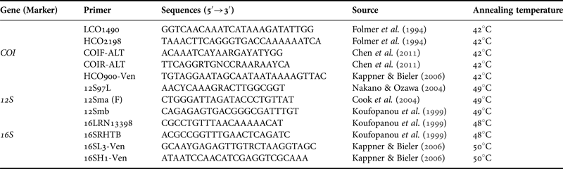
Live specimens of adults and larvae were preserved in 96% ethanol and deposited in the Museum of the Institute of Marine Biology, National Scientific Center of Marine Biology, Russian Academy of Sciences, Vladivostok (catalogue numbers 33224, 33225, 33226, 33227, 33228). Total DNA was extracted from whole larvae or muscle tissues excised from the abdomen of adult individuals using the HotSHOT method (Alasaad et al., Reference Alasaad, Rossi, Maione, Sartore, Soriguer, Pèrez, Rasero, Zhu and Soglia2008). The mitochondrial COI, 12S and 16S genes were amplified and sequenced using the universal invertebrate primer pairs. Primers and their annealing temperatures are given in Table 1.
Primers were used in research.

The PCR (polymerase chain reaction) was performed in 25 µl of reaction volume containing 17.4 µl of water; 2.5 µl of 10× buffer (Evrogen); 2.0 µl of dNTP (concentration 2.5 mM of each, Evrogen); 1 µl of each forward and reverse PCR primer (5 pmol/µl); 0.1 µl of Taq DNA polymerase (5 units/µl, Evrogen); and 1 µl of template DNA solution. The cycling parameters were an initial denaturation step at 94°С (3 min), followed by 30 cycles of denaturation for 45 s at 94°С, annealing for 90 s at 42–49°С (Table 1), and extension for 120 s at 72°С; and a final extension phase at 72°С for 5 min.
Amplification products were applied as templates for sequencing employing the same primers as for PCR and ‘Big Dye Terminator Cycle Sequencing Kit’ v. 3.1 (Applied Biosystems, USA) following the manufacturer's protocol. The cycling parameters were an initial denaturation step at 96°С (1 min), followed by 25 cycles of 10 s at 96°С, 10 s at 42–49°С and 4 min at 60°С. Sequencing reaction products were purified by ethanol precipitation and analysed on an ABI-3130 genetic analyser (Applied Biosystems, ABI, USA).
The contigs were obtained and edited using ChromasPro v. 1.7.6 (http://www.technelysium.com.au/chromas.html). The new sequences determined from larvae and adults of L. amurensis and B. princeps were deposited in GenBank under accession numbers KU707205–KU707215 (COI), KU707181–KU707192 (12S) and KU707193–KU707204 (16S). To compare our dataset with other species of Axiidea deposited in GenBank (NCBI, http://www.ncbi.nlm.nih.gov/) the genetic distances were calculated to construct the phylogenetic trees. To root the trees, the complete mitochondrial sequence of Alpheus distinguendus was used as outgroup (GenBank accession number: GQ892049). In addition, we submitted the information about our samples to the BOLD (project – LEO) (Ratnasingham & Hebert, Reference Ratnasingham and Hebert2013). Both species that we registered got unique BINs (Barcode Index Numbers). This approach clusters DNA barcodes to calculate operational taxonomic units (OTUs) that closely correspond to species (Ratnasingham & Hebert, Reference Ratnasingham and Hebert2013).
Sequences were aligned using Muscle (Edgar, Reference Edgar2004) implemented in the program MEGA 5 (Tamura et al., Reference Tamura, Peterson, Peterson, Stecher, Nei and Kumar2011). The final dataset after alignment, elimination of highly divergent regions (for, 12S and 16S) and trimming to the same length was 637, 311 and 318 bp, for COI, 12S and 16S respectively. Pairwise genetic distances (p-distances) were calculated using MEGA 5. Kimura's two-parameter model (K2P) (Kimura, Reference Kimura1980) was used for the COI gene; uncorrected genetic distances were calculated for the two other genes. All estimations of sequences and distances were performed excluding the outgroup. Phylogenetic trees for single genes were constructed using Bayesian (BI) and maximum likelihood (ML) methods. Bayesian trees were constructed using MrBayes v. 3.1 (Huelsenbeck & Ronquist, Reference Huelsenbeck and Ronquist2001). The HKY + G model was selected to generate Bayesian trees. The parameters set for the BI-tree were random trees = 2; mcmc ngen = 2,000,000; samplefreq = 100; sump burnin = 5000. The models of nucleotide substitution for ML-trees were selected using jModelTest v. 2.1.4 (Darriba et al., Reference Darriba, Taboada, Doallo and Posada2012) under the Hierarchial Likelihood Ratio Test. For 12S and 16S genes, the best-fit model was T3P + G (Tamura 3-Parameter Model). The ML-tree was built using the online software RAxML-HPC v. 8 on XSEDE (Stamatakis, Reference Stamatakis2014) on the web-server CIPRES Science Gateway (http://www.phylo.org/) (Miller et al., Reference Miller, Pfeiffer and Schwartz2010). The neighbour joining approach was also applied to construct trees (NJ-trees) in MEGA 5. The NJ-tree for COI was produced using a K2P model and T3P + G model was used for 12S and 16S trees. Nodal support was measured in RAxML using 1000 bootstrap replicates (Felsenstein, Reference Felsenstein1985).
The sequence data of the three genes was combined and trees constructed using the BI, ML and NJ methods. In these combined analyses, we included only samples for which there were sequences of at least two genes. The sequences were combined using SequenceMatrix v. 1.7.8 (Vaidya et al., Reference Vaidya, Lohman and Meier2011). A tree based on the combined dataset of three mtDNA genes did not show any significant differences with trees based on single genes so the combined trees are not shown here.
Laboratory larval culture
The first zoeae of Leonardsaxius amurensis collected from plankton samples were transferred to 2 l glass vessels with filtered and ultraviolet (UV) sterilized seawater and reared to the megalopal stage at a temperature of 22–23°C and a salinity of 32‰, comparable with the natural conditions. The density of larvae was about 10 specimens per litre. To determine the exact number of zoeal stages, a subset of the larvae was reared individually. The water in the vessels was changed daily. For this purpose, the larvae were concentrated at the edge of the vessel using a direct light source and transferred to another vessel by a pipette. The zoeal stages were fed with newly hatched Artemia sp. twice a day. The mortality of larvae of L. amurensis at earlier stages was not high (about 15% in zoea I); adhesion to water surface noted earlier for Nihonotrypaea petalura (see Konishi et al., Reference Konishi, Quintana and Fukuda1990; Kornienko et al., Reference Kornienko, Korn and Golubinskaya2015) was not observed. The larvae of L. amurensis were fixed in 4% formaldehyde for light microscopy and deposited in the Museum of the Institute of Marine Biology, National Scientific Center of Marine Biology, Russian Academy of Sciences, Vladivostok (catalogue number 31964).
Morphological descriptions
The outlines of the larva and its appendages were drawn using an Olympus CX41 microscope fitted with a camera lucida. Respective measurements were made with an ocular micrometer. The total length of zoea and megalopa (TL) was measured from the tip of the rostral spine to the posterior midpoint of the telson, carapace length (CL) was measured from the tip of the rostral spine to the posterior midpoint of the carapace. Description guidelines by Clark et al. (Reference Clark, Calazans and Pohle1998) were generally followed. Five specimens of each of the first, second and third zoeal stages and three of the fourth and fifth zoeal stages were dissected for morphological investigation. Long plumose setae on the exopods of the maxillipeds and pereiopods are drawn truncated and without setules.
RESULTS
Molecular identification
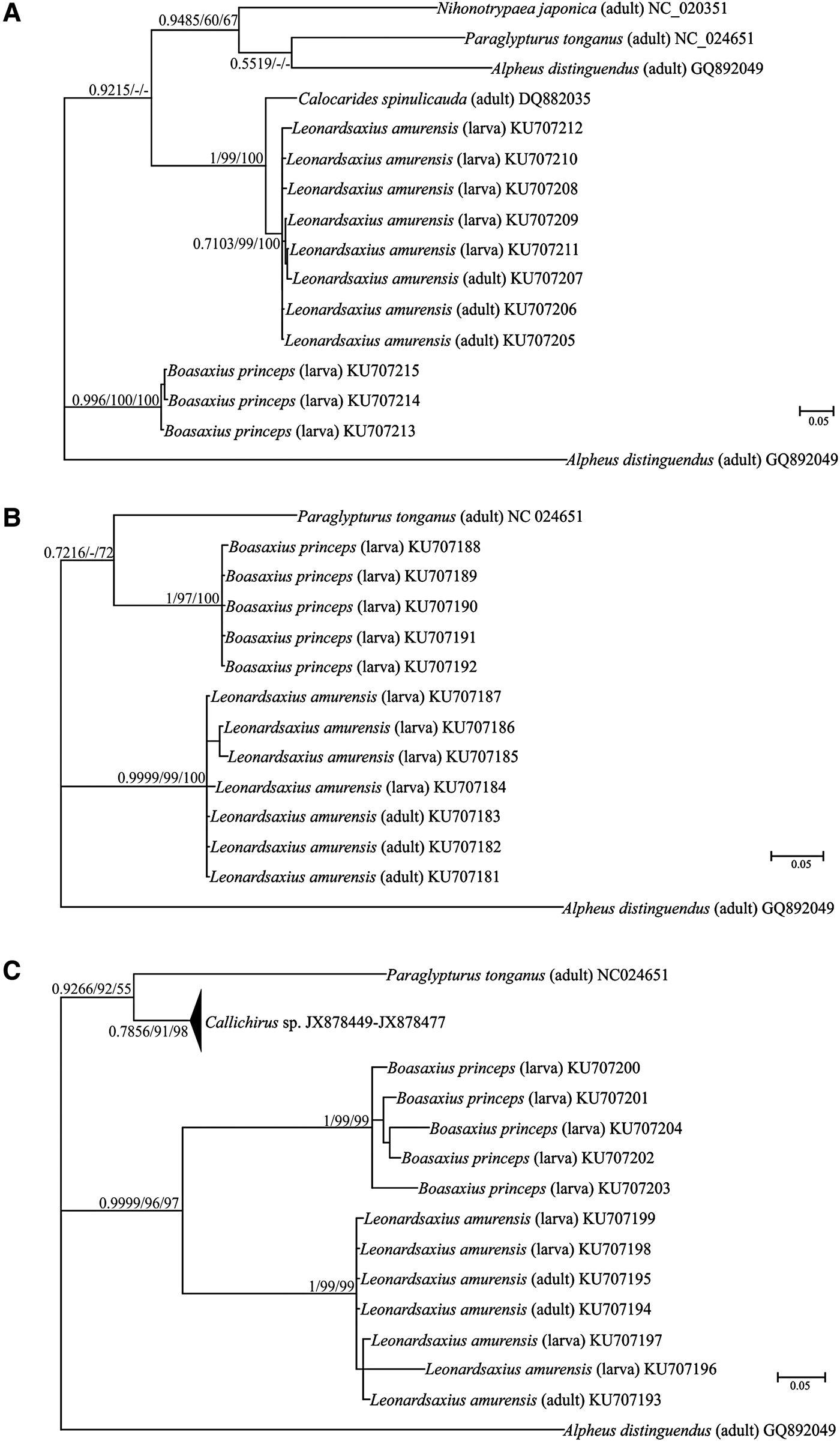
The molecular genetic analysis based on comparison of COI, 12S and 16S mtDNA sequences confirmed that axiid larvae found in the plankton and adult Leonardsaxius amurensis collected in the same area belong to the same species (Tables 2–5, Figure 1).
BA-trees analysis of the COI (A), 12S (B) and 16S (C) sequences for the infraorder Axiidea. Number above or under the branches are Bayesian posterior probabilities and bootstrap values of ML and NJ analysis.

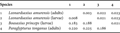
COI uncorrected genetic distances between species belonging to infraorder Axiidea. Above the diagonal is the SD.

12S uncorrected genetic distances between species belonging to infraorder Axiidea. Above the diagonal is the SD.

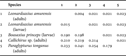
16S uncorrected genetic distances between species belonging to infraorder Axiidea. Above the diagonal is the SD.

Genetic distances within species of infraorder Axiidea. For all sequences uncorrected distances were calculated (distance ± SD).

Average interspecific and intraspecific distances based on three genes are shown in Tables 2–5. The comparison of pairwise genetic distances indicates stronger differences between species of infraorder Axiidea. The interspecifc divergence of each analysed molecular marker is always 10 times higher than the intraspecific genetic distances. Therefore the ‘barcoding gap’ (Meyer & Paulay, Reference Meyer and Paulay2005) can be observed for all the studied taxa. So, according to our data the larvae and adult specimens of L. amurensis belong to the same species. The BIN approach, applied to the COI fragment, confirmed the species identification through BOLD-IDS.
These clear differences between the sequences of each species were the basis for accurate identification of species. In addition, identification was confirmed by BI, ML and NJ analyses with high support values between different species for each marker (Figure 1).
Description of larval stages
SYSTEMATICS
Family Axiidae Huxley, 1879
Genus Leonardsaxius Sakai, Reference Sakai2011
Leonardsaxius amurensis (Kobjakova, 1937)
(Table 6, Figures 2–9)
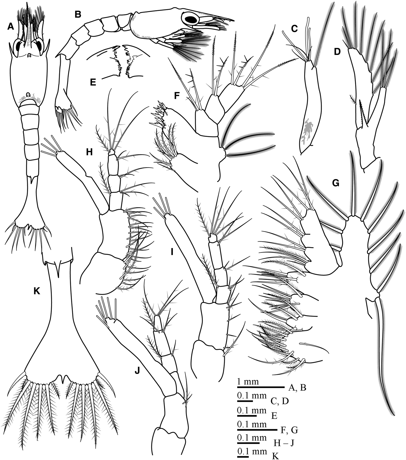
First zoea (Figure 2)
Leonardsaxius amurensis (Kobjakova, 1937), first zoea: (A) Dorsal view; (B) lateral view; (C) antennule; (D) antenna; (E) mandibles; (F) maxillule; (G) maxilla; (H) first maxilliped; (I) second maxilliped; (J) third maxilliped; (K) telson, dorsal view.

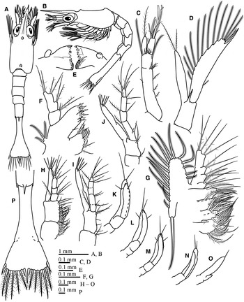
Leonardsaxius amurensis (Kobjakova, 1937), second zoea: (A) Dorsal view; (B) lateral view; (C) antennule; (D) antenna; (E) mandibles; (F) maxillule; (G) maxilla; (H) first maxilliped; (I) second maxilliped; (J) third maxilliped; (K) first pereiopod; (L) second pereiopod; (M) third pereiopod; (N) fourth pereiopod; (O) fifth pereiopod; (P) telson, dorsal view.

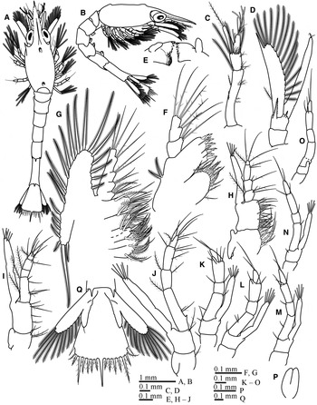
Leonardsaxius amurensis (Kobjakova, 1937), third zoea: (A) Dorsal view; (B) lateral view; (C) antennule; (D) antenna; (E) mandibles; (F) maxillule; (G) maxilla; (H) first maxilliped; (I) second maxilliped; (J) third maxilliped; (K) first pereiopod; (L) second pereiopod; (M) third pereiopod; (N) fourth pereiopod; (O) fifth pereiopod; (P) pleopod; (Q) telson and uropods, ventral view.

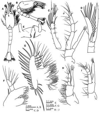
Leonardsaxius amurensis (Kobjakova, 1937), fourth zoea: (A) Dorsal view; (B) lateral view; (C) antennule; (D) antenna; (E) mandibles; (F) maxillule; (G) maxilla; (H) first maxilliped; (I) second maxilliped; (J) third maxilliped.

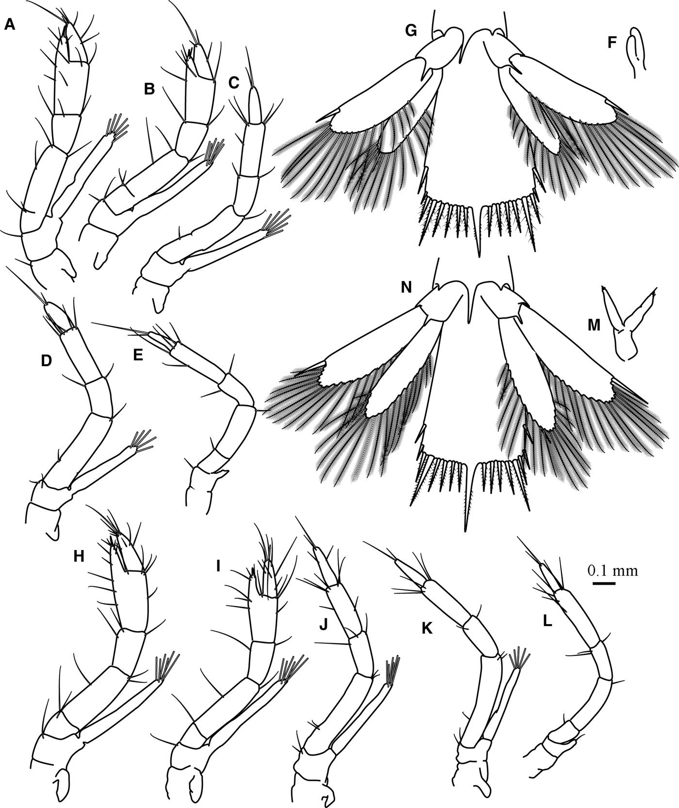
Leonardsaxius amurensis (Kobjakova, 1937), fourth zoea (A–G) and fifth zoea (H–N): (A) first pereiopod; (B) second pereiopod; (C) third pereiopod; (D) fourth pereiopod; (E) fifth pereiopod; (F) pleopod; (G) telson and uropods, ventral view; (H) first pereiopod; (I) second pereiopod; (J) third pereiopod; (K) fourth pereiopod; (L) fifth pereiopod; (M) pleopod; (N) telson and uropods, ventral view.

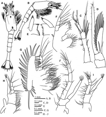
Leonardsaxius amurensis (Kobjakova, 1937), fifth zoea: (A) Dorsal view; (B) lateral view; (C) antennule; (D) antenna; (E) mandibles; (F) maxillule; (G) maxilla; (H) first maxilliped; (I) second maxilliped; (J) third maxilliped.

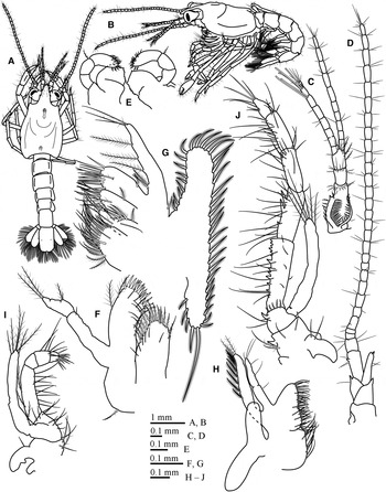
Leonardsaxius amurensis (Kobjakova, 1937), megalopa: (A) Dorsal view; (B) lateral view; (C) antennule; (D) antenna; (E) mandibles; (F) maxillule; (G) maxilla; (H) first maxilliped; (I) second maxilliped; (J) third maxilliped.

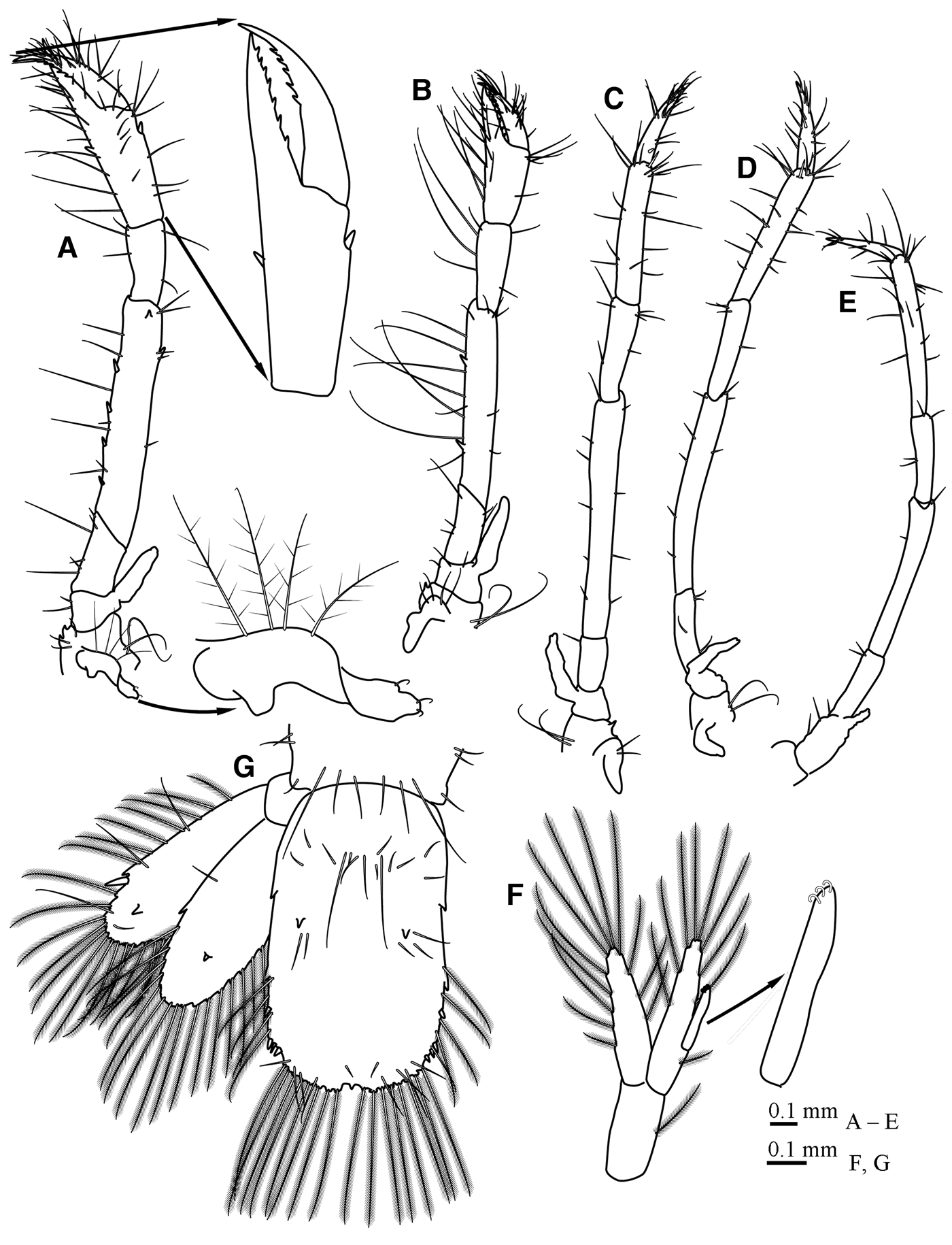
Leonardsaxius amurensis (Kobjakova, 1937), megalopa: (A) First pereiopod; (B) second pereiopod; (C) third pereiopod; (D) fourth pereiopod; (E) fifth pereiopod; (F) pleopods; (G) telson and uropods, ventral view.

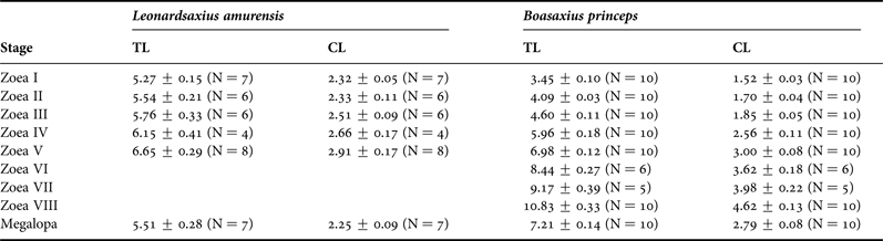
Measurements (mm) of larvae of Leonardsaxius amurensis (present data) and Boasaxius princeps (Kornienko et al., Reference Kornienko, Korn and Golubinskaya2014) (mean values ± SD).

TL, total length; CL, carapace length.
Minimum duration. Unknown.
Carapace (Figure 2A, B) Longer than wide, frontal margin with a pair of long lateroventral antennal spines, anteroventral margins serrated, anterior and posterior dorsomedian tubercles present; long straight rostral spine slightly shorter than carapace, serrated in distal part; eyes sessile; cervical groove not visible, linea thalassinica absent.
Antennule (Figure 2C) Elongated, uniramous, unsegmented, with 3 terminal aesthetascs, 3 terminal simple setae of variable length and 1 subterminal long densely plumose seta.
Antenna (Figure 2D) Peduncle with a distal serrated spine near the base of scaphocerite; endopod fused with peduncle, unsegmented, about half of exopod, with 3 long terminal plumose setae; scaphocerite with a distal short simple seta, 8 inner long plumose setae and 1 outer short sparsely plumose seta.
Mandibles (Figure 2E) Without palps.
Maxillule (Figure 2F) Coxal endite with 8–9 plumodenticulate setae; basial endite with 9 marginal plumodenticulate cuspidate setae and 3 submarginal plumodenticulate setae; endopod 3-segmented, with 3, 2, 2 + 2 plumodenticulate setae; exopod as a small projection, with 3 densely plumose setae.
Maxilla (Figure 2G) Coxal endite distinctly bilobed; proximal lobe with 4 marginal plumodenticulate setae and 2 submarginal plumodenticulate + 9–10 plumose setae, distal lobe with 1 marginal plumodenticulate + 1 simple setae and 1–2 submarginal plumodenticulate + 2 plumose setae; basial endite distinctly bilobed, proximal lobe with 3 marginal plumodenticulate + 2–3 simple setae and 1 submarginal plumodenticulate + 1 plumose setae, distal lobe with 3 marginal plumodenticulate + 1–2 simple setae and 1 submarginal plumodenticulate + 1 plumose setae; endopod unsegmented, with 4 + 2 + 2 + 5 setae (1 proximal seta plumose, others plumodenticulate); scaphognathite with 10–12 marginal densely plumose setae (proximal seta two times longer than others).
First maxilliped (Figure 2H) Coxa with 8–9 plumodenticulate setae; basis with 14–16 plumodenticulate setae; endopod shorter than exopod, 4-segmented, with 10 plumodenticulate and 2 plumose setae arranged 3 + 1, 2, 2, 3 + 1; exopod unsegmented, with 4 terminal plumose natatory setae.
Second maxilliped (Figure 2I) Coxa with 1 plumodenticulate seta; basis with 6 plumodenticulate setae arranged 1 + 1 + 1 + 3; endopod shorter than exopod, 4-segmented, with 11 plumodenticulate and 2 plumose setae arranged 3 + 1, 2, 2, 4 + 1; exopod unsegmented, with 4 terminal plumose natatory setae.
Third maxilliped (Figure 2J) Coxa unarmed; basis with 2 (long plumodenticulate and short simple) setae; endopod shorter than exopod, 4-segmented, with 7 plumodenticulate and 2 plumose setae arranged 0, 1 + 1, 3, 3 + 1; exopod unsegmented, with 6 terminal plumose natatory setae.
Pereiopods. Not visible.
Pleon (Figure 2A, B) Five pleonites, pleonite 5 with rather short smooth posterodorsal spine.
Pleopods. Absent.
Uropod. Absent.
Telson (Figure 2K) Fan-shaped, longer than wide; posterior margin with 7 + 1 + 7 processes: outermost spine-like seta with serrated inner margin, plumose ‘anomuran (thalassinidean) hair’, 4 plumodenticulate long and 1 short setae, short smooth central spine; central spine fused with telson, other processes articulated, process 4 the longest; anal spine absent.
Colour. Rostrum, antennules, posterior and ventral carapace margins with large orange chromatophores.
Second zoea (Figure 3).
Minimum duration. 5 days.
Carapace (Figure 3A, B) Rostrum flattened, lateral rostral margins serrated, anterodorsal carina evident; eyes stalked; otherwise unchanged.
Antennule (Figure 3C) Biramous; peduncle incompletely 2-segmented, proximal segment with 7 outer short sparsely plumose setae arranged 1 + 1 + 1 + 4 and 0–1 inner sparsely plumose seta, distal segment with 2 distal long plumose setae and 5–6 distal short sparsely plumose setae; endopod articulated from protopod, with 1 long plumose and 1 short simple seta; exopod articulated from protopod, with 6 terminal aesthetascs of different length, 1 subterminal short aesthetasc and 3 terminal simple setae.
Antenna (Figure 3D) Peduncle with 1 (rarely 2) spines, the second short, smooth; endopod slightly longer than half of exopod, with 2–3 terminal plumose setae; scaphocerite with a distal smooth spine, 13–14 inner long plumose and 1 outer short sparsely plumose setae.
Mandibles (Figure 3E) With palp buds.
Maxillule (Figure 3F) Coxal endite with 10–12 plumodenticulate and simple setae; basial endite with 12 marginal plumodenticulate cuspidate setae, 3 submarginal sparsely plumodenticulate and 1 lateral plumodenticulate setae; endopod 2-segmented with 3, 2 + 2 + 2 plumodenticulate setae; exopod unchanged.
Maxilla (Figure 3G) Proximal lobe of coxal endite with 6 marginal plumodenticulate setae and 3 submarginal plumodenticulate + 10–11 plumose setae, distal lobe with 1 marginal plumodenticulate + 1 simple setae and 2 submarginal plumodenticulate + 2 plumose setae; proximal lobe of basial endite with 3–4 marginal plumodenticulate + 4 simple setae and 1 submarginal plumodenticulate + 1 plumose setae, distal lobe with 3 marginal plumodenticulate + 2–3 simple setae and 1 submarginal plumodenticulate + 1 plumose setae; endopod unchanged; scaphognathite with 16–19 marginal densely plumose setae.
First maxilliped (Figure 3H) Coxa unchanged; basis with 18–20 plumodenticulate and simple setae; endopod unchanged; exopod with 5 terminal plumose natatory setae.
Second maxilliped (Figure 3I) Coxa with 2–3 plumodenticulate setae; basis unchanged; endopod incompletely 5-segmented, with 11 plumodenticulate and 3 plumose setae arranged 3 + 1, 2, 1, 2, 4 + 1; exopod with 6 terminal plumose natatory setae.
Third maxilliped (Figure 3J) Coxa with 1 plumodenticulate seta; basis unchanged; endopod longer than exopod, with 10–11 plumodenticulate and 3 plumose setae arranged 2 + 1, 1 + 1, 3–4, 4 + 1; exopod unchanged.
Pereiopods (Figure 3K–O) Biramous, unsegmented; exopods and endopods of pereiopods 1–4 nearly equal, exopod of pereiopod 5 considerably shorter than endopod; endopods of pereiopods 1–3 with 2 terminal and 2 subterminal setae, endopod of pereiopod 4 with 2 terminal setae, endopod of pereiopod 5 with 1–2 terminal setae.
Pleon (Figure 3A, B) Unchanged.
Pleopods. Absent.
Uropod. Absent.
Telson (Figure 3P) Posterior margin with 8 + 1 + 8 processes; central spine became longer.
Third zoea (Figure 4)
Minimum duration. 4 days.
Carapace (Figure 4A, B) Lateroventral antennal spines shorter than in previous stages, otherwise unchanged.
Antennule (Figure 4C) Peduncle 2-segmented, proximal segment with 10 outer short sparsely plumose setae arranged 2 + 4 + 4 and 3 inner plumose setae of different lengths arranged 1 + 1 + 1, distal segment with 3–4 long plumose and 2 + 5 short sparsely plumose setae; endopod with 1 long plumose and 2 short simple setae; exopod with 6 terminal, 1–2 subterminal aesthetascs and 3 terminal simple setae.
Antenna (Figure 4D) Peduncle 2-segmented; endopod nearly equal scaphocerite, with 2–3 terminal simple setae; scaphocerite with a distal smooth spine, 15–16 inner long plumose and 1 outer short sparsely plumose setae.
Mandibles (Figure 4E) Palps increased in size.
Maxillule (Figure 4F) Coxal endite with 12–15 plumodenticulate and simple setae; basial endite with 13–14 marginal plumodenticulate cuspidate setae, 3 submarginal and 1 lateral plumodenticulate setae; endopod unchanged; exopod projection with 1–2 densely plumose setae.
Maxilla (Figure 4G) Proximal lobe of coxal endite with 6–7 marginal plumodenticulate setae and 3 submarginal plumodenticulate + 10–11 plumose setae, distal lobe with 1 marginal plumodenticulate + 1 simple setae and 2 submarginal plumodenticulate + 2 plumose setae; proximal lobe of basial endite with 4 marginal plumodenticulate + 4–5 simple setae and 1 submarginal plumodenticulate + 1 plumose setae, distal lobe with 4 marginal plumodenticulate + 2–3 simple setae and 1 submarginal plumodenticulate + 1 plumose setae; endopod unchanged; scaphognathite with 20–23 marginal densely plumose setae.
First maxilliped (Figure 4H) Coxa unchanged; epipod bud present; basis with 20–22 plumodenticulate and simple setae; endopod and exopod unchanged.
Second maxilliped (Figure 4I) Coxa and basis unchanged; epipod bud present; endopod 4-segmented, with 11–13 plumodenticulate and 4 plumose setae arranged 4–5 + 2, 1, 3, 4–5 + 1; exopod unchanged.
Third maxilliped (Figure 4J) Coxa and basis unchanged; epipod bud present; endopod with a 1 (rarely 2) spine on proximal segment, 12–14 plumodenticulate and 3 plumose setae arranged 2–3 + 1, 1 + 1, 5, 4–5 + 1; exopod unchanged.
First pereiopod (Figure 4K) Chelate, with rare simple setae as shown; endopod longer than exopod, 4-segmented, fixed finger considerably shorter than dactylus; exopod unsegmented, with 6 plumose natatory setae.
Second pereiopod (Figure 4L) Chelate, with rare simple setae as shown; endopod longer than exopod, 4-segmented, fixed finger as a bud; exopod unsegmented, with 6 plumose natatory setae.
Third pereiopod (Figure 4M) Not chelate, with rare simple setae; endopod longer than exopod, 4-segmented; exopod with 5 plumose natatory setae.
Fourth pereiopod (Figure 3N) Not chelate, with rare simple setae; endopod longer than exopod, 4-segmented; exopod with 5 plumose natatory setae.
Fifth pereiopod (Figure 4O) Not chelate; endopod incompletely 5-segmented, with rare simple setae; exopod as a bud.
Pleon (Figure 4A, B) Six pleonites; pleonite 6 with short medial posterodorsal spine and a pair of posteroventral spines at the base of uropods; pleonites 2–5 with pleopod buds.
Pleopods (Figure 4B, P) Biramous buds.
Uropod (Figure 4Q) Biramous; endopod and exopod fused with protopod; endopod with 0–1 short simple seta; exopod with 10–11 long plumose setae.
Telson (Figure 4Q) Trapeziform, height slightly longer than posterior margin; long central spine nearly equal fourth processes.
Minimum duration. 6 days.
Carapace (Figure 5A, B) Unchanged.
Antennule (Figure 5C) Proximal segment of peduncle with 12 outer short sparsely plumose setae arranged 2 + 5 + 5 and 5 inner plumose setae of different lengths arranged 1 + 1 + 1 + 1 + 1; distal segment with 5 long plumose and 2 + 5 short sparsely plumose setae; endopod unchanged; exopod with 6 terminal and 2 subterminal aesthetascs, 3 terminal and 1 subterminal simple setae.
Antenna (Figure 5D) Peduncle unchanged; endopod slightly longer than scaphocerite, with short simple setae; scaphocerite with a distal smooth spine, 15–17 inner long plumose and 1 outer short sparsely plumose setae.
Mandibles (Figure 5E) Palps increased in size.
Maxillule (Figure 5F) Coxal endite with 15 plumodenticulate and simple setae; basial endite with 14–15 marginal plumodenticulate cuspidate setae, 3 submarginal and 1 lateral plumodenticulate setae; endopod unchanged; exopod projection reduced, setae unchanged.
Maxilla (Figure 5G) Proximal lobe of coxal endite with 7–8 marginal plumodenticulate setae and 3 submarginal plumodenticulate + 11–12 plumose setae, distal lobe with 1–2 marginal plumodenticulate + 1 simple setae and 2 submarginal plumodenticulate + 2 plumose setae; proximal lobe of basial endite with 4 marginal plumodenticulate + 5–6 simple setae and 1 submarginal plumodenticulate + 1 plumose setae, distal lobe with 4 marginal plumodenticulate + 4–5 simple setae and 1 submarginal plumodenticulate + 1 plumose setae; endopod unchanged; scaphognathite with 25–30 marginal densely plumose setae.
First maxilliped (Figure 5H) Coxa and epipod unchanged; basis with 24–25 plumodenticulate and simple setae; endopod and exopod unchanged.
Second maxilliped (Figure 5I) Coxa, epipod and basis unchanged; endopod with 13–16 plumodenticulate and 4 plumose setae arranged 6–7 + 2, 1, 3–4, 4–5 + 1; exopod unchanged.
Third maxilliped (Figure 5J) Coxa with 1–2 plumodenticulate setae; epipod unchanged; basis with 3–5 plumodenticulate setae; endopod 4-segmented, proximal segment with 2–3 spines and 17–22 plumodenticulate and 3 plumose setae arranged 5–7 + 1, 2 + 1, 6–8, 4–5 + 1; exopod unchanged.
First pereiopod (Figure 6A) Epipod bud present; endopod 2 times longer than exopod, incompletely 5-segmented; fixed finger slightly shorter than dactylus; exopod unchanged.
Second pereiopod (Figure 6B) Epipod bud present; endopod 2 times longer than exopod, incompletely 5-segmented, fixed finger shorter than dactylus; exopod unchanged.
Third pereiopod (Figure 6C) Epipod bud present; endopod 2 times longer than exopod, 4-segmented; exopod unchanged.
Fourth pereiopod (Figure 6D) Epipod bud present; endopod 2 times longer than exopod, 4-segmented; exopod unchanged.
Fifth pereiopod (Figure 6E) Endopod 5-segmented; exopod unchanged.
Pleon (Figure 5A, B) Pleonite 6 with an additional medial posteroventral spine.
Pleopods (Figures 5B & 6F) Increased in size.
Uropod (Figure 6G) Endopod and exopod separated from protopod; protopod with 1 ventral and 1 lateral spines; endopod with 9–13 long plumose setae; exopod with 14–17 long plumose setae and an articulated distal spine.
Telson (Figure 6G) Height 1.5 times longer than posterior margin; outermost spine-like seta moved to lateral telsonal margins, plumose ‘thalassinidean hair’ turned into smooth spine-like seta, central spine serrated.
Minimum duration. 5 days.
Carapace (Figure 7A, B) Unchanged.
Antennule (Figure 7C) Unchanged.
Antenna (Figure 7D) Peduncle unchanged; endopod unsegmented, considerably longer than scaphocerite, with numerous short simple setae; scaphocerite with 15–19 inner long plumose and 1 outer short sparsely plumose setae.
Mandibles (Figure 7E) Each palp with 2–5 setae.
Maxillule (Figure 7F) Coxal endite with 17 plumodenticulate and simple setae; basial endite with 15–17 marginal plumodenticulate cuspidate, 3 submarginal and 1 (rarely 2) lateral plumodenticulate setae; endopod and exopod unchanged.
Maxilla (Figure 7G) Proximal lobe of coxal endite with 7–8 marginal plumodenticulate setae and 3 submarginal plumodenticulate + 12–14 plumose setae, distal lobe with 2–3 marginal plumodenticulate + 1 simple setae and 3 submarginal plumodenticulate + 2 plumose setae; proximal lobe of basial endite with 4 marginal plumodenticulate + 5–6 simple setae and 1 submarginal plumodenticulate + 1 plumose setae, distal lobe with 4 marginal plumodenticulate + 6–7 simple setae and 1 submarginal plumodenticulate + 1 plumose setae; endopod unchanged; scaphognathite with 30–33 marginal densely plumose setae.
First maxilliped (Figure 7H) Coxa with 8–10 plumodenticulate setae; basis with 26–33 plumodenticulate setae; epipod, endopod and exopod unchanged.
Second maxilliped (Figure 7I) Coxa with 3–4 plumodenticulate setae; basis with 6–7 plumodenticulate setae; endopod with 16–19 plumodenticulate and 4 plumose setae arranged 7–9 + 2, 1, 4–5, 5 + 1; epipod and exopod unchanged.
Third maxilliped (Figure 7J) Coxa, epipod and basis unchanged; endopod incompletely 5-segmented, proximal segment with 2–3 spines, 20–26 plumodenticulate and 3 plumose setae arranged 3, 3–5 + 1, 2 + 1, 7–10, 5–6 + 1; exopod unchanged.
First pereiopod (Figure 6H) Endopod 5-segmented; otherwise unchanged.
Second pereiopod (Figure 6I) Endopod 5-segmented; otherwise unchanged.
Third pereiopod (Figure 6J) Unchanged.
Fourth pereiopod (Figure 6K) Unchanged.
Fifth pereiopod (Figure 6L) Unchanged.
Pleon (Figure 7A, B) Unchanged.
Pleopods (Figure 6M & 7B) Increased in size; exopod and endopod with terminal minor setae.
Uropod (Figure 6N) Endopod with 15–21 long plumose setae; exopod with 17–21 long plumose setae and an articulated distal spine.
Telson (Figure 6N) Nearly rectangular, height about 2 times longer than posterior margin; central spine longer than others.
Minimum duration. Unknown.
Carapace (Figure 8A, B) Rostral spine with pointed tip, slightly curved ventrally, longer than eye stalks, with 2 longitudinal rows of small sparsely plumose setae on the surface, lateral rostral margins each with 3–5 spines; carapace with a pair of minor short antennal spines and with about 30 sparsely plumose setae along posteroventral margins, anterior and posterior dorsomedian tubercles present. Cervical groove distinctly visible, linea thalassinica present.
Antennule (Figure 8C) Biramous; peduncle 3-segmented, proximal segment with 10–11 distal sparsely plumose setae, 8–14 lateral sparsely plumose setae, 1 lateral spine on outer margin and 17 + 11 plumose setae covering statocyst cavity, middle segment with 6–7 distal sparsely plumose and 4 simple setae, distal segment with 3–4 lateral plumose and 6–7 distal sparsely plumose setae of different lengths; endopod (inner flagellum) 10–11-segmented, each segment with 0–6 distal simple setae; exopod (outer flagellum) 8-segmented, each segment with 0–6 simple setae, sixth segment with 2 aesthetascs, penultimate segment with 4 aesthetascs of different lengths.
Antenna (Figure 8D) Peduncle 5-segmented, proximal segment unarmed, second segment with 1 spine, third with 1 spine and 1 simple seta, fourth with 1 lateral spine and with 1 sparsely plumose and 1 simple seta, fifth with 5–6 simple setae; flagellum 26–31-segmented, each with 0–8 distal simple setae as shown; reduced exopod with 1 spine and 2 simple setae.
Mandibles (Figure 8E) Palp 3-segmented, proximal segment unarmed, middle segment with 1 sparsely plumodenticulate seta, distal segment with 20–21 short plumodenticulate setae.
Maxillule (Figure 8F) Coxal endite with 23 plumodenticulate setae; basial endite with 20–21 marginal plumodenticulate cuspidate, 5–6 submarginal and 4 lateral plumodenticulate setae; endopod 2-segmented, proximal segment with 0–1 seta, distal segment with 2–3 plumose and 0–1 simple setae; exopod plumose setae absent, but 1–2 plumodenticulate setae at the base of maxillule present.
Maxilla (Figure 8G) Coxal endite bilobed, proximal lobe with 9–14 marginal plumodenticulate setae and 3 submarginal plumodenticulate + 17–20 plumose setae, distal lobe with 2–3 marginal plumodenticulate + 1–2 simple setae and 3 submarginal plumodenticulate + 4 lateral plumose setae; basial endite bilobed, proximal lobe with 6 marginal plumodenticulate + 6–8 simple setae and 3 submarginal plumodenticulate + 1 plumose (not always) setae, distal lobe with 6–7 marginal plumodenticulate + 9–10 simple setae and 1 submarginal plumodenticulate + 1 plumose (not always) setae; endopod unsegmented, with 5–6 lateral plumose and 2–3 terminal plumodenticulate setae; scaphognathite with 38–49 marginal plumose setae.
First maxilliped (Figure 8H) Coxa with 11–14 plumodenticulate setae; large epipod bilobed, unarmed; basis with 43–51 plumodenticulate setae; endopod slightly shorter than exopod, incompletely 2-segmented, with 4–9 simple setae; exopod unsegmented, with 3–4 apical plumose setae and 8–9 lateral densely plumose setae.
Second maxilliped (Figure 8I) Coxa with 3–4 plumodenticulate setae; epipod with 0–1 plumodenticulate setae; basis with 9–11 plumodenticulate setae; endopod longer than exopod, 4-segmented, ischium-merus with 9–12 plumodenticulate and 2 plumose setae, carpus unarmed, propodus with 7–9 plumodenticulate setae of different lengths, dactylus with 4 plumodenticulate and 4–5 simple setae; exopod unsegmented, with 6–9 plumose setae of different lengths.
Third maxilliped (Figure 8J) Coxa with 3–4 plumodenticulate setae and 1 spine; epipod with 2–3 plumodenticulate setae; basis with 1 spine, 3–4 lateral plumodenticulate setae and 5–6 distal sparsely plumose setae; endopod 5-segmented, ischium with crista dentata (13 spines) and 11–15 plumodenticulate setae, merus with 13–17 plumodenticulate setae and 3 spines of different lengths, carpus with 11–12 plumodenticulate setae and 1 distal spine, propodus with 7–9 and dactylus with 8–11 plumodenticulate setae of different lengths; exopod unsegmented, with 5–6 plumose setae.
First pereiopod (Figure 9A) Biramous, chelate, covered with mostly simple setae, right and left pereiopods nearly equal; coxa with 2 long flexible plumodenticulate setae and 2 spines; epipod with 3–4 proximal sparsely plumose setae and 2–3 distal hook-like setae; basis without spines; endopod 5-segmented, ischium without spines, merus with 4 inner, 1 outer and 1 distal spines, carpus without spines; propodus with 2 lateral spines (outer and inner), movable and fixed fingers nearly equal, cutting margin of fixed finger with 6–7 spines, cutting margin of movable finger with 4–5 spines; exopod small, reduced, with 0–3 setae.
Second pereiopod (Figure 9B) Biramous, chelate, covered with mostly simple setae; coxa with 2 long flexible plumodenticulate setae and 1–2 spines; epipod with 2–3 proximal sparsely plumose setae and 2–3 distal hook-like setae; basis without spines; endopod 5-segmented, ischium without spines, merus with 2–3 inner spines, carpus and propodus without spines, movable and fixed fingers nearly equal, cutting margin of fixed finger with 5–6 spines, cutting margin of movable finger with 3 spines; exopod small, reduced, with 0–3 setae.
Third pereiopod (Figure 9C) Biramous, covered with mostly simple setae; coxa with 2 flexible plumodenticulate setae and 1 spine; epipod with 2–3 proximal sparsely plumose setae; basis without spines; endopod 5-segmented, ischium, merus and carpus without spines, propodus with 1 distal spine-like serrated seta, dactylus with 2 inner spine-like setae; exopod small, reduced, with 0–3 seta.
Fourth pereiopod (Figure 9D) Biramous, covered with mostly simple setae; coxa with 2 flexible plumodenticulate setae; epipod unarmed; basis unarmed; endopod 5-segmented, ischium, merus and carpus without spines, propodus with 1 distal spine-like serrated seta, dactylus with 2 inner spine-like setae; exopod small, reduced, with 0–3 setae.
Fifth pereiopod (Figure 9E) Biramous, covered with mostly simple setae; coxa and basis without spines; epipod absent; endopod 5-segmented; exopod reduced, unarmed.
Pleon (Figure 8A, B) Consists of six pleonites and telson; dorsal surface covered with numerous simple setae of different lengths; pleonites 2–5 with well-developed pleopods, pleonite 6 with uropods.
Pleopods (Figures 8B & 9F) Biramous; endopod and exopod nearly equal, separated from protopod; protopod with 1 densely plumose seta; endopod with 8–10 marginal densely plumose setae and enlarged appendix interna bearing 3 cincinnuli; exopod with 12–13 marginal densely plumose setae.
Uropod (Figure 9G) Biramous; endopod and exopod nearly equal, separated from protopod; endopod with 1 outer lateral spine, 24–26 marginal densely plumose setae, 1 spine and 3–5 simple setae on the surface; exopod with 2 outer lateral spines, 28–36 marginal densely plumose setae, 1 spine and 3–4 simple setae on the surface.
Telson (Figure 9G) Two times longer than wide; dorsal surface covered with simple setae of different lengths and 1 pair of spines, lateral margins with 3–4 pairs of spines and 4–8 plumose setae, distal margin with 3 pairs of spines, 16–18 plumose setae and minor smooth central spine.
DISCUSSION
The larvae of the family Axiidae Huxley, 1879, a family of 112 described species (Ahyong et al., Reference Ahyong, Lowry, Alonso, Bamber, Boxshall, Castro, Gerken, Karaman, Goy, Jones, Meland, Rogers, Svavarsson and Zhang2011), are mostly poorly known (Pohle et al., Reference Pohle, Santana, Jansen and Greenlaw2011; Pohle & Santana, Reference Pohle, Santana, Martin, Olesen and Høeg2014). Partial descriptions based on laboratory-reared and plankton-collected material exist for two species from the East Atlantic: Axius stirynchus and Calocaris macandreae (Gurney, Reference Gurney1942; Barnich, Reference Barnich1996). Only the first zoea of Axiopsis serratifrons from Brazil has been obtained in the laboratory from an ovigerous female (Rodrigues, Reference Rodrigues1994). A detailed description of the complete larval development of Axius serratus from the North-west Atlantic was performed on the plankton-collected material (Pohle et al., Reference Pohle, Santana, Jansen and Greenlaw2011). Recently, we have, for the first time, carefully described the complete larval development of Boasaxius princeps (Boas, 1880) from the west Pacific obtained from ovigerous females (Kornienko & Korn, Reference Kornienko and Korn2012; Kornienko et al., Reference Kornienko, Korn and Golubinskaya2014).
Our results show that the development of recently re-described axiid species Leonardsaxius amurensis includes five zoeal stages and a single megalopa, as opposed to the related sympatric species B. princeps having eight zoeal stages (Kornienko et al., Reference Kornienko, Korn and Golubinskaya2014). At 22–23°C, the first megalopa of L. amurensis was attained 20 days after the beginning of the experiment. The first zoea and evidently the eggs of L. amurensis are larger than those of B. princeps. However, the fifth stages of both species obtained in the laboratory are nearly equal in size but megalopa of B. princeps is even longer than that of L. amurensis (Table 6).
The first zoea of L. amurensis is characterized by the presence of one short posterodorsal spine on the fifth pleonite whereas zoea I of B. princeps has four slightly long posterodorsal spines on pleonites 2–5. The shape of the telson of both species is very similar, however, B. princeps has 7 + 7 while L. amurensis 7 + 1 + 7 posterior processes in the first zoeal stage. Telson of the first zoea of B. princeps is similar to that of A. serratifrons (see Rodrigues, Reference Rodrigues1994). The telson of the first zoea in other known axiid species possesses numerous posterior processes: 17 + 1 + 17 in A. serratus (see Pohle et al., Reference Pohle, Santana, Jansen and Greenlaw2011), 14–16 + 1 + 14–16 in A. stirynchus, 17 + 17 in C. macandreae (see Gurney, Reference Gurney1942; Barnich, Reference Barnich1996), and 23–25 + 1 + 23–25 in Calocarides coronatus (see Elofsson, Reference Elofsson1959).
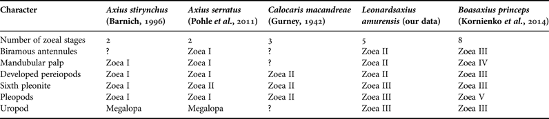
The number of zoeal stages in the family Axiidae varies widely as it does in the families Callianassidae and Upogebiidae. Most known axiids have an abbreviated development (Table 7). The larvae of A. stirynchus with two zoeal stages and of C. macandreae with three zoeal stages hatch with pereiopods and pleopods (Gurney, Reference Gurney1942; Barnich, Reference Barnich1996). In A. stirynchus, mandibular palps and pereiopods appear already in the first zoea (Gurney, Reference Gurney1942) and the sixth pleonite separates from the telson also in the first stage (Barnich, Reference Barnich1996). Axius serratus also possesses two zoeal stages with biramous antennules, mandibular palps, pereiopods and pleopods in the first stage, but the sixth pleonite separates from the telson in the second stage (Pohle et al., Reference Pohle, Santana, Jansen and Greenlaw2011). In B. princeps, having eight zoeal stages, biramous antennules appear in the third zoea, mandibular palp buds in the fourth zoea, unsegmented pereiopod buds in the second zoea, but biramous pereiopods in the third zoea, the sixth pleonite separates from the telson in the third zoea, pleopod buds appear only in the fifth zoea (Kornienko et al., Reference Kornienko, Korn and Golubinskaya2014).
The number of zoeal stages and the stage correspondent to the appearance of main morphological characters in axiid species.

In comparison with B. princeps, the development of L. amurensis is advanced and occupies an intermediate position between A. stirynchus, A. serratus or C. macandreae with abbreviated development and B. princeps with long pelagic development. In L. amurensis, biramous antennules, mandibular palp buds and biramous pereiopods appear in the second zoea, the sixth pleonite separates from the telson in the third zoea, pleopod buds appear also in the third zoea.
The number of zoeal stages in A. serratifrons is still unknown. Rodrigues (Reference Rodrigues1994) believed that the absence of pereiopods and pleopods and the very simple morphology of the third maxilliped, with few setae and unsegmented endopod, are indications of a long larval development of this species, probably with five or more stages. However, L. amurensis having five zoeal stages possesses a more advanced first larva with a well developed third maxilliped and considerably more setaceous appendages. At the same time, the first zoea of A. serratifrons is nearly identical to the first larva of B. princeps including the morphology of the third maxilliped. Therefore, we assume that larval development of A. serratifrons includes more than five zoeal stages.
In L. amurensis, maxillules of the first and the second zoeae possess a small unsegmented exopod projection with 3 plumose setae gradually reducing to 1–2 plumose setae in zoeae IV and V. A single exopod plumose seta is found also in the maxillule of the second zoea of B. princeps (Kornienko et al., Reference Kornienko, Korn and Golubinskaya2014). Among Decapoda, the presence of an unsegmented maxillular exopod with four long, plumose setae was noted in the protozoeal stage of penaeidean shrimps (suborder Dendrobranchiata) (Knight & Omori, Reference Knight and Omori1982; Ronquillo et al., Reference Ronquillo, Saisho and McKinley2006). Among Pleocyemata, the maxillular exopod is usually absent and represented by a single plumose seta; for example, in some Caridea (see Haynes, Reference Haynes1985; Terossi et al., Reference Terossi, Cuesta, Wehrtmann and Mantelatto2010; Guerao et al., Reference Guerao, Hernández and Urzúa2011) and in the late stages of numerous Brachyura. In hairy crabs Telmessus cheiragonus and Erimacrus isenbeckii, one maxillular exopod seta appears in the first zoea, two in the second zoea and three from the third to the fifth zoea (Kornienko & Korn, Reference Kornienko and Korn2010).
Earlier, we assumed that zoeae with one posterodorsal spine on the fifth pleonite, which often occur in the plankton of Peter the Great Bay and which were described by Kurata (Reference Kurata1965) for the coastal waters of Hokkaido as Axius sp. A, are indeed the larvae of L. amurensis (Kornienko et al., Reference Kornienko, Korn and Golubinskaya2014). Our data have only confirmed this hypothesis in regards to the larvae from Peter the Great Bay. The morphological data of Kurata are fragmentary but the described first zoea of Axius sp. A, like the larva of L. amurensis, has one pair of long antennal spines on the carapace, one posterodorsal spine on the fifth pleonite, and the telson with 7 + 1 + 7 processes on the posterior margin; the armament of the antenna is also identical in both L. amurensis and Axius sp. A. Kurata, just as we did, noted the presence of a small exopod with 3 plumose setae on the maxillule of zoea I, but the endopod setal formula in Axius sp. A is 2, 2, 2 + 2, while it is 3, 2, 2 + 2 in our larva. Moreover, the first zoeal stage from coastal waters of Hokkaido are slightly smaller (TL = 4.8, CL = 2.1 mm). In the second zoeal stage, a pair of processes adds on the posterior telsonal margin and biramous pereiopods appear in both species. In the third zoeal stage, biramous pleopods, missing in Axius sp. A, are present in L. amurensis. In addition, the posterodorsal spine on the sixth pleonite is bifurcated in Axius sp. A, but pointed in L. amurensis.
The differences between larval morphology in related species are often insignificant. For example, the first zoeae of sympatric species Nihonotrypaea japonica and N. petalura differ only by the presence of one seta on the coxa of the first maxilliped (Konishi et al., Reference Konishi, Quintana and Fukuda1990; Miyabe et al., Reference Miyabe, Konishi, Fukuda and Tamaki1998). So, the suggestion by Kornienko et al. (Reference Kornienko, Korn and Golubinskaya2014) that the larvae of Axius sp. A belong to L. amurensis may be erroneous.
It is pertinent to note that in contrast to the larvae of numerous brachyurans and anomurans reared in our laboratory, the number of zoeal stages in axiidean species with long development may vary in different populations of the same species and/or under moderate differences in laboratory conditions. The appearance of the additional larval stage may be a result of suboptimal rearing conditions and may not occur in the natural habitat (Kornienko et al., Reference Kornienko, Korn and Golubinskaya2015). In our experiment, two of 30 fourth zoeal stage larvae of L. amurensis moulted directly to megalopa instead of the fifth zoeal stage.
CONCLUSION
A molecular genetic analysis based on comparison of partial mitochondrial COI, 12S rDNA and 16S rDNA sequence data confirmed the identity of axiid larvae found in the plankton and L. amurensis adults collected in the same area. The larval development of L. amurensis includes five zoeal stages and a single megalopa. Zoeae I of L. amurensis are characterized by the presence of one short posterodorsal spine on the fifth pleonite in contrast to the larvae of related sympatric species B. princeps having four posterodorsal spines on pleonites 2–5. Leonardsaxius amurensis occupies an intermediate position between lobster shrimps with abbreviated pelagic development (2–3 zoeal stages) and species with long development (up to 8 zoeal stages). Thus, the number of zoeal stages in the family Axiidae varies widely, similarly to that in the families Callianassidae and Upogebiidae.
ACKNOWLEDGEMENTS
We are greatly indebted to Dr I. N. Marin (A.N. Severtzov Institute of Ecology and Evolution RAS, Moscow) for the collection and identification of Leonardsaxius amurensis. We are grateful to anonymous referees for their comments and suggestions that clearly improved the manuscript.
FINANCIAL SUPPORT
The collection of plankton samples and rearing of larvae were supported by the ‘Far East’ Program, Far Eastern Branch, Russian Academy of Sciences (grant number 15-I-6-007); the description of larvae was supported by the Russian Foundation for Fundamental Researches (grant number 16-34-00059); molecular analysis was supported by the ‘Far East’ Program, FEB RAS (grant number 15-I-6-014 o).