Introduction
Stigmidium Trevis. is a genus of ascomycete fungi, comprising mostly lichenicolous taxa, currently included in the Mycosphaerellaceae (Lumbsch & Huhndorf Reference Lumbsch and Huhndorf2007). The generic type S. schaereri (A. Massal.) Trevis., is lichenicolous on the sterile thalli of a species of Solorina. Stigmidium s. str. and was revised by Roux & Triebel (Reference Roux and Triebel1994). The genus is distinguished by perithecioid ascomata with punctiform ostioles with a hamathecium of external periphyses (sensu Roux & Triebel Reference Roux and Triebel1994), with periphysoids, lacking interascal filaments, and by hyaline 1-septate ascospores (rarely turning brown in a few species) (Roux & Triebel Reference Roux and Triebel1994; Calatayud & Triebel Reference Calatayud and Triebel2003). The periphysoids originate from the wall of the upper cavity of the pseudothecia are never attached to the bottom of the cavity and are referred to as pseudoparaphyses of which two types are recognized (Roux & Triebel Reference Roux and Triebel1994). Stigmidium s. str. has ‘Type a’ pseudoparaphyses which are narrow and two-celled. It currently comprises 22 species including the species to be described in this paper (Roux & Triebel Reference Roux and Triebel1994; Roux & Navarro-Rosinés Reference Roux and Navarro-Rosinés1994; Roux et al. Reference Roux, Triebel, Bricaud and le Coeur1995; Calatayud & Triebel Reference Calatayud and Triebel2001, Reference Calatayud and Triebel2003; Triebel & Cáceres Reference Triebel, Cáceres, Nash, Ryan, Diederich, Gries and Bungartz2004; Zhurbenko & Triebel Reference Zhurbenko and Triebel2008). A second group of species currently included in Stigmidium differ from Stigmidium s. str. in having ‘Type b’ pseudoparaphyses sensu Roux & Triebel (Reference Roux and Triebel1994) which are long and relatively large, made up of 3–5 more or less equal cells, and comprise the S. placynthii group which currently includes five species (Roux & Triebel Reference Roux and Triebel1994 & Reference Roux and Triebel2005; Roux & Navarro-Rosinés Reference Roux and Navarro-Rosinés1994; Kocourková & Knudsen Reference Kocourková and Knudsen2009a, Reference Kocourková and Knudsenb).
The genus Stigmidium includes at least 90 binominals (Etayo Reference Etayo2008; Mycobank 2009; Zhurbenko Reference Zhurbenko2009; Kocourková & Knudsen Reference Kocourková and Knudsen2009a, Reference Kocourková and Knudsenb). The majority of the species, 65 or more depending on validity of synonymies, make up Stigimidium s. lat. and are a heterogeneous group. Species have been transferred or described in Stigmidium using genera concepts differing from those in Roux & Triebel (Reference Roux and Triebel1994). Some species have interascal filaments (see Triebel Reference Triebel1989, Calatayud & Triebel Reference Calatayud and Triebel2003), others have a hamathecium not described in detail using the same concepts as Roux's & Triebel's taxonomy (see Halici & Hawksworth Reference Halici and Hawksworth2007), or a hamathecium has not been observed (see Zhurbenko Reference Zhurbenko2009), or with observations uncertain because of limited type material (see Etayo Reference Etayo2008). Kocourková & Knudsen (Reference Kocourková and Knudsen2009b) summarize the situation: “Due to a lack of standardized morphological nomenclature coupled with differences in taxonomic descriptions and generic concepts of Stigmidium by different authors, many Stigmidium species have overlapping ascospore and ascomata size, forcing keys to be structured according to host (Halici Reference Halici2008; Ihlen & Wedin Reference Ihlen and Wedin2008) rather than by individual morphological characters”. We expect that some species in Stigmidium s. lat. may actually belong in Stigmidium s. str. or in the closely related S. placynthii group with ‘Type b’ pseudoparaphyses. But other species will need to be segregated into other genera.
A further problem in Stigmidium, as in other lichenicolous genera, is that many species concepts were influenced by a host-based taxonomy. This problem was recognized already before the work of Roux & Triebel (Reference Roux and Triebel1994). Hawksworth (Reference Hawksworth1986) wrote “Species concepts in the genus Stigmidium Trevisan require a thorough re-appraisal as the taxonomy is currently largely host-based”. Stigmidium species appear to be narrowly host specific to particular species, genera, or families. On some host genera one species of Stigmidium s. lat. may comprise two or more species differing, for instance, in having ‘Type a’ or ‘Type b’ pseudoparaphyses, but with statistically narrow ascospore differences.
In this paper we describe a new species of Stigmidium on a new host species and genus, Thelenella muscorum (Fr.) Vain. (syn. Chromatochlamys muscorum (Fr.) H. Mayrhofer & Poelt), in a new host family Thelenellaceae O. E. Erikss. ex H. Mayrhofer. The species is a member of Stigmidium s. str. and is compared with other species with ‘Type a’ pseudoparaphyses.
Material and Methods
Specimens were examined using a Nikon SMZ 600 stereomicroscope. Hand-cut sections of ascomata were studied in distilled water, lactophenol cotton blue and KOH. The amyloid reactions were tested in iodine-potassium iodide solution (1%) (=Lugol's iodine solution), directly (I) and after a KOH pre-treatment (K/I). A 1 % solution of Brilliant Cresyl blue (BCr) was used for testing metachromatic reactions of the endoascus, epispore and vegetative hyphae. Samples were observed under a Nikon Eclipse 80i microscope fitted with ‘Nomarski’ differential interference constrast and a Nikon DS-Fi1 digital image system. All measurements were made in water, and taken by means of Nikon Image Analyze System®. Microscopic pictures were taken by mean of this system; habitus picture was taken by mean of a Nikon Image System® fitted to a Leica dissecting microscope. For ascospore and ascus size measurements, the highest and lowest values are given within parentheses, with the average together with ± standard deviation in italics; values are approximated to the nearest 0·5 μm.
The Species
Stigmidium humidum Pérez-Ortega, Halici & K. Knudsen sp. nov
MycoBank No.: MB 513453
Fungus lichenicola in thallo Thelenella muscorum parasiticus. Ascomata perithecioidea, subglobosa, atra, 75–140 μm diametro, semiimmersa. Peridium olivaceum, textura angulari, inaequaliter pigmentatum, K+ brunneo-caeruleus. Gelatina hymenialis I−, K/I−. Filamenta interascalia observata: pseudoparaphyses ‘typo a’. Asci saccati vel clavati (24–)41 ± 6(–54) × (9–)14·5 ± 2(–17) μm magni, 8-spori, endoascus pariete BCr+ cyanea, I−, K/I−. Ascosporae incoloratae, anguste pediformes, ad septum non vel laeviter constrictae, halonatae, (12–)14·5 ± 1·5(–18) × (3.5–)5 ± 0·5(–6·5) [cellula supera] × (3–)4 ± 0·5(–5) [cellula infera] μm magnae, longitudinis/latitudinis [cellulae superae] ratio: (2·5–) 3 ± 0·5(–4), epiplasma BCr+ cyanea. Conidiomata non observata.
Typus: Turkey, Afyon, Sandıklı, Akdağ Tabiat Parkı, Başalan Yaylası, 38° 20″ 37″ N, 30° 01′ 24″ E, on Thelenella muscorum, on mosses, alt. 1477 m, 03 June 2008, M. Candan (ANES 12281—holotypus).
(Fig. 1)
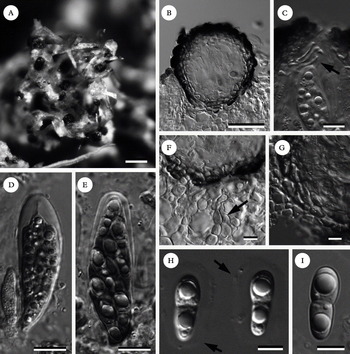
Stigmidium humidum (MAF 16095). A, habitus; B, ascomata in cross section; C, detail of hamathecial filaments: ‘Type a’ pseudoparaphyses; D, young ascus; E, mature asci with 8 ascospores; F. detail of vegetative hyphae (arrow) in host thallus; G, ascomatal wall, detail of cells in the lower part of the ascomata; H, young halonate ascospores, arrows pointing to halo; I, mature ascospore. B & F in lactophenol blue; D & E in Lugol's; C, G, H & I in water. Scales: A = 250 μm; B = 50 μm; C– G = 10 μm; H & I = 5 μm.

Vegetative hyphae hyaline to pale brown, composed of elongate to rectangular cells, c. 5 μm diam., immersed in the host tissues, BCr−.
Ascomata perithecioid, black, ± shiny, in dense groups, sessile or more often 1/3 to 2/3 immersed in the host thallus, 75–140 μm (n = 11). In transverse section, globose to subglobose, frequently with a depression around the ostiole. Outer part of the pseudothecium with an irregular rugose appearance. Wall pseudoparenchymatous, of textura angularis, brown, olive greenish in K, c. 10–20 μm wide, usually thinner at the base than near the ostiole, with 2–6 layers of cells; inner part light brown to hyaline, outer part dark brown. Cells of the ascomatal wall ± rectangular and compressed in transverse sections of the ascomata, and ± rounded-polygonal in superficial view, mostly 4–8 μm diam., I−; cells in the outer part of the ascomata forming an uneven surface. External periphyses poorly developed. Hymenial gel I−, K/I−. Ostiolar filaments present, 4–6 μm long, hamathecium composed of pseudoparaphyses of ‘type a’ (Roux & Triebel Reference Roux and Triebel1994) 5–10 μm long. Asci bitunicate, sessile to distinctly stalked at the base, clavate to saccate, thickened at the apex, 8-spored, ascospores often arranged distichiously, (24–)41 ± 6(–54) × (9–)14·5 ± 2(–17) μm (n = 28), endoascus BCr+ violet. Ascospores 1-septate, constricted at the septum, obovate, lower cell narrower and longer than the upper one, hyaline, halonate, surface smooth, not pseudotetrablastic (with a large oil droplet in each cell), inner ascospore wall BCr+ violet, (12–)14·5 ± 1·5(–18) × (3·5–)5 ± 0·5(–6·5) (upper cell) × (3–)4 ± 0·5(–5) (lower cell) μm [n = 61; length/breadth ratio upper cell (2·5–)3 ± 0·5(–4)].
Etymology. The name refers to the occurrence of the species in moist microhabitats on Thelenella muscorum among bryophytes and it may eventually be discovered on related host genera that also occur in these microhabitats.
Ecology and distribution. The species is known from three localities in central Spain and one in western Turkey. The species seems to be relatively common on T. muscorum (syn. Chromatochlamys muscorum, see Fryday & Coppins Reference Fryday and Coppins2004 for a recent discussion of the taxonomy of the host) thalli in higher elevation areas of Sistema Central and Sistema Ibérico. The species does not produce clear necrosis or decolouration in host thalli, so we assume a parasymbiotic state for the species. However, thalli infected with S. humidum show a smaller number of host perithecia than those thalli not infected, which might indicate a negative effect of this taxon on its host. More careful observations are needed to clarify this question.
Additional specimens examined. Spain: Castilla y León: Segovia, Honrubia de la Cuesta, siliceous outcrops close to the village, 41° 30′ 45″ N, 3° 42′ 8″ W, on Thelenella muscorum on mosses, alt. 968 m, 06 August 2007, S. Pérez-Ortega (MAF 16094); Burgos, Barbadillo de Pez, siliceous outcrops close to the road, 5 km N Bardadillo, 42° 8′ 13·2″ N, 3° 11′ 33″ W, alt. 1122 m, 22 May 2006, S. Pérez-Ortega (MAF 16095).
Discussion
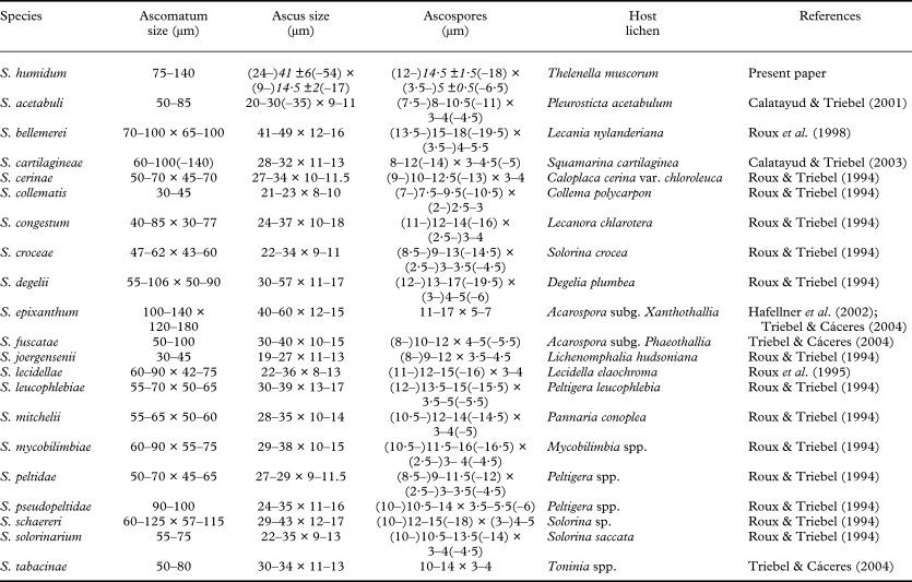
Stigmidium humidum is the first species in the genus to be recognized on the lichen host genus Thelenella and belongs to Stigmidium s. str., having ‘type A’ periphysoids or pseudoparaphyses (sensu Roux & Triebel Reference Roux and Triebel1994). Within the genus 22 species are known with ‘type a’ periphysoids including S. humidum, and several characters of these species are compared in Table 1.
Comparison of Stigmidium species showing ‘Type a’ pseudoparaphyses

Stigmidium humidum is most similar to S. bellemerei Cl. Roux & Nav.-Ros., S. degelii R. Sant., S. leucophlebiae Cl. Roux & Triebel, S. mycobilimbiae Cl. Roux, Triebel & Etayo, S. schaereri (A. Massal.) Trevis and S. stereocaulorum Zhurb. & Triebel, but, apart from a different unrelated lichen host, also differs in some other characters. Stigmidium bellemerei, a species recognized on Lecania nylanderiana has relatively longer ascospores [(13·5–)15–18(–19·5) × (3·5–)4–5·5 μm] which are moreellipsoid and pseudotetrablastic (having 2 large oil droplets per ascospore cell) and relatively smaller ascomata [70–100 × 65–100 μm] (Roux et al. Reference Roux, Navarro-Rosinés and Tranchida1998). Stigmidium degelii, a species recognized on Degelia plumbea, has ascospores rather similar in size to those of S. humidum [(12–)13–17(–19·5) × (3–)4–5(–6)], but they are pseudotetrablastic, and the ascomata are relatively smaller [55–106 × 50–90] (Roux & Triebel Reference Roux and Triebel1994). Stigmidium leucophlebiae, a species recognized on Peltigera leucophlebia, also has rather similar sized but pseudotetrablastic ascospores [(12–)13·5–15 (–15·5) × 3·5–5 (–5·5) μm], but this species has immersed ascomata in the dying parts of the host thallus which are relatively smaller [55–70 × 50–65 μm], while S. humidum has semi-immersed to superficial ascomata (Roux & Triebel Reference Roux and Triebel1994). Stigmidium mycobilimbiae, a species recognized on muscicole Mycobilimbia spp., has relatively shorter and narrower ascospores than S. humidum and they are also pseudotetrablastic and non-halonate [(12–)13·5–15(–15·5) × 3·5–5(–5·5) μm]. This species also has smaller ascomata [60–90 × 50–75 μm] (Roux & Triebel Reference Roux and Triebel1994). Stigmidium schaereri, a species recognized on Solorina sp., has rather similar sized ascospores to S. humidum [(10–)12–15(–18) × (3–)4–5 μm] which are pseudotetrablastic and non-halonate, but the endoascus and inner perispore of S. schaereri give a negative reaction in BCr, whereas S. humidum shows violet reactions in the endoascus and inner perispore (Roux & Triebel Reference Roux and Triebel1994). Stigmidium stereocaulorum, a species recognized on Stereocaulon spp., also has rather similar sized ascospores to S. humidum [(10–)12–15(–17) × (3–)4–4·5(–5·5) μm], but they are non-halonate. Besides, the ascomata of S. stereocaulorum are relatively smaller [50–75(–100) μm] (Zhurbenko & Triebel Reference Zhurbenko and Triebel2008).
Capronia minutosetosae Halici, D. Hawksw. A. O. Türk & Candan was described on Thelenella muscorum, and this species is also known from Turkey and Spain (Halici Reference Halici2008; Halici et al. Reference Halici, Hawksworth, Candan and Türk2010). It differs from Stigmidium humidum in having dark brown setae arising from the upper part of ascomata and 3-5-septate brown ascospores.
Pharcidia haesitans Nyl. ex Vouaux (Vouaux Reference Vouaux1912; Santesson et al. Reference Santesson, Moberg, Nordin, Tonsberg and Vitikainen2004; Ihlen & Wedin Reference Ihlen and Wedin2008) has reportedly a similar ascospore size of 14–17 × 6–7 μm and occurs on Protothelenella sphinctrinioides (Nyl.) Mayrhofer & Poelt in the family Protothelenellaceae Vĕzda, H. Mayrhofer & Poelt, which is related to the Thelenellaceae. The type species of P. haesitans as well as the sample collected by K. Kalb in Sweden (Santesson et al. Reference Santesson, Moberg, Nordin, Tonsberg and Vitikainen2004; Ihlen & Wedin Reference Ihlen and Wedin2008) do not seem to belong to Stigmidium s. str. as they show K/I+ blue hymenial gel, long periphysoids with 3–5 cells, and ascospores larger than previously published and will be revised separately by Kocourková and Knudsen.
We thank Harrie Sipman for checking our Latin. SPO is very obliged to the project ANT-COMPLEX (CGL2006-12179-CO2-01, MEC, Spain) for support. The studies of MC were financially supported by TÜBÌTAK (107T605 coded project).