Implications
The price of wool is largely dependent on fibre diameter and yield (Coelli and Atkins, Reference Coelli and Atkins1995), and wool producers have used selective breeding of sheep with desirable genetic traits to produce sheep with low-fibre diameter and high wool follicle densities (Gregory, Reference Gregory1982), with improvements generally seen over many generations. In this study, we altered maternal thyroid hormone status coinciding with the initial stages of foetal primary wool follicle development and showed that wool follicle parameters were altered in sheep at birth and 12 months later. This shows that transient manipulations in the maternal environment can result in altered wool follicle characteristics in progeny within a single generation.
Introduction
Wool follicle development begins during distinct times in the prenatal period (Carter and Hardy, Reference Carter and Hardy1947; Hardy and Lyne, Reference Hardy and Lyne1956). The fleece of Merino sheep is composed of three populations of wool follicles. Primary wool follicles (associated with sweat glands) initiation occurs from day 60 of gestation (D60); secondary wool follicles initiate from D80 and secondary wool follicle branching starts from D100 (Carter, Reference Carter1939; Hardy and Lyne, Reference Hardy and Lyne1956). The fine fibre properties of Merino sheep are attributed to the secondary wool follicle branching phenomenon, not commonly seen in other breeds of sheep (Moore et al., Reference Moore, Jackson, Isaacs and Brown1996; Rogers, Reference Rogers2006). In addition, there is a negative correlation between wool follicle density and fibre diameter (Hynd et al., Reference Hynd, Ponzoni, Grimson, Jaensch, Smith and Kenyon1996; Moore et al., Reference Moore, Jackson, Isaacs and Brown1998), and hence the larger the degree of secondary wool follicle branching, the finer the fleece. Fleece characteristics are influenced during these periods of foetal development. Although traits such as wool follicle densities are highly heritable (Carter and Clarke, Reference Carter and Clarke1957; Jackson et al., Reference Jackson, Nay, Turner and Newton1975; Hancock et al., Reference Hancock, Mayo and Brady1979), the foetal and maternal environments can also contribute to fleece characteristics (Short, Reference Short1955; Gregory, Reference Gregory1982).
Thyroid hormones are involved in normal foetal development, with thyroid hormone status playing important roles in the development and function of other organs that originate from epithelial–mesenchyme interactions, such as the lungs (Smith and Sabry, Reference Smith and Sabry1983). The importance of thyroid hormones during foetal development was best described by Hopkins and Thorburn (Reference Hopkins and Thorburn1972), who performed foetal thyroidectomy between D81 and D96, resulting in increased gestation, lower BW and high perinatal mortality.
Thyroid hormones are also important during wool and hair follicle development and cycling and thyroid hormone receptors are expressed in most compartments of the human hair follicle, such as the outer root sheath, dermal papilla cells and hair matrix and throughout the epithelium (Billoni et al., Reference Billoni, Buan, Gautier, Gaillard, Mahe and Bernard2000). When thyroidectomy surgery was performed between D81 and D96, wool follicle initiation was not affected. However, wool follicle maturation was perturbed in athyroid foetal lambs, evidenced by the absence of fibres in wool follicles (Hopkins and Thorburn, Reference Hopkins and Thorburn1972). Both primary and secondary wool follicles were present, but the proportion of wool follicles containing fibres was reduced. Furthermore, potential (immature) secondary/primary wool follicle (S/P) ratios were not altered, but there was a marked decrease in mature S/P ratios and sebaceous gland development was also delayed (Hopkins and Thorburn, Reference Hopkins and Thorburn1972). Conversely, administration of thyroxine (T4) to foetal sheep at D90 accelerated the development of primary wool follicles and sebaceous glands (Hopkins and Thorburn, Reference Hopkins and Thorburn1972).
The timing of the thyroidectomy largely affects the degree of retardation of skin development; the earlier the thyroidectomy was performed, the greater was the retardation of the wool follicles, sweat glands and sebaceous glands (Chapman et al., Reference Chapman, Hopkins and Thorburn1974; Wallace et al., Reference Wallace, Simpson-Morgan and McCullagh1994). Postnatal wool follicle development was also affected, since lambs that underwent thyroidectomy at birth had perturbed in secondary wool follicle maturation (Ferguson et al., Reference Ferguson, Schinckel, Carter and Clarke1956). Conversely, administration of exogenous thyroid hormones restored the maturity of secondary follicles in athyroid lambs (Ferguson et al., Reference Ferguson, Schinckel, Carter and Clarke1956). Thyroidectomy surgery performed in adult Merino sheep resulted in the induced shutdown of large numbers of wool follicles (Hynd, Reference Hynd1994).
To date, the effects of long-term manipulation of thyroid hormone status late in gestation on wool follicle development have been studied by Hopkins and Thorburn (Reference Hopkins and Thorburn1972) and Chapman et al. (Reference Chapman, Hopkins and Thorburn1974). However, the influences of short-term manipulations of thyroid hormone status earlier in gestation are unknown. We hypothesise that transient manipulation of circulating thyroid hormone status during times coinciding with the initial stages of foetal primary wool follicle development (D55 to D64) in Merino sheep will influence fleece characteristics such as wool follicle densities and maturity; specifically, increased maternal thyroid hormone status would lead to increased maturity of wool follicles in postnatal lambs and higher yields in adult sheep. In this study, pregnant Merino sheep were treated with exogenous thyroid hormones T4 or an inhibitor of thyroid hormone synthesis propylthiouracil (PTU) for a 10-day period and fleece characteristics at birth and in 1-year-old sheep were determined.
Material and methods
Unless indicated, all chemicals were purchased from Sigma Aldrich (St Louis, MO, USA). Experiments were approved by the University of Adelaide Animal Ethics Committee and were conducted in accordance with the National Medical and Research Council ‘Australian code of practice for the care and use of animals for scientific purposes’.
South Australian Merino ewes 3 years of age originating from a single mob and with an average wool fibre diameter of 21 μm were synchronised using CIDRS impregnated with 0.3 g progesterone (Eazi-Breed, Pharmacia & Upjohn Pty Ltd, Rydalmere, New South Wales, Australia) 2 weeks before insemination. The CIDRS were removed 24 h before insemination; the ewes were stimulated with an intramuscular injection (i.m.) of 400 IU pregnant mare serum gonadotrophin (Folligon, Intervet Australia Pty Ltd, Bendigo East, Victoria, Australia) and were exposed to testosterone-treated wethers (200 mg Durateston, Intervet Australia Pty Ltd, Bendigo East, VIC, Australia) for the 24 h leading up to artificial insemination. The synchronised ewes (n = 294) were artificially inseminated by laparoscopy with freshly collected semen from a single South Australian Merino sire, with a fibre diameter of 17 μm. The day on which artificial insemination was performed was deemed as D0.
Ultrasound scans were performed on D49 to determine the pregnancy status of the ewes and singleton-bearing ewes were randomly allocated to treatment groups by stratified randomisation based on live weight.
The influence of altering thyroid hormone status during primary wool follicle initiation was determined by treating pregnant ewes daily between D55 and D64, coinciding with primary wool follicle development, with (i) control (27 ml of 1 N NaOH, pH 8.9, oral (i.o.)); (ii) PTU (40 mg/kg live BW PTU in 1 N NaOH, pH 8.9, i.o.); or (iii) T4 (1 mg l-thyroxine in 60% ethanol, i.m.). In total, 12 ewes were allocated to each treatment group.
Blood samples were collected from the ewes on the first treatment day, D55 and on D63 to determine maternal plasma total T4 and triiodothyronine (T3) levels using a radioimmunoassay (Diagnostic Systems Laboratories Inc., Webster, TX, USA). The detection range for the total T4 assay was 1 to 50 μg/dl and the sensitivity of the assay was 0.4 μg/dl. The detection range for the total T3 assay was 25 to 1000 ng/dl and assay sensitivity was 4.3 ng/dl. Samples were assayed within the same assay run and the intra-assay coefficients of variations for both assays were <6%. Ewes were regularly monitored during and after the treatment periods, including taking rectal temperatures daily and watching for signs of lethargy, loss of appetite and general behaviour. There were no observed differences in the general behaviour of pregnant ewes as a result of treatment with exogenous T4 or PTU.
Lambing
About 2 weeks before the end of gestation, ewes were separated into their respective treatment groups and housed in 20 × 20 m pens. Ewes were maintained on a diet of 1.0 kg lucerne hay and 0.5 kg grain mix (30% rolled lupins, 35% whole oat and 35% whole barley) per ewe per day.
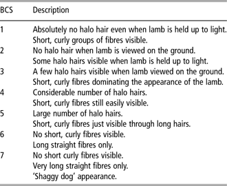
Within 12 h of birth, lambs were assigned a unique identification number and weighed. Birth coat scores (BCS) were taken using a visual score based on the South Australian Research and Development Institute selection demonstration flock project (Table 1), with a value of 1 assigned to lambs with no halo hair v. a maximum score of 7 assigned to lambs with very long, straight fibres and no short curly fibres visible. Assessments were carried out by one experienced operator at the same time of the day. A second BCS was taken on all the lambs on a single day and there were no variations from the initial scores.
Selection criteria used to assign BCS to newborn lambs
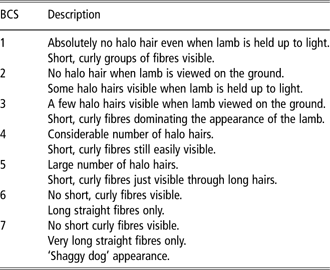
BCS = birth coat scores.
Skin histology
Mid-side skin samples were taken from the lambs after birth and again at 12 months of age using a 1-cm biopsy punch after subcutaneous injection of a local anaesthetic (Lignocaine 20 with adrenaline, Troy Laboratories Pty Ltd, Smithfield, New South Wales, Australia). Skin biopsies and thyroid glands were collected from deceased animals where possible and thyroid glands were weighed. All skin samples were fixed in 10% buffered formalin and processed by dehydration in increasing concentrations of ethanol and d-limonene (Histosolve; HD Scientific, Wetheril Park, New South Wales, Australia). Explants were embedded in paraffin wax in transverse orientation, and 7 μm serial sections were cut and stained using a modified SACPIC staining protocol (Nixon, Reference Nixon1993).
S/P ratios were determined in 10 non-overlapping views at ×10 objective using a light microscope. Wool follicle and sebaceous gland densities were measured by counting the number of wool follicles or sebaceous glands in 10 non-overlapping screens at ×20 objective. All measurements were corrected for shrinkage during sample processing by measuring the circumference of the skin section following staining and applying a shrinkage factor to each measurement (shrinkage factor = circumference post staining/circumference of the biopsy punch). Primary wool follicles were defined as wool follicles associated with a sweat gland; mature secondary follicles were wool follicles with a fibre present and immature secondary follicles were wool follicles in which fibre production had not commenced (Hardy and Lyne, Reference Hardy and Lyne1956). All of the measurements were taken using Video Pro 32 video image analysis software (Leading Edge, Adelaide, South Australia, Australia).
Wool measurements
The lambs were tip shorn at 3 months of age to remove the birth coat and shorn 12 months later, when the animals were 15 months old (hence 12 months of wool growth). Wool samples were also collected from 27-month-old animals (second year of growth). Wool samples were collected from the mid-side of animals and yield, fibre diameter (μ), micron deviation, CV and s.d. of fibre diameter, percentage of fibres >30 μm, curvature and spinning fineness were determined using an optical fibre diameter analyser (Classings Pty Ltd, Murray Bridge, South Australia, Australia). Staple lengths and staple strength were measured using a staple breaker (model 2; Agritest Ptd Ltd, Clontarf, New South Wales, Australia).
Statistical analyses
Plasma T4 and T3 levels were analysed using a general linear model, with treatment and time at which the sample was taken as fixed effects. Birth data (gestational length, birth weight and BCS), skin data (wool follicle densities, S/P ratios and sebaceous gland densities) and wool data were analysed using a linear mixed model with treatment as the fixed effect and lamb identification number and dam as random effects. Pair-wise comparisons were determined using a Bonferroni test and P-values <0.05 (P < 0.05) were deemed significant. Data are presented as means ± s.e.m. and all statistical analyses were performed using SPSS version 15 statistical software (SPSS Inc., Chicago, IL, USA).
Results
Maternal T4 and T3 levels
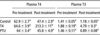
Maternal total T4 and T3 levels were measured in plasma samples collected on D55 (the first treatment day) and on D63 (second to last day of treatment) to determine the influence of exogenous T4 and PTU administration (Table 2). Although there were no changes in plasma total T4 and T3 levels of dams after treatment with control and PTU, treatment with exogenous T4 led to a 3.3-fold increase in plasma total T4 levels and 2.5-fold increase in plasma total T3 levels compared with pre-treatment levels (Table 2, P < 0.001). Furthermore, the increased plasma T4 and T3 levels of dams after treatment with exogenous T4 were significantly higher than the levels seen for the dams treated with control or PTU (Table 2, P < 0.001).
Blood plasma thyroid levels T4 and T3 of pregnant ewes before and after treatment with exogenous T4 or PTU, an inhibitor of thyroid hormone synthesis and activation

T4 = thyroxine; T3 = triiodothyronine; PTU = propylthiouracil.
Data are presented as means ± s.e.m.
a,b,x,yMeans within a row with different superscripts within plasma T4 or T3 are significantly different at P < 0.001.
Lambing
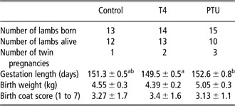
Between the 17 and 25 June 2005, a total of 42 lambs were born from 36 ewes, with the control group having one set of twins, the T4 group having two sets of twins and the PTU group having three set of twins (Table 3). This was despite efforts to select for singleton pregnancies at the D49 pregnancy scan. As a result, multiple pregnancies were accounted for during the statistical analyses. Although there were no significant differences in the gestational length of lambs from the control and PTU groups, administration with exogenous T4 led to significantly shorter gestational lengths compared with ewes treated with PTU (Table 3, P = 0.003).
Lambing outcomes from ewes treated with exogenous T4 or an anti-thyroid drug PTU during periods corresponding with foetal wool follicle initiation

T4 = thyroxine; PTU = propylthiouracil.
Data are presented as means ± s.e.m.
a,bMeans within a row with different superscripts are significantly different at P = 0.003.
Manipulation of thyroid hormone status during periods coinciding with foetal primary wool follicle development did not affect lamb birth weight or BCS (a measure of fleece length and curvature) taken within 12 h of birth (Table 3, P > 0.05).
Thyroid glands were collected whenever possible from lambs that died (control = 1, T4 = 1 and PTU = 3 lambs) and were fixed in 10% buffered formalin. Total thyroid gland weight from the control and T4 groups were 0.67 and 0.8 g, respectively, compared with 1.16, 2.18 and 1.11 g from the lambs exposed to PTU (1.49 ± 0.35 g). Although the sample number was low, thyroid glands derived from the PTU group appeared heavier than the other treatment groups.
Skin measurements from thyroid-manipulated lambs (1 week old)
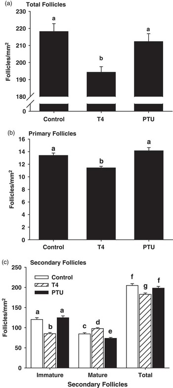
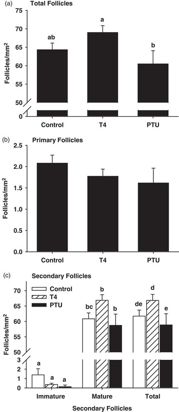
Skin biopsies were taken from 1-week-old lambs and the wool follicle morphology was determined. Total wool follicle density was significantly lower in lambs exposed to exogenous T4 in utero, compared with the control and PTU groups (Figure 1a, P < 0.05), and this was attributed to primary (Figure 1b) and total secondary follicle densities (Figure 1c) being lower in the T4 treatment group. Interestingly, T4 treatment in utero led to a decrease in the density of immature secondary follicles and an increase in mature secondary follicle density, compared with the control and PTU-treated lambs (Figure 1c).
(a) Total wool follicle, (b) primary wool follicle and (c) secondary wool follicle densities of 7-day-old lambs exposed to exogenous thyroxine (T4) or propylthiouracil (PTU) between gestational days 55 and 64. Immature = immature secondary follicles, mature = mature secondary follicles and total = immature and mature secondary follicles. Data are presented as means ± s.e.m. and bars with different letters (a, b, c, d, e, f and g) indicate significant differences (P < 0.05).

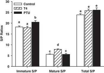
In regard to S/P ratios, there were no significant differences between total S/P ratios (Figure 2). However, there were significant differences within the immature and mature S/P ratios, with PTU administration between D55 and D64 leading to an increase in the immature S/P ratio and T4 administration leading to lambs having increased mature S/P ratios (Figure 2, P < 0.05).
Secondary to primary follicle (S/P) ratios of 7-day-old lambs that were exposed to exogenous thyroxine (T4) or propylthiouracil (PTU) between gestational days 55 and 64, coinciding with primary wool follicle initiation. Immature S/P = immature secondary/primary follicle ratio, Mature S/P = mature secondary/primary follicle ratio, Total S/P = total secondary/primary follicle ratio. Data are presented as means ± s.e.m. and bars with different letters (a, b, c, d and e) are significantly different (P < 0.05).

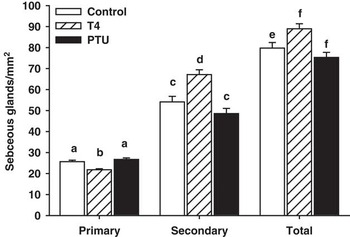
Sebaceous gland densities (number of glands per mm2) were significantly higher in lambs exposed to exogenous T4, compared with the control and PTU-administered groups (Figure 3, P < 0.05). This was largely due to a significant increase in the density of sebaceous glands associated with secondary follicles (Figure 3).
Sebaceous gland density of wool follicles of 7-day-old lambs that were exposed to exogenous thyroxine (T4) or propylthiouracil (PTU) between gestational days 55 and 64, coinciding with primary wool follicle initiation. Data are presented as means ± s.e.m. and bars with different letters (a, b, c, d, e and f) are significantly different (P < 0.05).

Skin measurements from thyroid-manipulated lambs (12 months old)
Skin biopsies were taken from the sheep 1 year after birth and wool follicle parameters were determined. Sheep exposed to exogenous T4 in utero had significantly higher total follicle densities compared with the PTU treatment group (Figure 4a, P < 0.007). The difference in total wool follicle density was largely attributed to significant increases in mature and total secondary follicle densities (Figure 4c, P < 0.05), as there were no differences in primary wool follicles between treatments (Figure 4b). Sebaceous gland densities were also determined, and there were no significant differences between treatment groups (data not shown).
(a) Total wool follicle, (b) primary wool follicle and (c) secondary wool follicle densities of 12-month-old sheep that were exposed to exogenous thyroxine (T4) or propylthiouracil (PTU) between gestational days 55 and 64. Immature = immature secondary follicles, mature = mature secondary follicles and total = immature and mature secondary follicles. Data are presented as means + s.e.m. and bars with different letters (a, b, d, e, ab, bc and de) indicate significant differences (P < 0.05).

Sheep were also weighed and there were no significant differences between treatment groups with regard to BW (control = 82.0 ± 3.0 kg; T4 = 76.1 ± 2.4 kg and PTU = 75.8 ± 4.5 kg; P > 0.05).
Wool measurements (first and second year of growth)
Lambs were tip shorn 3 months after birth to remove the birth coat and were shorn 12 months later (first year of growth, 18-month-old animals). A total of 26 hoggets were shorn (control = 9, T4 = 10, PTU = 7). There were no significant differences between treatments in unskirted fleece weight, skirted fleece weight, yield (μ), micron deviation, CV and s.d. of fibre diameter, percentage of fibres >30 μm, curvature and spinning fineness. However, there was a trend for T4 administration to increase the percentage of fibres >30 μm, compared with the control and PTU treatments (P = 0.07). Administration of exogenous thyroid hormones or PTU did not influence staple length, staple strength or point of break.
Sheep were shorn 12 months later (second year of growth, 30-month-old animals) and subjected to the same measurements. There were no significant differences in any of the parameters measured (P > 0.05).
Discussion
The influence of long-term manipulation of thyroid hormone status on the development of wool follicles late in gestation has previously been reported (Ferguson et al., Reference Ferguson, Schinckel, Carter and Clarke1956; Hopkins and Thorburn, Reference Hopkins and Thorburn1972; Chapman et al., Reference Chapman, Hopkins and Thorburn1974), as has the influence of thyroid hormones on the function of postnatal wool and hair follicles (Ferguson et al., Reference Ferguson, Schinckel, Carter and Clarke1956; Williams et al., Reference Williams, Thornberry and Gilmore1990; Villar et al., Reference Villar, McMillen, Dicks and Rhind2000). However, the long-term effects of transient manipulation of thyroid hormone status coinciding with primary wool follicle initiation on wool follicle development and function are unknown. We hypothesise that increased maternal thyroid hormone status would lead to increased maturity of wool follicles in postnatal lambs and higher yields in adult sheep. In this study, pregnant ewes were treated with exogenous T4 or PTU, an inhibitor of T4 synthesis, and conversion to the active form of the hormone, between gestational days 55 to 64, and the wool follicle and fibre parameters of the resultant lambs were determined over a 2-year period.
Analysis of the skin taken 7 days after birth showed that lambs exposed to exogenous T4 in utero had significantly lower total wool follicle densities compared with the control and PTU treatment groups and this decrease in wool follicle density was attributed to both lower primary and secondary follicle densities. Despite the T4 treatment group having lower wool follicle densities, the maturity of secondary wool follicles was higher than in the other treatments, as shown by an increase in the density of mature secondary follicles (wool follicles in which a fibre was present, Figure 1c), higher densities of sebaceous glands associated with secondary follicles (Figure 3) and the fact that total S/P ratios were not affected, but T4 treatment led to higher mature S/P ratios (Figure 2).
The decrease in postnatal wool follicle densities in this study was surprising as previous studies have shown that exogenous T4 treatment during gestation did not influence wool follicle initiation (Ferguson et al., Reference Ferguson, Schinckel, Carter and Clarke1956). However, the observations that suggest secondary wool follicle maturation were enhanced by exogenous T4, which agrees with the reported literature (Ferguson et al., Reference Ferguson, Schinckel, Carter and Clarke1956). Differences between the studies are likely to be due to differences in the timing of the treatment, where we treated pregnant ewes for a short period of time (D55 to D64), v. Ferguson et al. treating lambs post partum (Ferguson et al., Reference Ferguson, Schinckel, Carter and Clarke1956). This is significant considering that different populations of wool follicles develop at different times pre- and postnatally (Hardy and Lyne, Reference Hardy and Lyne1956), with our treatments coinciding with the initial stages of primary wool follicles development compared with postnatal treatment with T4, during secondary follicle development.
After 1 year, the wool follicle profiles of the sheep changed, with sheep exposed to exogenous T4 in utero displaying higher total wool follicle densities (Figure 4) compared with sheep treated with PTU, and this was largely attributed to increased total and mature secondary follicle densities. This is the opposite of what was seen at birth and more in line with postnatal induction of a hyperthyroid state leading to increased wool follicle activity, considering that mature secondary follicles are defined as wool follicles in which a fibre is present (Hardy and Lyne, Reference Hardy and Lyne1956). The changes in wool follicle densities in the 12 months after birth may be due to numerous factors, such as changes in the BW of animals or attrition and shutdown of secondary wool follicles. As wool follicle densities do not take into account the surface area of the sheep (hence BW), it is possible that the changes in wool follicle densities could have reflected changes in BW. This was not the case as there were no significant differences in the birth weight of lambs or the BW of adult sheep taken at shearing.
Despite treatment with exogenous T4 and PTU being applied during a period coinciding with primary wool follicle initiation, the long-term effects on wool follicle parameters were seen in the secondary wool follicles, namely the increased total and secondary wool follicle densities. Perinatal and post-partum attrition of the secondary wool follicle has previously been reported (Hocking Edwards, Reference Hocking Edwards1999; Toland Thompson et al., Reference Toland Thompson, Hebart, Penno and Hynd2007) and it is likely that immature secondary follicles would be more susceptible to attrition than mature secondary wool follicles that have already commenced wool fibre production. Soon after birth, animals exposed to exogenous T4 had significantly higher proportions of mature secondary follicles compared with the other treatment groups, as reflected by higher secondary follicle densities and mature S/P ratios. In comparison, animals within the PTU treatment group had higher numbers of immature wool follicles. It is possible that thyroid hormone status during wool follicle initiation determines the susceptibility of secondary wool follicles to undergo attrition, with increased thyroid hormone levels leading to more secondary follicles remaining active. This resulted in the increased total and secondary follicle densities at 1 year of age as seen in the T4 group compared with animals exposed to PTU in utero.
The influence of inducing hyperthyroidism or the hypothyroidism state in adult animals is well documented, with treatment with exogenous thyroid hormones leading to increases in fibre growth, length growth rate and, in some cases, decreases in fibre diameter (Williams et al., Reference Williams, Thornberry and Gilmore1990; Hynd, Reference Hynd1994; Puchala et al., Reference Puchala, Prieto, Banskalieva, Goetsch, Lachica and Sahlu2001). Conversely, athyroid animals have a higher degree of secondary follicle shutdown compared with control animals (Hynd, Reference Hynd1994). Hypothyroidism in adult sheep influences fibre production by decreasing the proportion of proliferating cells within the follicular bulb that enter the fibre cortex, and hence more cells are used to make the inner root sheath (Hynd, Reference Hynd1994). This, in turn, results in lower production ratios (the ratio of fibre area/fibre area plus the inner root sheath). This agrees with the observation that thyroid hormone status during foetal wool follicle development is important for the later development and maturation of follicles, rather than wool follicle initiation, suggestive of a role in promoting cellular differentiation within the follicle required for follicle maturation and fibre formation (Ferguson et al., Reference Ferguson, Schinckel, Carter and Clarke1956; Hopkins and Thorburn, Reference Hopkins and Thorburn1972). Hence, thyroid hormone status plays an important role in the ability of a follicle to produce fibre, through the promotion of follicle maturity, attrition rates of secondary follicles (both in the perinatal period) or complete shutdown of adult wool follicles.
Although foetal thyroid hormone status was not determined following treatment with either PTU or T4, we are confident that lambs were exposed to hyperthyroid and hypothyroid conditions in utero. First, maternal T4 and T3 plasma levels were elevated following treatment, and given that the placenta is permeable to maternal endogenous T4 and T3 until late in gestation (Atkins et al., Reference Atkins, Cohen and Phillips2000), it is likely that the lamb was exposed to exogenous T4 in utero. Second, in the limited number of animals that died during the perinatal period, the thyroid glands of lambs exposed to PTU in utero appeared to have larger thyroid glands (very low sample numbers prevented valid statistical analyses), suggestive of a negative feedback response. PTU has been shown to readily cross the human placenta throughout the entirety of gestation (Atkins et al., Reference Atkins, Cohen and Phillips2000), and when used late in gestation, approximately 10% of foetuses/neonates displayed either hypothyroidism or hyperthyroidism but thyroid hormone status was resolved within 1 month of birth or cessation of treatment (Cheron et al., Reference Cheron, Kaplan, Larsen, Selenkow and Crigler1981; Rosenfeld et al., Reference Rosenfeld, Ornoy, Shechtman and Diav-Citrin2009). The fact that we saw enlarged thyroid glands in lambs exposed to PTU in utero may be due to this study using doses 1.5- to 2.0-fold higher than the recommended maintenance doses for humans. Lastly, the doses of exogenous T4 and PTU used in this experiment were similar to those previously used that have resulted in altered thyroid hormone status in foetal sheep.
In conclusion, transient manipulation of thyroid hormone status coinciding with foetal primary wool follicle initiation altered the wool follicle characteristics of animals soon after birth. Specifically, treatment with exogenous thyroid hormones led to decreases in primary, secondary and total wool follicle densities. However, it appeared that these same animals had higher total wool follicle densities and more mature secondary follicles at 12 months of age. Differences in wool follicle densities did not translate to changes in fibre yield or diameter. This shows that transient manipulation of maternal hormone status coinciding with foetal wool follicle development can have long-term effects on skin characteristics.
Acknowledgements
The authors thank Dr Margie Trowbridge from Brecon Breeders for help with the insemination, Skye Rudiger for the collection of semen and the numerous people who helped with the insemination, treatment and lambing periods. This study was funded by the Sheep Genomics Program (Australian Wool Innovation Pty Ltd and Meat and Livestock Australia).