Introduction
Fat deposition in pigs is very similar to that in human beings, so the pig is considered to be an ideal model in which to study obesity, diabetes mellitus and cardiovascular disease in humans (Bellinger et al., Reference Bellinger, Merricks and Nichols2006). Lipid deposition is influenced by the combined effects of pre-adipocyte proliferation, differentiation and apoptosis. Investigating the mechanism of lipid deposition is meaningful for both animal science and human medicine. As members of the nuclear hormone receptor family, retinoid X receptors (RXRs), including α, β, and γ types, are transcription factors of ligands that when activated regulate many aspects of cell activity. Tontonoz and colleagues (Reference Tontonoz, Graves and Budavari1994) first proved that heterodimers formed from RXRα and peroxisome proliferator activated receptors gamma (PPARγ) could promote the differentiation of adipocytes. Chawla and colleagues (Reference Chawla, Schwarz, Dimaculangan and Lazar1994) also found that, after activation by its ligand, PPARγ could combine with RXRα to form a heterodimer, this then forming a ternary complex with a PPARγ ligand that was able to induce differentiation of pre-adipocytes. Lin and colleagues (2007b) reported that RXRα could promote pre-adipocyte differentiation, but the relationships between RXRα, apoptosis and pre-adipocyte differentiation, as well as their different mechanisms, have not been reported to date. In the present study, semi-quantitative reverse transcriptase polymerase chain reaction (semi-qRT-PCR), cell transfection, fluorescein stain with acridine orange (AO) and Hoechst 33342, and flow cytometry technology were applied to study variation in porcine pre-adipocyte apoptosis after treatment with RXRα 9-cis retinoic acid (9-cisRA), RXRα over-expression and RXRα silencing. This study therefore provides further insight into the influence of RXRα on porcine pre-adipocyte differentiation and apoptosis, and also suggests some new methods for the study of lipogenesis.
Materials and methods
Materials
One 3-day-old healthy Landrace pig was obtained from the experimental farm at Northwest A & F University (Yangling, Shaanxi, China).
Methods
Primary culture of porcine pre-adipocytes
The primary culture of porcine pre-adipocytes was carried out using the methods of Li and colleagues (Reference Li, Yang, Lu and Sun2005) . Adipose tissue was removed from the dorsal subcutaneous fat deposit in the neck. The adipose tissue was minced to achieve a volume of 1 mm3, followed by digestion with 1 g/l type I collagenase (DMEM/F12+20 g/l bovine serum albumin added to 1 g/l type I collagenase; Invitrogen, USA) for 60–70 min at 37°C. The digested tissue suspension was then filtered through a 200-μm nylon mesh and centrifuged at 1500 rpm for 3 min to separate the fat cake and the stromal-vascular (SV) fraction. Erythrocyte lysate (using NH4Cl 154 mmol/l+KHCO3 10 mmol/l+EDTA 0.1 mmol/l) was maintained for 10 min at room temperature, then centrifuged at 1000 rpm for 5 min. The cells were washed twice with DMEM/F12, and centrifuged at 1000 rpm for 5 min after each wash. Finally, the cells were suspended in DMEM/F12 and 10% FBS (folium ilicis chinensis, acquired in Hangzhou). Pre-adipocyte density was measured using Trypan blue and blood cell counting slides. After counting the pre-adipocytes, they were seeded in plating medium. The seeded cells were cultured at 25°C in 5% CO2 in a humidified atmosphere. The medium was changed every 2 days. The pre-adipocytes were subjected to the following experiments at 50% confluence. A single batch of porcine pre-adipocytes from an individual pig was used for all replicates.
Experimental design
Three culture groups were investigated: (1) an RXRα ligand 9-cisRA (10 nmol/l and 10 μmol/l; Sigma-Aldrich, St Louis, MD, USA) treatment group; (2) a pEGFPC2–RXRα transfection group, rebuilt from pYFP–RXRα (obtained from Professor Gonzalez, National Institutes of Health, New York, USA); (3) an RXRα–siRNA transfection group. RXRα–siRNA was synthesized according to the RXRα gene sequence (GenBank accession no. NMDQ279926); negative siRNA was synthesized by Guangzhou RiboBio Co., Ltd (Guangzhou, Guangdong, China).
Transfection was carried out according to the protocol of the Lipofectamine 2000 Kit (Invitrogen). Twenty-four hours before transfection, treatment groups were placed in a culture solution without double-antibody, and 1 h before transfection were placed into an OptiMEM I (Sigma) culture solution. We diluted Lipofectamine 2000 with OptiMEM I, and diluted pEGFPC2–RXRα, pEGFPC2 zero carrier, RXRα–siRNA and negative siRNA into 2 μg/ml, 2 μg/ml, 50 nmol/l and 50 nmol/l solutions, respectively. OptiMEM I was then mixed with DNA–Lipofectamine 2000 and RNA–Lipofectamine 2000, then allowed to stand for 20 min at room temperature. The mixture of DNA–Lipofectamine 2000 and RNA–Lipofectamine 2000 was added into culture solution drop by drop, according to the following mixture ratio: culture solution=1:10 (v/v). Following culture for 6 h in an incubator at 37°C and 5% CO2, new medium was substituted for the old medium.
Semi-qRT-PCR
After 72 h transfection, the transfection group cells were collected and total RNA isolated using Trizol (Fermentas, Life Science Hanover, MD, USA). The quality and purity of the RNA was checked using agarose gel electrophoresis and ultraviolet spectrophotometry.
Specific primers of RXRα and β-actin were designed according to GenBank (accession nos DQ279926 and AY550069) and synthesized by Invitrogen Co., Ltd., Shanghai Branch (Table 1). The reverse transcript of the control was completely designed along Fermentas guidelines, and 1 μl RNA and random hexamer primer were used to prepare the first chain of cDNA. The cDNA was prepared to obtain a volume of 25 μl. The PCR amplification reaction system consisted of 2 μl dNTP mix (2.5 mmol/l), 2.5 μl PCR buffer (NH)2SO4, 1.5 μl MgCl2 (25 mmol/l), 1 μl cDNA, 0.5 μl primer I (20 μmol/l), 0.5 μl primer II (20 μmol/l), 0.125 μl rDNA synthase (0.5 U/μl), and dH2O added to obtain a total reaction volume of 25 μl. Amplification conditions were 95°C for 2 min, 94°C for 1 min, 55.7/58°C for 45 s and 72°C for 1 min, with a final 10 min extension phase at 72°C. The reaction product was detected by 1% agarose gel electrophoresis and the electrophoresis picture analysed using the Dolphin-DOC gel imaging system. The result was checked against an identical initial quantity of β-actin and expressed with a correlation value for the transfection group and control group.
Oligonucleotide PCR primers

Hoechst 33342 staining
Hoechst 33342 (Sigma) was added to the culture medium in the experimental group to a final concentration of 10 μg/ml and incubated at 37°C for 30 min. The culture medium was then removed and the adipocytes fixed with 4% paraformaldehyde for 5–10 min. The excitation fluorescence microscopy process mode use of UV excitation filter wedge films, with a blocking wedge of 400–500 nm.
AO staining
An adipocyte suspension (1×10⁷/ml) was prepared and AO added (Sigma) at a ratio of 10:1, and observations made on the variation of morphology and cell number count. Every field of observation had 500 cells and was repeated 3–5 times. Apoptotic rates were calculated.
Flow cytometry
Adipocytes were collected in triplicate after treatment for 72 h. The density of cells was regulated to 1×10⁶/ml and data were collected using an Elite flow cytometer. After staining with Annexin V/PI, the data were analysed using Cell Quest, and the apoptotic rates were obtained by counting cell numbers in the stage of apoptosis.
Statistical analysis
Experimental data were expressed as mean±s.d. Analysis of variance and significance tests were conducted using SPSS version 11.5 and one-way ANOVA, with P<0.05 indicating significant differences.
Results
RXRα mRNA expression after transfection
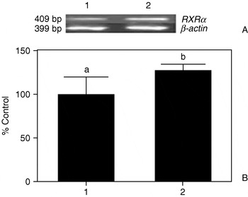
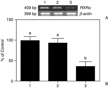
According to the semi-qRT-PCR results, the expression of RXRα mRNA was significantly higher than in the control group (P<0.05; Fig. 1). This result suggests successful RXRα over-expression. However, the expression of RXRα mRNA was significantly lower after transfection with RXRα–siRNA when compared with the non-transfected group and the group transfected negatively with RXRα–siRNA (P<0.05; Fig. 2).
RXRα mRNA expression in porcine differentiating adipocytes transfected by pEGFPC2–RXRα (A) and the significance test (B). Lanes: 1, control (empty vector pEGFPC2 transfection); 2, pEGFPC2–RXRα. Error bars represent standard error. Different letters indicate significant differences between treatments (P<0.05); the same letters indicate no significant difference (P>0.05).

RXRα mRNA expression in porcine differentiating adipocytes transfected by RXRα–siRNA (A) and the significance test (B). Lanes: 1, non-transfection; 2, negative siRNA; 3, RXRα–siRNA. Error bars represent standard error. Different letters indicate significant differences between treatments (P<0.05); the same letters indicate no significant difference (P>0.05).

Hoechst 33342
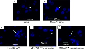
Each group presented with both normal and apoptotic cells as observed using fluorescence microscopy after staining with Hoechst 33342. A series of morphologic changes in the nuclei of apoptosic cells and normal cells appeared normal and had the same staining (see Fig. 3A, arrow). The apoptotic cells displayed cytoblast condensing, chromosome agglutination (see Fig. 3B and C, arrows), chromosome marginalization (see Fig. 3D, arrow) and apoptotic bodies (see Fig. 3E, arrow).
Hoechst 33342 staining results of porcine adipocytes in different treatment groups. (A) Control group, arrow indicates the normal nucleus; (B) 10 nmol/l 9-cisRA group, arrow indicates cytoblast condensing and chromosome agglutination; (C) 10 μmol/l 9-cisRA group, arrow indicates apoptotic bodies; (D) pEGFPC2–RXRα transfection group, arrow indicates chromosome marginalization; (E) RXRα–siRNA transfection group, arrow indicates apoptotic bodies. Scale bars,100 μm. (See online for a colour version of this figure.)

Acridine orange
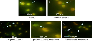
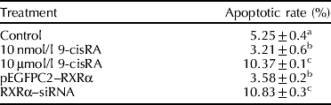
Following AO staining, the nuclei of normal cells appeared homogeneously yellow or green under fluorescence and the cytoplasm appeared as homogeneous jacinth or saffron yellow under fluorescence. Most cells appeared to have the same size (see Fig. 4A, arrow). The nuclei of apoptotic cells appeared green or dark green under fluorescence, the volume of the cells decreased, and in some cells cytoblasts had condensed (see Fig. 4B–E, arrows). The apoptotic rate was calculated by noting variations in cell morphology. The results are shown in Table 2. Compared with the control group, the apoptotic rates significantly decreased in the 10 µmol/l 9-cisRA and pEGFPC2–RXRα treated groups (P<0.05), while those in the 10 μmol/l 9-cisRA and RXRα–siRNA treated groups were significantly higher than the control group (P<0.05).
Acridine orange (AO) staining results of porcine adipocytes in different treatment groups. (A) Control group, arrow indicates the normal nucleus; (B) 10 nmol/l 9-cisRA group, arrow indicates apoptotic nucleus; (C) 10 μmol/l 9-cis RA group, arrow indicates apoptotic nucleus; (D) pEGFPC2–RXRα transfection group, arrow indicates apoptotic nucleus; (E) RXRα–siRNA transfection group, arrow indicates apoptotic nucleus. Scale bars,100 μm. (See online for a colour version of this figure.)

Influence of different treatment on porcine pre-adipocyte apoptotic rate determined by fluorescence staining
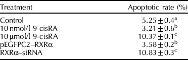
Different letters indicate significant differences between treatments (P<0.05); the same letters indicate no significant difference (P>0.05).
Flow cytometry
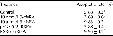
The results of flow cytometry are shown in Table 3. Not only did the apoptotic rate significantly decrease in the 10 nmol/l 9-cisRA and pEGFPC2–RXRα treated groups (P<0.05), but the apoptotic rates in the 10 µmol/l 9-cisRA and RXRα-siRNA treated groups were significantly higher than the control group (P<0.05).
Effects of different treatment on the apoptotic rate of porcine pre-adipocyte using a flow cytometry detection method
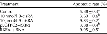
Different letters indicate significant differences between treatments (P<0.05); the same letters indicate no significant difference (P>0.05).
Discussion
Apoptosis, together with proliferation and differentiation, plays an important role in controlling the number of fat cells. Miller and colleagues (Reference Miller, Faust, Goldberger and Hirsch1983) suggested that an apparent reduction in weight not only results from a decrease in the volume of fat cells, but also a reduction in their number. However, there have been no reports describing the mechanism of adipocyte loss. Prins and colleagues (Reference Prins, Walker, Winterford and Cameron1994) first reported that human adipocytes undergo apoptosis following growth factor deprivation or mild heat injury in vitro, thus demonstrating a cellular mechanism by which normal adipocyte loss could occur. However, due to the characteristics of fat cells (relatively small nuclear plasma, presence of a large number of fat drops, thus preventing changes in apoptosis characteristics), few studies have been carried out in this regard. Prins and colleagues (Reference Prins, Niesler and Winterford1997) and Lin and colleagues (2004a) proved that TNF-α could induce apoptosis of human or rat pre-adipocytes in vitro. Although some more in-depth studies on adipocyte apoptosis using 3T3 cell lines and mouse or human primary cultured adipocytes have been conducted (Valladares et al., Reference Valladares, Alvarez, Ventura, Roncero, Benito and Porras2000; Reusch and Klemm, Reference Reusch and Klemm2002; Fischer-Posovszky et al., Reference Fischer-Posovszky, Tornqvist, Debatin and Wabitsch2004) little was known about the molecular mechanism of porcine adipocyte apoptosis.
As a ligand-activated transcription factor, RXRα plays an important role in the processes of cell development, particularly in cell proliferation and apoptosis. Zhong and colleagues (Reference Zhong, Yang, Huang, Cohen and Roy-Burman2003) noted retarded cell growth performance and apoptosis after over-expression of RXRα in CWR22R prostate cell lines originally lacking the RXRα gene. Lee and colleagues (Reference Lee, Ma, Yan, Liu, Zhang and Cohen2005) indicated that IGFBP3 in the RXRα knockout mouse could not induce apoptosis. Chen and colleagues (Reference Chen, Chen and Pan2008) reported the involvement of RXRα in the apoptosis of hippocampus neurons. Nevertheless, there were no reports about the effect of RXRα on adipocyte apoptosis.
As a derivative of vitamin A, 9-cisRA not only plays an important role in embryo development, cell differentiation, apoptosis and the environmental stability of biological bodies, but is also involved in the proliferation and differentiation of adipocytes (Bonet et al., Reference Bonet, Ribot, Felipe and Palou2003; Villarroya et al, Reference Villarroya, Iglesias and Giralt2004). Studies have shown that 9-cisRA can induce apoptosis by inhibiting neuroblastoma cell line proliferation (Zhao et al., Reference Zhao, Shi, Wei, Wang and Zhang2007) as well as lung cancer cell apoptosis (Xiang et al., Reference Xiang, Yao, Huang and Wan2007). However, there have been no reports about its effects on adipocyte apoptosis. The present study found that 9-cisRA inhibited porcine pre-adipocyte apoptosis at a level of 10 nmol/l, but induced it at a level of 10 μmol/l. Because 9-cisRA can specifically combine with the homologous dimer RXR/RXR and heterodimers such as RXR/RAR, PPAR, vitamin D and so on (Levin et al., Reference Levin, Sturzenbecker and Kazmer1992), the main biological effect of 9-cisRA is related to its RXRα receptor (Hellemans et al., Reference Hellemans, Verbuyst and Quartier2004). Nagy and colleagues (Reference Nagy, Thomazy and Shipley1995) found that 9-cisRA induced apoptosis of the myeloid leukaemia cell line through RXRα (HL60). In our study, the fact that 9-cisRA affected porcine pre-adipocyte apoptosis suggests that RXRα might be involved in this process. RXRα influences the differentiation of adipocytes. Imai and colleagues (Reference Imai, Jiang, Chambon and Metzger2001) noted that, after selectively knocking out RXRα, adipocyte differentiation was inhibited. Lin and colleagues (2007b) reported that RXRα gene silencing inhibited porcine pre-adipocyte differentiation. In order to further study the effect of RXRα on adipocytes and particularly its role in apoptosis, on the basis of ligand treatment by transfection with pEGFPC2–RXRa or siRNA, our study causes over-expression of or interfered RXRα in the porcine pre-adipocyte.
Finally, we found that RXRα has the function of inhibiting the apoptosis of porcine pre-adipocytes, and confirmed the results using ligands. RXRα can form a dimer with other nuclear receptors to induce cell apoptosis. For instance, liver X receptor (LXR)/RXR can inhibit Islet B cell proliferation and mediate its apoptosis; RXRα may induce apoptosis by assisting the transport of orphan nuclear receptor TR3. RXRα and orphan nuclear receptor Nur77 can form a heterodimer, as Nur77 shifts from the nucleus to mitochondria and then combines with Bcl2 to trigger apoptosis (Lin et al., Reference Lin, Zhao and Chen2004b; Wente et al., Reference Wente, Brenner, Zitzer, Gromada and Efanov2007; Zhang, Reference Zhang2007). Whether RXRα combines with other nuclear receptors to form a dimer to regulate porcine pre-adipocyte apoptosis, and also its mechanism, still remain to be determined. Lin and colleagues (2007a) reported that 10 nmol/l 9-cisRA promoted porcine pre-adipocyte differentiation by up-regulating the mRNA expression levels of RXRα and PPARγ, whereas 10 μmol/l 9-cisRA exerted the totally opposite effect. The apoptosis rates at these two concentrations were significantly difference, with the apoptosis rate high in the inhibited differentiation process, and declining in the promoted differentiation process. This is consistent with the differentiation and apoptosis of porcine pre-adipocytes after RXRα over-expression and gene silencing. Therefore, it can be seen that the differentiation and apoptosis of porcine pre-adipocytes is affected by adjusting RXRα, and the promoting effect of RXRα on porcine pre-adipocyte differentiation not only relates to the regulation of adipocyte transcription factor, but also the inhibition of adipocyte apoptosis. This result provides information for further research exploring the development of molecular mechanisms in porcine pre-adipocytes.
Acknowledgements
The authors would like to thank Dr Hong Ji for his kind help in reviewing the manuscript. This work was supported by the National Natural Sciences Foundation of China (no. 30471239).