INTRODUCTION
Kudoid parasites are microscopic, spore-forming myxosporean parasites that infect a wide variety of fish and can cause significant losses in the seafood industry either through direct mortality or by spoiling fish meat (Reference EgusaEgusa, 1986b; Moran et al. Reference Moran, Whitaker and Kent1999). Myxosporean species undergo a biphasic life cycle between a vertebrate and invertebrate host (Lom and Dyková, Reference Lom and Dyková2006). To date, the invertebrate host for myxosporeans within the Kudoidae is unknown, and consequently, the morphology of the actinospores within this life stage are also unknown. Therefore, kudoids are defined by their myxospore life cycle stage that occurs in the vertebrate host (mostly fish) and researchers have no recourse to conduct cross-transmission studies.
In the Kudoidae, species boundaries are based on a combination of tissue tropism; on spore morphology (including a recognized extent of variation in spore size); and more recently, on ribosomal DNA sequences (typically small subunit (SSU) regions) which have proven useful to determine relationships between species and to identify subtle species differences (Blaylock et al. Reference Blaylock, Bullard and Whipps2004; Diamant et al. Reference Diamant, Ucko, Paperna, Colorni and Lipshitz2005; Holzer et al. Reference Holzer, Blasco-Costa, Sarabeev, Ovcharenko and Balbuena2006; Whipps and Kent, Reference Whipps and Kent2006; Burger and Adlard, Reference Burger and Adlard2010). Links between infections in closely related host species are less informative since several species of kudoids are known to infect a number of unrelated fish hosts (see Blaylock et al. Reference Blaylock, Bullard and Whipps2004; Diamant et al. Reference Diamant, Ucko, Paperna, Colorni and Lipshitz2005; Whipps and Kent, Reference Whipps and Kent2006; Burger et al. Reference Burger, Barnes and Adlard2008).
One of the most obvious diagnostic features of kudoids is the number of shell valves and polar capsules in the spores. Kudoa species are characterized by having more than 3 shell valves and an equal number of polar capsules. The majority of species have 4 polar capsules, but species with 5–13 have also been reported. Some species have multiple spore morphotypes within a single plasmodium, where the number of polar capsules is not uniform amongst spores originating from a single primordial cell. Usually only the dominant morphotype is used for identification and characterization.
The phenomenon of multiple spore morphotypes is common in spores with radial symmetry, particularly those with 5 or more spore valves and polar capsules, including species such as: K. monodactyli Gunter et al. Reference Gunter, Cribb, Whipps and Adlard2006 and K. neurophila (Grossel et al. Reference Grossel, Dykova, Handlinger and Munday2003), which have spores with 5 polar capsules dominant; Kudoa lethrini Burger et al. Reference Burger, Cribb and Adlard2007 and K. yasunagai (see Reference EgusaEgusa, 1986a), which have spores with 7 polar capsules dominant; Kudoa chaetodoni Burger et al. Reference Burger, Cribb and Adlard2007 that has spores with almost even dominance of spores with 8 and 9 polar capsules; and K. permulticapsula Whipps et al. (Reference Whipps, Adlard, Bryant and Kent2003a) that has spores with 13 polar capsules that dominate each infection. The cause of this morphological plasticity is unknown, but seems to happen less often in species characterized by 4 valves and polar capsules (reported only from 5 of 70 four-valved species).
In 2007 a Kudoa sp. with a dominant 5 polar capsule (5pc) morphotype and a minor 6pc morphotype was found in the brains of 2 whiting species, Sillago ciliata and S. maculata off North Stradbroke Island in Queensland, Australia (Burger et al. Reference Burger, Cribb and Adlard2007). This species was characterized by a combination of morphological and small subunit ribosomal DNA (18S rDNA) data with a surprising result. The Kudoa species from whiting was genetically identical to a morphologically distinct species, K. yasunagai. Kudoa yasunagai infects the brains of several fish species, Lateolabrax japonicus, Oplegnathus fasciatus, Seriola quinqueradiata (see Yasunaga et al. Reference Yasunaga, Hatai, Ogawa and Yasumoto1981), and Paralichthys olivaceus (see Whipps et al. Reference Whipps, Grossel, Adlard, Yokoyama, Bryant, Munday and Kent2004) from Japan and Plotosus lineatus from the Philippines (see Cheung and Nigrelli, Reference Cheung and Nigrelli1990). Kudoa yasunagai has a dominant 7pc morphotype and a minor 6pc (and rarely 8pc) morphotype. Despite the morphological difference and geographical separation the authors took the conservative approach and were reluctant to officially describe the Kudoa species from whiting due to its identical SSU rDNA sequence to K. yasunagai. Burger et al. (Reference Burger, Cribb and Adlard2007) hypothesized that the reason for the genetic similarity between these two species was either that SSU rDNA was too conserved to allow species discrimination, or that these two species were actually conspecific and phenotypic variation caused by their occurrence in different hosts produced different dominant spore morphotypes.
During subsequent parasitological surveys (2008), a new kudoid with 6pc and 7pc morphotypes was collected from the brain of monocle bream, Scolopsis monogramma. This specimen was superficially similar to Kudoa yasunagai and prompted the authors to re-examine the hypotheses suggested by Burger et al. (Reference Burger, Cribb and Adlard2007). Here we investigate morphological and genetic relationships between kudoid species with multiple morphotypes that infect the brain. In addition to re-examining the material from Burger et al. (Reference Burger, Cribb and Adlard2007) (i.e. Kudoa sp. from whiting, K. chaetodoni from butterfly fish, and K. lethrini from emperor fish) we analysed the sample from monocle bream and a new specimen similar to K. neurophila from the brain of yellowtail kingfish, Seriola lalandi.
MATERIALS AND METHODS
Sample collection
Most of the fish were collected from Queensland, Australia: Heron Island (23° 27′S; 151° 55′E) and Lizard Island (14° 39′S; 145° 27′E) on the Great Barrier Reef; and North Stradbroke Island (27° 29′S; 153° 24′E) in Moreton Bay. The fish were caught using line, spear, or seine net and were euthanized by prolonged immersion in clove oil. The Seriola lalandi samples were collected from Jurien Bay (30° 18′S; 115° 2′E) in Western Australia and donated by Dr Fran Stephens.
The fish were examined for kudoid infection in the brain tissue by taking a subsample of brain tissue (approximately 1/3 to 1/2 of the brain depending on size) which was placed on a glass plate (76×50 mm), moistened with vertebrate saline (0·85% salt), and macerated with scalpel blades. A second glass plate was placed on top and the liquid squeezed out into a 2 ml centrifuge tube. An aliquot (∼15 μl) of the suspension from the bottom of the tube was examined for kudoid spores under an Olympus BH2 microscope at 400× magnification. When kudoid spores were detected, a subsample of brain was preserved in 10% neutral buffered formalin for histological analysis, and the remainder of the brain and the saline preparation were preserved by freezing for morphological analysis and DNA extraction of spores. Where material was limited, priority went to frozen samples from which both morphological information and DNA could be recovered.
Histology
Sections (5 μm thick) were cut from the 10% formalin-fixed brain sample from Scolopsis monogramma using standard histological techniques. Slides were stained with Giemsa and eosin, and cover-slips applied using DePeX (BDH, England). Digital, light photomicrographs of the sections were taken at 400× magnification with an Olympus BH2 microscope using a Nikon Digital Sight DS-L1 (Nikon Corporation, Japan). The histology photomicrographs from the other samples were taken from slides deposited at the Queensland Museum, registration numbers: G464863 Kudoa chaetodoni, G463712 K. neurophila, and G464871 Kudoa sp. ex Sillago ciliata.
Morphometric analysis
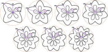
Spore suspensions in saline were prepared as described in the sample collection methods from fish samples listed in Table 1. The 6pc morphotype from Kudoa lethrini was not included in the analysis since it occurred at a low intensity thus sufficient sample size for analysis could not be obtained. Photomicrographs were taken of the spores at 400× magnification with an Olympus BH2 microscope under phase-contrast lighting using Nikon Digital Sight DS-L1 digital camera (Nikon, Tokyo). Spore measurements were taken from the photomicrographs using the measuring tool in the Nikon NIS Elements imaging software (Nikon, Tokyo) that was calibrated using a stage micrometer. Comprehensive morphometric characterization of the spores from the new isolates in Scolopsis monogramma and Seriola lalandi were conducted in accordance with Burger and Adlard (Reference Burger and Adlard2010) to allow comparison with existing species. Only the Valve Width, Suture Radius, Apical Polar Capsule Length, and Apical Polar Capsule Width were used for comparison of spore parameters between different morphotypes (see Fig. 1). Where possible, measurements of at least 30 spores from each morphotype in apical view were used to represent each isolate. However, only 16 spores for the minor 4pc morphotype from the Se. lalandi sample and 6 spores of the minor 4pc morphotype from the Sillago ciliata sample were able to be obtained.
Schematics of measurements used to characterize the transition between spore morphotypes. VW, valve width; SR, suture radius; APCL, apical polar capsule length; APCW, apical polar capsule width.

Origin of kudoid samples used for assessment of morphometric progression with polar capsule number increase

* Only 16 spores were measured; # only 6 spores were measured.
The apical view morphological data were subjected to a principal component analysis using the software PAST version 1.82b (Hammer et al. Reference Hammer, Harper and Ryan2001) to observe overall morphological differences between the different morphotypes from the different host isolates. Scatter plots with 95% confidence ellipses were generated using variant-covariant matrices. Correlations between morphometric characters and regression analysis of each morphometric apical-view character against polar capsule number were conducted using Stata™ 10.0 (StataCorp, 2008).
DNA analysis
DNA was extracted from spore suspensions that had been examined under the microscope for morphological analysis. Additional spore suspensions of Kudoa lethrini from 2 lethrinid fishes: Gymnocranius audleyi and Lethrinus harak (from Burger et al. (Reference Burger, Cribb and Adlard2007)); and Kudoa sp. ex Sillago maculata (previously identified as the same species as Kudoa sp. ex Sillago ciliata from Burger et al. (Reference Burger, Cribb and Adlard2007)), were used to generate additional LSU sequences to examine intra-specific variation within the gene. The suspensions were concentrated by centrifugation for 6 min at 4750 g and the supernatant fraction discarded. The DNA from the pellet was extracted using a QIAgen DNeasy Kit (QIAGEN Inc., Valencia, Ca, USA) according to the manufacturer's instructions. Small subunit (SSU) rDNA was amplified using the PCR primers KudMB1f 5′-GCCATGGATAACTGTGGTAAATCTAGAGC-3′ (see Burger et al. Reference Burger, Barnes and Adlard2008) and universal 18R 5′-CTACGGAAACCTTGTTACG-3′ (see Whipps et al. Reference Whipps, Adlard, Bryant, Lester, Findlay and Kent2003b). Large sub-unit (LSU) rDNA was amplified using PCR primer combination Kt28S1f 5′-CAAGACTACCTGCTGAAC-3′ and 28S1R 5′-GTGTTTCAAGACGGGTCG-3′ (see Whipps et al. Reference Whipps, Grossel, Adlard, Yokoyama, Bryant, Munday and Kent2004). PCR reactions, cycling parameters, and DNA sequence generation were as described by Burger et al. (Reference Burger, Barnes and Adlard2008).
SSU and LSU rDNA sequence data were edited using BioEdit (Hall, Reference Hall1999) and aligned using ClustalW (Thompson et al., Reference Thompson, Higgins and Gibson1994) with other kudoid sequences (the 3 most closely related species (Burger and Adlard, Reference Burger and Adlard2010) to those that infect brain tissue for which both SSU and LSU sequences were available: Kudoa grammatorcyni; K. monodactyli; and K. thalassomi, see Burger and Adlard (Reference Burger and Adlard2010). Neighbour-joining analyses were conducted using PAUP* 4.0b10 (Swofford, 2002) and bootstrap confidence values are based on 1000 replicates.
Pairwise differences were analysed in PAUP* 4.0b10 to determine total nucleotide distance and percentage differences between the sequences.
RESULTS
Histology
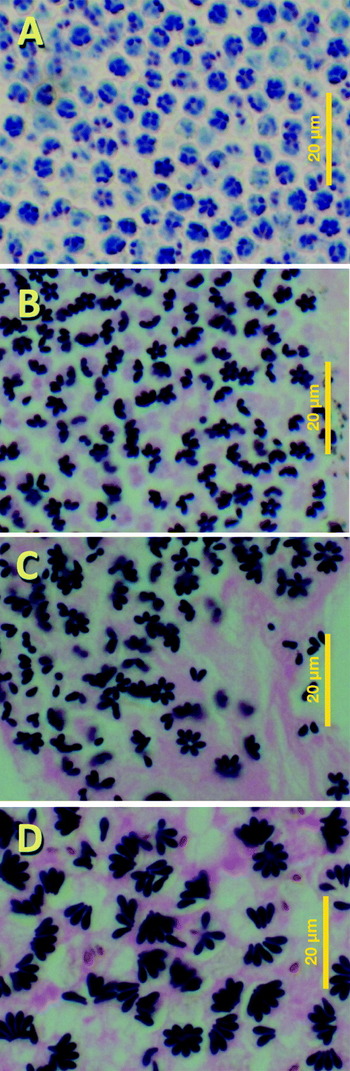
In Fig. 2, the spores within pseudocysts of K. neurophila ex Latris lineata (Fig. 2A, from the type slide, G463712, Queensland Museum), Kudoa sp. ex Sillago ciliata (Fig. 2B, from voucher specimen, G464871), Kudoa sp. from Scolopsis monogramma (Fig. 2C) and K. chaetodoni (Fig. 2D, from the type slide, G464863, Queensland Museum) display plasticity in the number of valves and polar capsules.
Stained histological sections of infected brain tissue indicating multiple morphotypes in a single plasmodium. (A) Kudoa neurophila ex Latris lineata (stained with Giemsa), (B) Kudoa sp. ex Sillago ciliata (stained with Giemsa and eosin (G & E)), (C) Kudoa sp. ex Scolopsis monogramma (G & E), (D) K. chaetodoni ex Chaetodon unimaculatus (G & E). Scale bar=20 μm.

Each of the samples used in this study had 2 or more spore morphotypes within individual plasmodia. For the purpose of this manuscript, we define a dominant morphotype as being above 40% of the spore population, minor morphotypes as between 5 and 40%, and rare morphotypes as occurring in less than 5% of spores.
In the K. neurophila section (Fig. 2A), 4pc and 5pc spore morphotypes are visible; the Kudoa sp. from Si. ciliata (Fig. 2B) has 5pc and 6pc morphotypes visible, the Kudoa sp. from Scolopsis monogramma (Fig. 2C) has 5 to 8pc morphotypes visible in the field, and the K. chaetodoni sample (Fig. 2D), has the dominant 8pc and 9pc morphotypes visible in the field, but unfortunately the minor 7pc morphotype was not seen in this section.
Genetic variation
Two new SSU rDNA sequences of Kudoa infection in brain tissue were generated for this study: one from Scolopsis monogramma and the other from Seriola lalandi. The SSU rDNA alignment was a total of 1449 nucleotides in length.
Seven new LSU rDNA sequences were generated in this study for a comparative analysis of intraspecific genetic variation provided by both SSU and LSU. LSU sequences from isolates of K. lethrini ex Lethrinus harak (from Lizard Island) and the Kudoa sp. ex Sillago maculata (from North Stradbroke Island) were included despite the absence of corresponding SSU sequences being available on GenBank. These 2 LSU sequences were identical to the LSU sequences previously reported for both kudoid infections and their alignment was a total of 734 nucleotides in length.
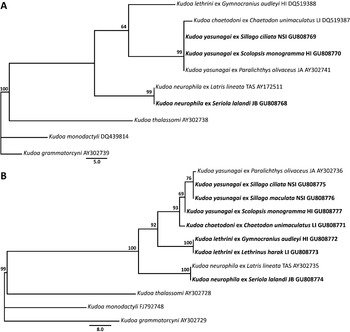
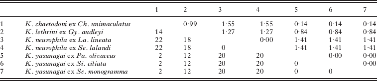
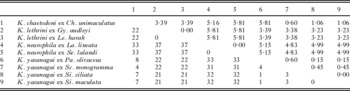
The genetic distances between isolates are shown in the Neighbour-joining trees in Fig. 3 and in the distance matrices in Tables 2 and 3. The SSU and LSU rDNA trees show similar topology, but vary in distance between the sequences. In SSU, the Kudoa sp. ex Seriola lalandi was 100% identical to K. neurophila ex Latris lineata while the Kudoa isolates from Scolopsis monogramma and Sillago ciliata were 100% identical to K. yasunagai ex Paralichthys olivaceus from Japan. In LSU Kudoa sp. ex Se. lalandi was 100% identical to K. neurophila ex La. lineata. However, the LSU sequences for Kudoa isolates from Sc. monogramma, Si. ciliata, and Si. maculata differed at a maximum of 4 nucleotides (maximum 0·6%, 1 and 4 nucleotide difference) to that of K. yasunagai ex Pa. olivaceus. This difference is equal to the difference between the Kudoa isolate from Sc. monogramma and K. chaetodoni from Chaetodon unimaculatus. On the basis of our genetic evidence the isolate from Seriola lalandi will be referred to as K. neurophila from here onwards. The isolates from Sc. monogramma, Si. ciliata and Si. maculata will be referred to as K. yasunagai (with evidence of their difference from K. chaetodoni to follow). Overall, from the 2 trees, the isolates of Kudoa yasunagai are most similar to K. chaetodoni, then to K. lethrini, and then to K. neurophila.
Neighbour-joining distance analysis of SSU rDNA (A) and LSU rDNA (B) from Kudoa species infecting brain tissue. Bootstrap support based on 1000 replicates shown at nodes. New sequences indicated in bold. Locality of new sequence isolates indicated prior to GenBank Accession number, HI, Heron Island; JA, Japan; JB, Jurien Bay; LI, Lizard Island; NSI, North Stradbroke Island; TAS, Tasmania.

SSU rDNA total nucleotide differences (lower triangle) and percentage differences (upper triangle) of isolates from Kudoa species infecting brain tissue
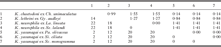
LSU rDNA total nucleotide differences (lower triangle) and percentage differences (upper triangle) of isolates from Kudoa species infecting brain tissue

Morphometrics
Kudoa neurophila
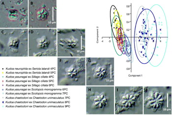
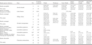
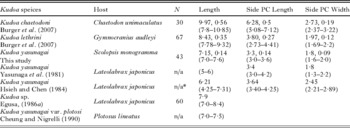
The Kudoa neurophila isolate from Seriola lalandi has a dominant 5pc morphotype, with a minor 4pc morphotype (see Fig. 4A, B). The spores with 5 polar capsules from Se. lalandi are similar in shape and size to those of Kudoa neurophila described from Latris lineata (see Grossel et al. Reference Grossel, Dykova, Handlinger and Munday2003). Tables 4 and 5 show the measurements of the spores from Se. lalandi, where Table 4 shows apical view measurements from spores with 4pcs and 5pcs and Table 5 shows a comparison of the side-view measurements with other species that infect brain tissue. The Width measurement from Grossel et al. (Reference Grossel, Dykova, Handlinger and Munday2003) is the distance from a valve tip to the sutural indent of a symmetrically bisected spore and is the equivalent to this study's Valve Width and Suture Radius measurements combined. The average width of the 5pc spores from Grossel et al. (Reference Grossel, Dykova, Handlinger and Munday2003) is 7·73 μm and the equivalent average from K. neurophila ex Se. lalandi spores is slightly larger at 8·51 μm. In the side-view measurements, those from Se. lalandi are similar, but smaller in length (mean 6·13 ex Se. lalandi cf 6·59 ex L. lineata), and larger in polar capsule size (mean 3·78 ex Se. lalandi cf 3·03 ex L. lineata in polar capsule length and 2·27 ex Se. lalandi cf 1·70 ex L. lineata in polar capsule width) than those from K. neurophila ex La. lineata.
Phase-contrast photomicrographs of the spore morphotypes from each isolate with corresponding Principal Components Analysis (including 95% ellipses) of morphometric data. Spore photos: (A and B) Kudoa neurophila ex Seriola lalandi; (C, D and E) K. yasunagai ex Sillago ciliata; (F and G) K. yasunagai ex Scolopsis monogramma; (H, I and J) K. chaetodoni ex Chaetodon unimaculatus. Scale bar=10 μm.

Apical view morphometrics from brain-infecting kudoid species that have multiple morphotypes in ascending order of dominant morphotype number

* Formalin-preserved spores. 1Spore Width in Grossel et al. (Reference Grossel, Dykova, Handlinger and Munday2003) is equal to the sum of Valve Width and Suture Radius in this study.
Side-view morphometric comparison of brain infecting kudoid species with a dominant 5 polar capsule morphotype

Kudoa yasunagai
The new K. yasunagai isolate from Scolopsis monogramma has a dominant 7pc morphotype, a minor 6pc morphotype (Fig. 4F, G), and rare 5pc and 8pc morphotypes. This isolate is compared to species with similar polar capsule numbers in Tables 4 and 6. The 6pc morphotype from Sc. monogramma compared to that from Si. ciliata had similar mean measurements, but the isolate from Sc. monogramma was generally slightly larger (mean spore width 11·45 ex Sc. monogramma cf 10·41 ex Si. ciliata; thickness 10·26 ex Sc. monogramma cf 9·25 ex Si. ciliata; valve width 5·73 ex Sc. monogramma cf 5·2 ex Si. ciliata; suture radius 4·03 ex Sc. monogramma cf 3·94 ex Si. ciliata; apical polar capsule length 2·49 ex Sc. monogramma cf 2·23 ex Si. ciliata; apical polar capsule width 1·64 ex Sc. monogramma cf 1·63 ex Si. ciliata). The side-view measurements between these 2 isolates are also very similar (see Table 5 and 6 for these measurements, length 7·15 ex Sc. monogramma cf 7·31 ex Si. ciliata; side polar capsule length 3·3 ex Sc. monogramma cf 3·5 ex Si. ciliata; side polar capsule width 1·8 ex Sc. monogramma cf 1·93 ex Si. ciliata).
Side-view morphometric comparison of brain infecting kudoid species with a dominant 7 polar capsule morphotype

* Formalin-preserved spores.
The other morphometric records of species with a 7pc morphotype vary. The most significantly different isolate of K. yasunagai to that from Sc. monogramma is from Lateolabrax japonicus recorded by Yasunaga et al. (Reference Yasunaga, Hatai, Ogawa and Yasumoto1981). It is significantly larger in spore width (15 μm compared to the mean of 11·6 μm from K. yasunagai ex Sc. monogramma). K. chaetodoni (Fig. 4H) is also much larger, particularly in width (mean 14·04 for K. chaetodoni cf 11·62 for K. yasunagai ex Sc. monogramma), length (mean 9·97 for K. chaetodoni cf 7·15 for K. yasunagai ex Sc. monogramma), and side polar capsule length (mean 6·28 for K. chaetodoni cf 3·3 for K. yasunagai ex Sc. monogramma).
Both the 4 and 5pc spore morphotypes from Kudoa yasunagai ex Si. ciliata are similar in size to those from K. neurophila ex Se. lalandi (Fig. 4C, D). However, the spores from Si. ciliata were larger in valve size (4pc morphotype mean 9·56:8·96; 5pc morphotype mean 9·53:9·17) and slightly larger in length (mean 7·31:6·59). The K. yasunagai isolate from Si. ciliata also has a minor 6pc morphotype (Fig. 4E), as was seen by Grossel et al. (Reference Grossel, Dykova, Handlinger and Munday2003) with K. neurophila, but was not seen in the K. neurophila sample in this study.
Principal components analysis
The multivariate analysis of the apical measurements (Fig. 4) from the samples used in this study show a distinct separation of the Kudoa chaetodoni spores from the other isolates. The general trend of this species is positive along the Component 2 (x) axis and consistent along the Component 1 (y) axis. The other isolates show a positive trend along the Component 2 axis and a negative trend along the Component 1 axis. Although the K. neurophila morphotypes sit similarly along the Component 2 axis of those from K. yasunagai ex Si. ciliata, they are more positive on the Component 1 axis. Due to the low sample number of the 4pc morphotype from Sillago ciliata, the 95% confidence ellipse is large; however, the measurements fit well with the trends of the other morphotypes from K. yasunagai.
Correlation and regression
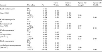
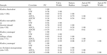
Polar capsule number has the highest correlation with Suture Radius for all of the 4 samples analysed (and highest overall correlation for the Kudoa yasunagai samples from Sc. monogramma and Si. ciliata), whereas apical polar capsule width has the lowest (see Table 7). For K. chaetodoni and K. neurophila, the highest correlation of measurements is between the suture radius and the valve width (0·93 and 0·85 respectively).
Correlation analysis between morphometric characters from the Kudoa samples measured
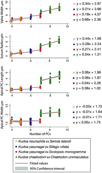
The regression analyses of the spore morphometric data indicate the general trends between different morphotypes (Fig. 5) for each of the measurements taken. Kudoa chaetodoni is again quite distinct from the other samples in this study. For each of its measurements, the slope of the trend line indicates a consistent increase in size with polar capsule number increase. The highest increase being from the valve width, with an increase of 0·66 μm per extra polar capsule. The other species morphotypes have an overall positive trend except for 1 measurement, Apical Polar Capsule Width, which has an average slightly negative to neutral correlation with polar capsule number increase (but note, when the 95 % confidence interval is included, the upper end of each interval is slightly positive).
Regression analysis of morphotype polar capsule number vs morphometric characters for the 4 isolates in the morphometric analysis. Linear equations listed for each isolate for each measurement. 95% confidence interval indicated with grey shading.

The K. neurophila and K. yasunagai isolates show similar trends between morphotypes, but K. neurophila has consistent slightly larger increases correlating with polar capsule number.
DISCUSSION
There are currently 6 described and 1 undescribed Kudoa species that specifically infect the brain/central nervous system: K. chaetodoni; K. lethrini; K. neurophila; K. paralichthys Cho and Kim, 2003; K. tetraspora Narasimhamurti and Kalavati, Reference Narasimhamurti and Kalavati1979; K. yasunagai; and Kudoa sp. (of Langdon, Reference Langdon1990). Of these 7, 4 have been noted to possess multiple morphotypes. It is noteworthy that Kudoa species that infect brain tissue which have spores with 4pcs have been characterized with a single morphotype only (K. paralicthys, K. tetraspora, and Kudoa sp. (of Langdon, Reference Langdon1990)) while species with a 5pc or more morphotype dominant in their infections (K. chaetodoni, K. lethrini, K. neurophila, and K. yasunagai) possess multiple morphotypes.
The original description of K. neurophila from striped trumpeter, Latris lineata has a dominant 5pc morphotype with a 6pc spore referred to as the only morphological anomaly (Grossel et al. Reference Grossel, Dykova, Handlinger and Munday2003). Nonetheless, careful scrutiny of the type specimen at the Queensland Museum revealed a minor morphotype of K. neurophila with 4pcs from La. lineata. The new isolate of K. neurophila ex Yellowtail kingfish, Seriola lalandi, is also characterized from fresh material as having a dominant 5pc morphotype and a minor 4pc morphotype (no histological section available).
Our rDNA results suggest that the same morphotype of some Kudoa species may not always be the dominant one of a particular species, a phenomenon first described by Yasunaga et al. (Reference Yasunaga, Hatai, Ogawa and Yasumoto1981) with their account of what was later described as K. yasunagai by Hsieh and Chen (Reference Hsieh and Chen1984). This is the opposite of the findings of several recent studies (Blaylock et al. Reference Blaylock, Bullard and Whipps2004; Diamant et al. Reference Diamant, Ucko, Paperna, Colorni and Lipshitz2005; Yokoyama and Itoh, Reference Yokoyama and Itoh2005; Burger et al. Reference Burger, Barnes and Adlard2008; Burger and Adlard, Reference Burger and Adlard2010), where rDNA analyses revealed that morphologically indistinguishable spores from different hosts often represent the same genetic species. In the brain of the original host, Japanese seabass, Lateolabrax japonicus, the kudoid species had a dominant 7pc and a minor 6pc morphotype, meanwhile in the Striped knifejaw, Oplegnathus fasciatus, the opposite kudoid morphotype was dominant (76·1% to 23·9% ratio of 6–7) (see Yasunaga et al. Reference Yasunaga, Hatai, Ogawa and Yasumoto1981). Reference EgusaEgusa (1986a) also found variation between the spore morphotype ratios in infections similar to those described by Yasunaga et al. (Reference Yasunaga, Hatai, Ogawa and Yasumoto1981), where there was differing dominance between the 6pc and 7pc morphotype (also including rare morphotypes with 4, 5, 8, 10, and 12pcs) in different fish species (La. japonicus, Pagrus major, Seriola quinqueradiata, Takifugu rubripes), in different individual fish, and even in different cysts within an individual fish. Because of the variation in morphotypes, Yasunaga et al. (Reference Yasunaga, Hatai, Ogawa and Yasumoto1981) and Reference EgusaEgusa (1986a) refrained from officially assigning a name to the infections they described.
The Coral catfish Plotosus lineatus (syn. Plotosus anguillarus) from the Philippines was reported by Cheung and Nigrelli (Reference Cheung and Nigrelli1990) to show the same abnormal swimming behaviour as that of La. japonicus reported by Yasunaga et al. (Reference Yasunaga, Hatai, Ogawa and Yasumoto1981). On examination, a kudoid infection in the brain of the Coral catfish had similar spores with 6pc and 7pc spore morphotypes, which was referred to by Cheung and Nigrelli (Reference Cheung and Nigrelli1990) as K. yasunagai var. plotosi after the host.
The K. yasunagai sample from Scolopsis monogramma is 100% identical in SSU rDNA and 99·4% similar (4 nucleotide differences) in LSU rDNA sequence to the K. yasunagai sequences from a halibut, Paralichthys olivaceus, from Japan. The morphological similarities (both have 7pcs) and previously reported broad host range and wide geographical distribution which includes Japan (Reference EgusaEgusa, 1986a; Yasunaga et al. Reference Yasunaga, Hatai, Ogawa and Yasumoto1981) and the Philippines (Cheung and Nigrelli, Reference Cheung and Nigrelli1990), of K. yasunagai provide convincing evidence that the Kudoa sample from Sc. monogramma indeed represents a new host of K. yasunagai. However, the Kudoa samples from Sillago ciliata and Si. maculata from North Stradbroke Island (27° 29′S; 153° 24′E) are genetically more similar (100% SSU; 99·85% LSU) to K. yasunagai sequences from Paralichthys olivaceus than the isolate from Sc. monogramma, but are morphologically distinct (5pc morphotypes dominant compared to 7pc). Could this also be K. yasunagai? In LSU rDNA, the difference between K. yasunagai ex P. olivaceus with the Kudoa samples from Sc. monogramma and Si. ciliata is 4 nucleotides (0·6%) and 1 nucleotide (0·15%), respectively. These differences are well within previously recognized levels of intra-specific variation of Kudoa species (SSU up to 8 nucleotides (0·6%), LSU up to 13 nucleotides (1·83%)); however, it should be noted that these levels do not include the higher variation found in K. thyrsites (see Burger and Adlard, Reference Burger and Adlard2010). This brings us back to the original dilemma faced by Burger et al. (Reference Burger, Cribb and Adlard2007) i.e. what level of genetic variation constitutes a species within Kudoa and does SSU rDNA provide sufficient resolution for specific diagnoses?
One of the aims of this study was to determine whether the Kudoa sp. from Sillago ciliata characterized by Burger et al. (Reference Burger, Cribb and Adlard2007) was K. yasunagai and whether SSU rDNA has reached its limit of resolution. LSU rDNA has been proven to have greater resolving power of Kudoa species than SSU; a phenomenon demonstrated by the clear separation of K. paraquadricornis and K. quadricornis (SSU difference 1 nucleotide, 0·1%; LSU difference 25 nucleotides, 5·6%) (see Burger and Adlard, Reference Burger and Adlard2010). The level of sequence variation in LSU between the isolates from Sc. monogramma and Si. ciliata and K. yasunagai are not significantly larger than that displayed in the SSU. To rely solely on rDNA would indicate that not only the isolates from Sc. monogramma and Si. ciliata were conspecific with K. yasunagai ex Pa. olivaceus, but that K. chaetodoni could also be referable to K. yasunagai with only 2 nucleotide differences in the SSU (0·14%) and 4 nucleotide differences (0·6%) in the LSU to the Sc. monogramma isolate (note that K. chaetodoni has a maximum distance to all K. yasunagai isolates of 8 nucleotides, 1·21% in LSU when compared with that from the Pa. olivaceus isolate). Species boundaries must be assessed on a case-by-case basis using a combination of genetic, morphological and other biological data (for example pathology, geographical locality and host habitat) because there is no yardstick for the level of molecular differences that defines a species (Nolan and Cribb, Reference Nolan, Cribb, Baker, Muller and Rollinson2005; Gunter and Adlard, Reference Gunter and Adlard2009; Burger and Adlard, Reference Burger and Adlard2010).
To determine the validity of the species K. chaetodoni and to further explore the relationship between the K. yasunagai isolates from Sc. monogramma and Si. ciliata, an in-depth morphological examination was conducted. When comparing the overall measurements, spores of K. chaetodoni are considerably larger than the other brain infecting species, except for some similarity in the apical polar capsule size between K. chaetodoni and the isolates of the K. yasunagai 7pc morphotype described in Reference EgusaEgusa (1986a) and Cheung and Nigrelli (Reference Cheung and Nigrelli1990). In the principal components analyses, 95% ellipses of the K. chaetodoni morphotypes are separate from all other morphotypes examined (including the separation between the 7pc morphotype from the equivalent 7pc morphotype from Sc. monogramma) and show a more neutral trend between the 3 morphotypes along the Component 1 axis. This separation was supported by the regression analyses, which showed K. chaetodoni to consistently have a greater positive progression in morphometric characters with increased morphotype polar capsule number. The morphometric analyses combined with the overall spore shape of K. chaetodoni suggest that when there are more polar capsules present, the polar capsules tend to be situated further away from the centre of the spore so that the slight increase in polar capsule size can be accommodated in the spore valve. The other isolates have shown an almost stable polar capsule size regardless of the number within the morphotype. The morphological differences between K. chaetodoni and these other isolates suggest that despite the close molecular affinity between K. chaetodoni and the new isolate from Sc. monogramma, K. chaetodoni is still considered a unique species.
Morphological data also support the genetic distinction between the 2 isolates studied with a dominant 5pc morphotype, K. yasunagai from Si. ciliata and K. neurophila from Seriola lalandi. The K. yasunagai isolate from Si. ciliata is similar to K. neurophila in spore size and trends between morphotypes, but it can be visually distinguished from K. neurophila, which has more rounded spore valves with a subtly smaller ratio between apical polar capsule length and suture radius compared to valve width. The species are 20 nucleotides (1·41%) different in SSU rDNA and 32 nucleotides (4·99%) in LSU; but also, there are 2 species, K. chaetodoni and K. lethrini that are more closely related to the K. yasunagai isolate from Si. ciliata than the 2 species are related to each other.
The similarities between shape and overall trends between the morphotypes of K. yasunagai from Sc. monogramma and Si. ciliata give support for their conspecificity. The slight average morphometric differences (note that the ranges overlap) between the 6pc morphotypes associated with the valve size may be consistent with intra-specific variation between different hosts (for example in K. alliaria (see Whipps and Diggles, Reference Whipps and Diggles2006) and K. iwatai (see Diamant et al. Reference Diamant, Ucko, Paperna, Colorni and Lipshitz2005)).
Unfortunately, there are no morphology records relating to the SSU and LSU rDNA sequences for the K. yasunagai isolate generated from the Japanese Paralichthys olivaceus to allow comparison of the morphology of the genetically similar isolates from Sc. monogramma and Si. ciliata. However, considering: that the Kudoa infection from Pa. olivaceus isolate was obtained from the type locality; this species has a broad host specificity; and that no other species has been described from that area that fit the morphological and tissue location description, we consider these sequences to be valid genetic representatives of K. yasunagai. Thus, the close genetic relationship between the isolates from Sc. monogramma and Si. ciliata with K. yasunagai allow these isolates to be proposed as new host-location combinations for K. yasunagai.
Making the connection between the Kudoa infection dominated by a 5pc morphotype in Si. ciliata and K. yasunagai that is characterized as having spores with 7pcs (and a few exceptions) is not intuitive. The new approach employed in this study that includes characterizing multiple morphotypes has allowed us to make these connections and show that while polar capsule number may vary between spores of a single species, the overall morphology ratios between spore variants are useful in species characterization. The variability in polar capsule number does not suggest that increasing polar capsule number is a distinct evolutionary line in the Kudoidae. The genetic relationships amongst multivalvulid species lead to the demise of the families and genera that were based on polar capsule number (Whipps et al. Reference Whipps, Grossel, Adlard, Yokoyama, Bryant, Munday and Kent2004). However, possibly the evolution of variability in polar capsule number has occurred. The benefit in multiple morphotypes may be a cost-benefit tactic instigated by the primordial cell where a higher number of polar capsules may give a higher success rate of attachment to the next host, but producing more polar capsules may be more costly in energy. With variation in polar capsule number these energy costs may be more balanced. In the future, when the alternate host in the life cycle of kudoids is discovered and infection experiments can be carried out, the effects of host on spore morphology can be assessed further and evolutionary links can be investigated.
The variability in spore morphotypes creates significant implications for future diagnosis. If phenotypic variation results in the production of different dominant morphotypes from different hosts and even from different individuals, there is clearly a risk of misdiagnosis. Unfortunately, this phenomenon does not facilitate easy specific diagnosis without a combination of host information with molecular and morphometric analysis of spores. However, the possibility of designing molecular probes (similar to the PCR assay developed by Grossel et al. (Reference Grossel, Handlinger, Battaglene and Munday2005) for K. neurophila) that correspond to species of interest will greatly simplify diagnostic procedure.
New Material
Phylum Myxozoa
Class Myxosporea
Order Multivalvulida
Family Kudoidae Meglitsch, 1960
Genus Kudoa Meglitsch, 1947
Kudoa neurophila (syn. Pentacapsula neurophila (Whipps, Grossel, Adlard, Yokoyama, Bryant, Munday and Kent, 2004) (Grossel, Dykova, Handlinger and Munday, 2003)).
Type host: Latris lineata (Forster, 1801), Striped trumpeter (Teleostei, Perciformes, Latridae).
Type locality: Crayfish Point, Derwent River, Tasmania, Australia (42° 52’ S; 147° 19′E).
Location in the host: Central nervous system, brain tissue.
Other hosts: Seriola lalandi Valenciennes, 1833, Yellowtail Kingfish (Teleostei, Perciformes, Carangidae).
Other locality: Jurien Bay, Western Australia, Australia (30° 18′S; 115° 2′E).
Type material: Syntypes G463712, located in the Queensland Museum, Brisbane, Australia (Haematoxylin and eosin-stained histological section).
New material deposited: GenBank sequences: SSU rDNA Accession number GU808768 and LSU rDNA GU808774 (see Fig. 3).
Plasmodia: occasionally surrounded by a membrane. Advanced cysts are surrounded by fibrous tissue and necrosis of spores is often evident.
Spore description: Spore shape fitting the amended description of the genus Kudoa. Dominant spore morphotype pentaradial with 5 equal rounded spore valves 5 polar capsules in apical view (Fig. 4A). Minor spore morphotype quadrate, tetramerous radial symmetry, with 4 equal rounded spore valves in apical view (Fig. 4B). Polar capsules convergent, pyriform. In side view, spores are pyramidal. Turns of the polar filament not discernable. Spore measurements shown in Tables 4 and 5.
Kudoa yasunagai (syn. Septemcapsula yasunagai) (Hsieh and Chen, Reference Hsieh and Chen1984).
Type host: Lateolabrax japonicus (Cuvier, 1828), Japanese seabass (Teleostei, Perciformes, Lateolabracidae).
Type locality: Goto, Nagasaki Prefecture, Japan (32° 42′N; 128° 50′E).
Location in the host: Brain tissue.
Other hosts: Oplegnathus fasciatus (Temminck and Schlegel, 1844) Striped knifejaw (Perciformes, Oplegnathidae); Pagrus major (Temminck and Schlegel, 1843) Red seabream (Perciformes, Sparidae); Plotosus lineatus (Thunberg, 1787) Coral catfish (Siluriformes, Plotosidae); Scolopsis monogramma (Cuvier, 1830) Monocle bream (Perciformes, Nemipteridae); Seriola quinqueradiata Temminck and Schlegel, 1845, Yellowtail (Perciformes, Carangidae); Sillago ciliata Cuvier, 1829, Sand whiting (Perciformes, Sillaginidae); Sillago maculata Quoy and Gaimard, 1829, Winter whiting (Perciformes, Sillaginidae); and Takifugu rubripes (Temminck and Schlegel, 1850) Japanese pufferfish (Tetraodontiformes, Tetraodontidae).
Other localities: New York Aquarium, host Plotosus lineatus collected from the Phillipine Islands (14° 35′N; 121° 0′E); Heron Island on the Great Barrier Reef, Queensland, Australia (23° 27′S; 151° 55′E); North Stradbroke Island in Moreton Bay, Queensland, Australia (27° 29′S; 153° 24′E).
Type material: from Hsieh and Chen (Reference Hsieh and Chen1984) preserved in the Institute of Hydrobiology, Academia Sinica (now the Chinese Academy of Sciences).
New material deposited: Giemsa and eosin-stained histological section voucher ex Scolopsis monogramma, registration number: G465421. GenBank sequences: SSU rDNA Accession numbers GU808769-GU808770 and LSU rDNA GU808775-GU808777 (see Fig. 3 for specific isolate numbers).
Other material: Vouchers previously deposited as Kudoa sp. from Sillago ciliata and Sillago maculata (see Burger et al. Reference Burger, Cribb and Adlard2007) in the Queensland Museum, South Brisbane, Australia registration numbers: G464871–G464876 (Giemsa and eosin-stained histological sections) and G464877–G464882 (H&E-stained histological sections).
Plasmodia: Intracerebral, generally without envelope (and in some cases cysts may be found with a fibrous membrane and fibrous structure within) or extracerebral cysts encased with a thin membrane.
Spore description: Spore shape fitting the amended description of the genus Kudoa Whipps, Grossel, Adlard, Yokoyama, Bryant, Munday and Kent, 2004. Dominant spore morphotype varies between having 5–7 polar capsules and spore valves. Radially symmetrical in apical view (Fig. 4). Minor spore morphotypes include those with 4, 8, 9, 10 and 12 polar capsules. Polar capsules convergent, pyriform. In side view, spores are pyramidal. Turns of the polar filament not discernable. Spore measurements are shown in Tables 4 and 6.
ACKNOWLEDGMENTS
The authors would like to thank Dr Fran Stephens for providing the infected Yellowtail kingfish specimens from Western Australia. For their assistance in fish collection throughout the project, we would like to thank: Dr Rod Bray, Dr Mal Bryant, Tony Byrne, Professor Tomas Cribb, Scott Cutmore, Abigail Downie, Ricky Gleeson, Dr Nicole Gunter, Dr Terrence Miller, Dr Matthew Nolan, Chris Rohner, Tane Sinclair-Taylor, and Jeremy Ullmann. For assistance in fish dissection we also thank Holly Heiniger. We are grateful to the staff and for the facilities of Heron and Lizard Island Research Stations. For access to molecular analysis equipment and assistance, we would like to thank Dr Andy Barnes and Dr Justice Baiano. For histology assistance, we would like to thank Chris Cazier from the Veterinary Pathology Laboratory, the University of Queensland. We appreciate Dr Nicole Gunter's comments on the manuscript.
FINANCIAL SUPPORT
We would like to thank the ARC/NHMRC Research Network for Parasitology for travel funds. This study is a contribution funded by the Australian node of the CReefs global research initiative, a partnership between the Australian Biological Resource Study (ABRS) and BHP Billiton, the Great Barrier Reef Foundation, the Australian Institute of Marine Science and the Alfred P. Sloan Foundation, Grant number: 209/29. CReefs is a field program of the Census of Marine Life.