Introduction
The inflammatory response around the Schistosoma mansoni egg in vertebrate host tissues, especially in the liver, is the morphological substrate of the infection. Raso et al. (Reference Raso, Bernardes, Tafuri, Bogliolo and Neves1978) considered that the granuloma constitution is affected by two features: the host response and the time of development of the pathological process.
Initially, granulomatous lesions are characterized by polymorphonuclear and lymphocytic cell infiltrates, where the inflammatory reaction is intense. Macrophages are the most common cells observed in chronic disease, and the eggs progressively acquire a growing fibrous component (Andrade & Warren, Reference Andrade and Warren1964; Carvalho et al., Reference Carvalho, Alvarenga and Melo1986; Silva et al., 2000; Tristão et al., Reference Tristão, Melo, Vasconcelos and Grossi2000).
The CD4+T-cell response induced by S. mansoni egg antigens regulates the development of granulomas during parasite infection (Pearce & MacDonald, Reference Pearce and MacDonald2002). However, the exact mechanism of recruitment of the leucocytes, mainly CD4+T cells, into granulomas is not clear. There is evidence that tumour necrosis factor-α (TNF-α) and other mediators play crucial roles in cell migration to the infection site (Calich et al., Reference Calich, Coppi-Vaz and Burger1985; Joseph & Boros, Reference Joseph and Boros1993; Goes et al., Reference Goes, Rezende, Gazzinelli and Doughty1994; Haseeb et al., Reference Haseeb, Sbirazian and Preis2001; Sandor et al., Reference Sandor, Weinstock and Wynn2003). Besides, Henri et al. (Reference Henri, Chevillard, Mergani, Paris, Gaudart, Camila, Dessein, Montero, Elwali, Saeed, Magzoub and Dessein2002) showed that high TNF-α levels were associated with an increased risk of periportal fibrosis in humans. On the other hand, in murine schistosomiasis, a decrease in hepatic granulomatous pathology and fibrosis is associated with a diminished release of cytokines produced by T-helper 2 cells (Th2) (Hoffmann et al., Reference Hoffmann, Cheever and Wynn2000; Pearce & MacDonald, Reference Pearce and MacDonald2002) and an increase in the production of the T-helper 1 cell (Th1) cytokines (Cheever et al., Reference Cheever, Xu, Macedonia, Cox, Hieny and Sher1992; Sandor et al., Reference Sandor, Weinstock and Wynn2003). Schistosoma mansoni acute infection in rodents is known to induce a strong Th2-type response as evidenced by an increased production of interleukin-4 (IL-4), interleukin-5 (IL-5) and interleukin-10 (IL-10) associated with a down-regulation of interleukin-12 (IL-12) and interferon-γ (IFN-γ) (Grzych et al., Reference Grzych, Pearce, Cheever, Caulada, Caspar, Heiny, Lewis and Sher1991; Pearce et al., Reference Pearce, Caspar, Grzych, Lewis and Sher1991; Sher et al., Reference Sher, Fiorentino, Caspar, Pearce and Mosmann1991). In chronic infection, a critical requisite for the amelioration of the pathological events caused by S. mansoni is the preventive down-regulation of the granulomatous inflammation around parasite eggs.
Pentoxifylline (PTX), a methylxanthine derivative with haemorheological properties, possesses several pharmacologic effects that result in increased fluidity of cell membranes, immunomodulatory and anticoagulant effects, stimulation of fibrinolysis and alteration of fibroblast function (Ward & Clissold, Reference Ward and Clissold1987; Samlaska & Winfield, Reference Samlaska and Winfield1994). Furthermore, PTX shows some anti-inflammatory activity, which is explained, at least in part, by an inhibition of the production or function of certain pro-inflammatory cytokines, mainly TNF-α, whose inhibition has been demonstrated in different immunopathological processes (Sáez-LIorens et al., Reference Sáez-LIorens, Ramilo, Mustafá, Mertsola, Alba, Hansen and McCracken1990; Tilg et al., Reference Tilg, Eibl, Pichl, Gächter, Herold, Brankova, Huber and Niederwieser1993; Bienvenu et al., Reference Bienvenu, Doche, Gutowski, Lenoble, Lepape and Perdrix1995; Heyderman et al., Reference Heyderman, Klein, Daramola and Levin1995; Navarro et al., Reference Navarro, Punzón, Pizarro, Fernández-Cruz, Fresno and Muñoz-Fernández1996; Ostrosky-Zeichner et al., Reference Ostrosky-Zeichner, Soto-Hernandez, Angeles-Morales, Teixeira, Nava-Ruiz, Rios, Solis and Sotelo1996; Marcinkiewicz et al., Reference Marcinkiewicz, Grabowska, Lauterbach and Bobek2000; Muniz-Junqueira, Reference Muniz-Junqueira2007; Jesus et al., Reference Jesus, Luna, Almeida, Machado and Carvalho2008). Tilg et al. (Reference Tilg, Eibl, Pichl, Gächter, Herold, Brankova, Huber and Niederwieser1993) showed, however, that the suppressive activity of PTX on TNF-α, in vitro, was similar to the effects of the treatment with anti-TNF-α, and that the addition of TNF-α receptors did not antagonize the inhibitory potential of the drug. However, the modulation induced by PTX is not restricted solely to the release of these cytokines. In fact, PTX is also able to inhibit the production of the anti-inflammatory cytokine IL-10 (Bienvenu et al., Reference Bienvenu, Doche, Gutowski, Lenoble, Lepape and Perdrix1995; Navarro et al., Reference Navarro, Punzón, Pizarro, Fernández-Cruz, Fresno and Muñoz-Fernández1996; Marcinkiewicz et al., Reference Marcinkiewicz, Grabowska, Lauterbach and Bobek2000).
The oogram technique described by Pellegrino & Faria (Reference Pellegrino and Faria1965) and utilized in the screening of anti-schistosomal drugs is a sensitive method for evaluating parasite oviposition, which provides immediate results in experimental schistosomiasis. Recently, oogram analysis has been utilized in immunization assays to verify reduction in the total number of parasite eggs in host tissues (Pacheco et al., Reference Pacheco, Zucconi, Mati, Garcia, Miyoshi, Oliveira, Melo and Azevedo2005). Besides, mice treated with a glucocorticoid during acute infection with S. mansoni have also shown alterations in the number of parasite eggs observed in the host's intestines. Although drugs of this category have a potent anti-inflammatory action that can block the passage of eggs to the intestinal lumen, they have no schistosomicidal action and do not modify egg-laying kinetics (Hermeto et al., Reference Hermeto, Bicalho, Silva, Melo and Pereira1994).
On the other hand, the treatment of rodents with PTX during infection with S. mansoni (Reis et al., Reference Reis, Ventura, Souza, Arana-Pino, Pelajo-Machado, Pereira, Lenzi, Conceição and Takyia2001; El-Lakkany & Nosseir, Reference El-Lakkany and Nosseir2007) and Schistosoma japonicum (Xiong et al., Reference Xiong, Zhu and Luo2003) has been evaluated. However, in these studies, the route of drug administration, times of onset and duration of treatment were different from those used in the present paper. Moreover, such an advanced chronic infection and the estimation of fecundity of female worms by oogram have not yet been considered. Thus, liver histological analysis and intestinal oogram of Swiss mice harbouring S. mansoni infection after short-term subcutaneous treatment with PTX (daily for 5 days) were carried out in order to obtain additional information about the effects of the drug on granuloma constitution and the oviposition kinetics of the parasite, both in acute and chronic infections.
Materials and methods
Mice and infection
Fifty albino Swiss mice, males weighing 25 ± 2 g, were infected by the subcutaneous route with about 50 S. mansoni cercariae of the LE strain (Belo Horizonte, Brazil) shed by laboratory reared and infected Biomphalaria glabrata. Rodents were afterward distributed into four different groups. First, there were two groups of 10 animals each where one group of mice was treated with PTX to evaluate the drug effects in acute S. mansoni infection and the other was designated the control group. The two other groups, each with 15 animals, represented the control and treated groups with chronic parasitic infection.
Drug, treatment and recovery of adult worms from the hepatic portal system
PTX [1-(5-oxyohexyl)-3,7-dimethylxanthine, 1-(5-oxyohexyl) theobromine] administration was initiated 42 and 140 days post-infection (DPI) for the acute and chronic infection groups, respectively. Treatment was carried out daily with 200 mg/kg (subcutaneous route) of the drug (Trental®) for five consecutive days. Rodents of both control groups did not receive PTX treatment.
Mice in the acute and chronic infection groups were killed by cervical dislocation at 49 DPI and 147 DPI, respectively, according to the local animal experimentation ethics committee (CETEA/UFMG). The recovery of the worms was performed through perfusion of the portal system according to Pellegrino & Siqueira (Reference Pellegrino and Siqueira1956), with minor modifications.
Histology and oogram analysis
Liver fragments were collected, fixed in 10% formalin, paraffin-embedded, sectioned and stained by haematoxylin-eosin histological technique. Six 4 μm-thick hepatic sections were examined for each animal. The average sizes of the granulomatous lesions were obtained with the aid of an ocular millimetre grid fitted to a light microscope. Two perpendicular diameters were considered for analysis. Only granulomas containing a preserved central miracidium were measured.
Fragments of the distal ileum from all animals were transferred to Petri dishes containing isotonic saline. They were opened lengthwise with scissors and the excess mucus was removed. Intestinal fragments measuring 1 cm were cut off, partially dried on absorbent paper, weighed and placed between a glass slide and a plastic coverslip. The preparations were then inverted and pressed on a rubber surface padded with absorbent paper following a technique described previously (Pellegrino & Faria, Reference Pellegrino and Faria1965).
For the qualitative oogram evaluation, the parasite eggs present on the slides were examined with a light microscope (total magnification = 100 × ), and all eggs were counted and classified as immature (first to fourth stage), mature or dead eggs (Pellegrino et al., Reference Pellegrino, Oliveira, Cunha and Siqueira1962). Quantitative oograms were obtained, using the formula: number of eggs/g of tissue = (number of eggs in intestinal fragment/weight of fragment in milligrams) × 1000 (Hermeto et al., Reference Hermeto, Bicalho, Silva, Melo and Pereira1994). The parameter ‘second stage of immature egg/female/g’ was used to verify the number of eggs per female parasite per day (Pacheco et al., Reference Pacheco, Zucconi, Mati, Garcia, Miyoshi, Oliveira, Melo and Azevedo2005). Although these are eggs laid recently by S. mansoni females, the coefficient of variation in their number has been shown to be lower than that of the other immature stages (unpublished results).
Data obtained were submitted to ANOVA and Tukey's post-hoc test.
Results
Portal system perfusion
Macroscopic examination of the abdominal viscera in mice treated with PTX revealed mesenteric vasodilatation. No difference was seen in the total number of worms recovered among all groups. There was also no significant variation in the gender of the parasites recovered from untreated versus treated mice (table 1). In acute infection groups, the mean percentages of adult worms recovered from the portal system of the mice were 30.8% and 25% in control and treated groups, respectively; in the chronic infection groups, these values were 20.6% and 18.2%, respectively.
Mean numbers and standard deviation of adult Schistosoma mansoni worms recovered from the portal systems of mice treated with pentoxifylline (PTX) in acute and chronic infections.

Histopathology and oogram analysis
Forty granulomas were evaluated per animal. Most acute hepatic lesions observed in the mice were exudative, and the main inflammatory reaction of the mixed type. Lymphocytes and polymorphonuclear neutrophils were abundant, and the eosinophils and epithelioid cells were less common. On the other hand, the predominant histological pattern observed in chronic infection was of smaller and fibrotic granulomatous lesions – almost the totality of hepatic granulomas. Macrophages were the most common cells observed in these lesions.
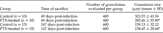
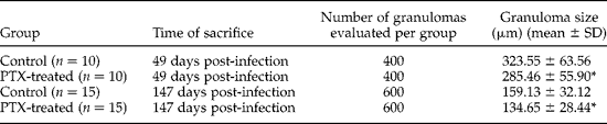
A significant reduction in mean diameter of the liver granulomas was observed in the groups of rodents treated with PTX, both in the acute and chronic infection, when compared to their respective control groups (P < 0.001), as shown in table 2. Indeed, an important decrease in the periovular inflammatory infiltrate was demonstrated in liver granulomas of mice treated with PTX, and these granulomas were less concentric and more irregular. Reduction in fibrous content of granulomas was also observed in mice that were treated with PTX, particularly in chronic infection.
Size (diameter) of 2000 granulomata observed in the livers of 50 mice treated with pentoxifylline (PTX) in acute and chronic Schistosoma mansoni infections.
* Significantly (P < 0.001) different from the respective control group.
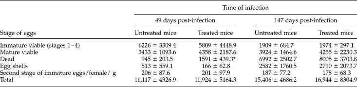
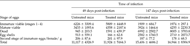
Data obtained by oogram analysis of intestine samples are presented in table 3. Mean total numbers of S. mansoni eggs/g of tissue in mice treated with PTX during acute and chronic disease (11,924 ± 5164.3 and 16,944 ± 8304.9, respectively) were not statistically different from values obtained for their respective control groups (11,117 ± 4326.9 and 15,406 ± 4686.2). Egg-laying kinetics was not notably modified by PTX treatment and the immature stage distribution of S. mansoni eggs showed minor intrinsic variations. There were also no statistical differences in the parameter second stage/female/g between untreated mice (206 ± 87.6 and 187 ± 77.2 in acute and chronic infection, respectively) and treated mice (201 ± 97.0 and 178 ± 68.3 in acute and chronic infections, respectively). The mean number of S. mansoni dead eggs was lower in the control group than in treated animals (P < 0.05), in the acute infection groups.
Mean numbers and standard deviation of Schistosoma mansoni eggs (immature viable, mature viable, dead, egg shells, total and second stage of immature eggs/female/g) observed with the oogram for intestinal fragments of mice treated with pentoxifylline in acute and chronic infections.
*P < 0.05.
Discussion
Although the haemorheological and immunomodulatory properties of PTX have been studied substantially in many different systemic disorders (Ward & Clissold, Reference Ward and Clissold1987; Sáez-LIorens et al., Reference Sáez-LIorens, Ramilo, Mustafá, Mertsola, Alba, Hansen and McCracken1990; Samlaska & Winfield, Reference Samlaska and Winfield1994; Bienvenu et al., Reference Bienvenu, Doche, Gutowski, Lenoble, Lepape and Perdrix1995; Louie et al., Reference Louie, Baltch, Franke, Ritz, Smith, Singh and Gordon1996; Ostrosky-Zeichner et al., Reference Ostrosky-Zeichner, Soto-Hernandez, Angeles-Morales, Teixeira, Nava-Ruiz, Rios, Solis and Sotelo1996; Marcinkiewicz et al., Reference Marcinkiewicz, Grabowska, Lauterbach and Bobek2000; Oliveira et al., Reference Oliveira, Neto, Martins, Rodrigues, Antonino and Magalhães2000; Muniz-Junqueira, Reference Muniz-Junqueira2007; Jesus et al., Reference Jesus, Luna, Almeida, Machado and Carvalho2008), knowledge of PTX effects in experimental schistosomiasis is limited (Reis et al., Reference Reis, Ventura, Souza, Arana-Pino, Pelajo-Machado, Pereira, Lenzi, Conceição and Takyia2001; Xiong et al., Reference Xiong, Zhu and Luo2003; El-Lakkany & Nosseir, Reference El-Lakkany and Nosseir2007).
The increase in red blood cell deformability within the microcirculation induced by treatment with PTX is not the only factor that determines the rheological properties of this drug. It stimulates fibrinolysis, decreases plasma fibrinogen levels, blocks red blood cell and platelet aggregations, and inhibits macromolecular extravasation in microcirculation and vasoconstriction (Ward & Clissold, Reference Ward and Clissold1987; Samlaska & Winfield, Reference Samlaska and Winfield1994; Seiffge et al., Reference Seiffge, Bissinger, Kremer, Laux and Schleyrbach1995). Thus, the mesenteric vasodilatation of the abdominal viscera observed macroscopically in the present study can be related to circulatory system changes caused by this drug. The absence of a significant difference in parasite burden in control and treated groups indicates no schistosomicidal effects of PTX, as expected. However, a slight worm reduction in infected mice after prolonged PTX treatment (8 weeks) has been reported (El-Lakkany & Nosseir, Reference El-Lakkany and Nosseir2007).
In untreated mice, histological analysis of liver fragments gave results similar to those found in the literature, where the average size of the acute hepatic granulomas was larger than that observed in chronic infection (table 2). The granulomatous lesions clearly showed an exudative reaction pattern and a substantial lymph–histiocytic inflammatory infiltrate in acute infections of murine schistosomiasis. With progression of the pathological process, modifications in the cell constitution and aspect of the granulomas occur, and they become more fibrous and concentric (Raso et al., Reference Raso, Bernardes, Tafuri, Bogliolo and Neves1978; Carvalho et al., Reference Carvalho, Alvarenga and Melo1986; Silva et al., 2000; Tristão et al., Reference Tristão, Melo, Vasconcelos and Grossi2000). This event, also observed in the present study, has been considered to be of benefit to the host as it shows a less intense inflammatory reaction, although more efficient in the chronic phase of schistosomiasis (Andrade & Warren, Reference Andrade and Warren1964).
A decrease in the inflammatory infiltrate around S. mansoni eggs observed in groups of mice treated with PTX, compared to their respective control groups in both acute and chronic infections, suggests that the drug interferes with mechanisms, not yet elucidated, that affect the recruitment of white blood cells. The less concentric and more irregular granulomas observed can be explained by the reduction in, or inhibition of, the migration of leucocytes to the site of hepatic damage. Indeed, the effects of PTX on leucocytes have been reported, mainly the interference with their adhesion to endothelial cells (Samlaska & Winfield, Reference Samlaska and Winfield1994; Seiffge et al., Reference Seiffge, Bissinger, Kremer, Laux and Schleyrbach1995). In experimental schistosomiasis, during the cercaria–schistosomulum transformation process, there was a delay in cell adhesion to S. mansoni larvae, possibly due to inhibition of neutrophil migration, suggesting that PTX could have some control over complement system components (A.L. Melo, unpublished results). Vascular permeability is induced by several inflammatory mediators, such as histamine and C4 leukotriene, as well as chemotactic factors such as the C5a complement peptide and B4 leukotriene, which are dependent on the presence of circulating and adherent leucocytes. Although PTX reduces leucocyte adhesion and vascular permeability to the macromolecules induced by the administration of lipopolysaccharide (LPS) in mice, it inhibits the synthesis of thromboxane and increases prostaglandin production (Seiffge et al., Reference Seiffge, Bissinger, Kremer, Laux and Schleyrbach1995). Nevertheless, other factors not related to leucocyte diapedesis may have been involved in the granuloma modifications observed in the animals treated with PTX. Tristão et al. (Reference Tristão, Melo, Vasconcelos and Grossi2000) demonstrated that the modulation of the inflammatory response around S. mansoni eggs is related to apoptosis. Wanchu et al. (2003) suggested that PTX may reduce this phenomenon and may improve CD4+ lymphocyte survival in patients infected with human immunodeficiency virus I (HIV-I).
As mentioned above, the action of PTX at the TNF-α level is already well known (Sáez-LIorens et al., Reference Sáez-LIorens, Ramilo, Mustafá, Mertsola, Alba, Hansen and McCracken1990; Samlaska & Winfield, Reference Samlaska and Winfield1994; Heyderman et al., Reference Heyderman, Klein, Daramola and Levin1995; Louie et al., Reference Louie, Baltch, Franke, Ritz, Smith, Singh and Gordon1996; Ostrosky-Zeichner et al., Reference Ostrosky-Zeichner, Soto-Hernandez, Angeles-Morales, Teixeira, Nava-Ruiz, Rios, Solis and Sotelo1996; Jesus et al., Reference Jesus, Luna, Almeida, Machado and Carvalho2008) and could be correlated with alterations observed on the fibrosis and the diameter of the granulomatous lesions. In fact, TNF-α correlates with periportal fibrosis in human schistosomiasis (Henri et al., Reference Henri, Chevillard, Mergani, Paris, Gaudart, Camila, Dessein, Montero, Elwali, Saeed, Magzoub and Dessein2002) and plays a fundamental role in cell migration to the infection site. The serum levels, as well as the respective soluble receptors (sTNF-RI and sTNF-RII), correlate with the beginning of S. mansoni oviposition and granuloma formation (Haseeb et al., Reference Haseeb, Sbirazian and Preis2001). Besides, mice treated with anti-TNF-α have shown a decrease in size of hepatic granulomas. The administration of recombinant murine TNF-α to mice chronically infected with S. mansoni was found to restore granuloma size to that observed in acute infection (Joseph & Boros, Reference Joseph and Boros1993). However, the immunomodulatory activity of PTX has been related to inhibition of pro-inflammatory cytokines besides TNF-α, such as interleukin-1 (IL-1), IL-12 and IFN-γ (Bienvenu et al., Reference Bienvenu, Doche, Gutowski, Lenoble, Lepape and Perdrix1995; Heyderman et al., Reference Heyderman, Klein, Daramola and Levin1995; Navarro et al., Reference Navarro, Punzón, Pizarro, Fernández-Cruz, Fresno and Muñoz-Fernández1996; Marcinkiewicz et al., Reference Marcinkiewicz, Grabowska, Lauterbach and Bobek2000), as well as IL-10, the anti-inflammatory cytokine that enhances Th1-type cell proliferation and reduces that of the Th2 type (Navarro et al., Reference Navarro, Punzón, Pizarro, Fernández-Cruz, Fresno and Muñoz-Fernández1996).
In fact, Xiong et al. (Reference Xiong, Zhu and Luo2003) observed that treatment with PTX in the early stage of the S. japonicum infectious process can reduce the levels of TGF-β1 and type I and III collagens in the liver, and suggested the potential utilization of PTX as an anti-fibrotic drug in combination with therapy against schistosomiasis japonica. Reduction in TGF-β1 and collagen in experimental schistosomiasis mansoni has been reported (El-Lakkany & Nosseir, Reference El-Lakkany and Nosseir2007), but further investigations are necessary. In the present study, a decrease in the fibrous component from liver granulomas due to treatment with PTX was also observed, confirming those previous results.
A significant reduction was observed in the number of eosinophils in granulomas after intraperitoneal treatment for 55 days (5 days/week) with 10 mg/kg of PTX started at 35 days of infection, but no significant differences were noted among the groups in relation to collagen synthesis (Reis et al., Reference Reis, Ventura, Souza, Arana-Pino, Pelajo-Machado, Pereira, Lenzi, Conceição and Takyia2001). It was suggested by the authors that this lack of effect on the collagen network was probably due, in part, to the beginning of drug administration after granuloma formation, when immune cells were already stimulated. In the study of Reis et al. (Reference Reis, Ventura, Souza, Arana-Pino, Pelajo-Machado, Pereira, Lenzi, Conceição and Takyia2001), liver sections were made in order to count the number of granulomas by light microscopy. A decrease in the total number of hepatic granulomas was observed, related to some toxic effect of the drug on egg release and/or adult worm fecundity. The results provided by El-Lakkany & Nosseir (Reference El-Lakkany and Nosseir2007) are similar to those of Reis et al. (Reference Reis, Ventura, Souza, Arana-Pino, Pelajo-Machado, Pereira, Lenzi, Conceição and Takyia2001); however, the larger daily dose of PTX (200 mg/kg) used in the present study may explain the notable effects on the constitution of granulomatous lesions, even in short-term treatment with PTX and after the stimulation of schistosomotic granuloma development. Indeed, the effects of administration of PTX may depend on the time of treatment, but are amplified when high doses are used (Tilg et al., Reference Tilg, Eibl, Pichl, Gächter, Herold, Brankova, Huber and Niederwieser1993; Marcinkiewicz et al., Reference Marcinkiewicz, Grabowska, Lauterbach and Bobek2000; Xiong et al., Reference Xiong, Zhu and Luo2003).
Despite the original purpose of the oogram method and its formerly described use in the evaluation of anti-schistosomal compounds (Pellegrino et al., Reference Pellegrino, Oliveira, Cunha and Siqueira1962; Pellegrino & Faria, Reference Pellegrino and Faria1965), this technique has also been employed to study the biology of the parasite, including egg release into the intestinal lumen (Hermeto et al., Reference Hermeto, Bicalho, Silva, Melo and Pereira1994) and the individual fecundity of S. mansoni (Valadares et al., Reference Valadares, Coelho, Pellegrino and Sampaio1981). Some authors have reported that TNF-α is directly connected to the fecundity of the parasite (Amiri et al., Reference Amiri, Locksley, Parslow, Sadick, Rector, Ritter and McKerrow1992; Haseeb et al., Reference Haseeb, Solomon and Palma1996). However, TNF-α alone does not appear necessary or sufficient to affect the fecundity of S. mansoni or S. japonicum females (Cheever et al., Reference Cheever, Poindexter and Wynn1999). Besides, the evaluation of second-stage immature S. mansoni eggs by the oogram technique utilized in the present work seems to be the most sensitive parameter to analyse alterations in the fecundity of the female parasite, since it permits the observation of newly laid eggs and shows few confounding factors, as suggested in a previous study (Pacheco et al., Reference Pacheco, Zucconi, Mati, Garcia, Miyoshi, Oliveira, Melo and Azevedo2005). No change in the parameter second stage of immature egg/female worm/g and in total number of parasite eggs was observed, indicating the absence of alterations in egg viability and also in S. mansoni oviposition (table 2), in contrast to the studies of Reis et al. (Reference Reis, Ventura, Souza, Arana-Pino, Pelajo-Machado, Pereira, Lenzi, Conceição and Takyia2001) and El-Lakkany & Nosseir (Reference El-Lakkany and Nosseir2007), who observed a reduction in egg number in rodent liver, as already mentioned.
It has been accepted that host cells seem to favour the passage of S. mansoni eggs to the organ lumen and that high numbers of parasite eggs trapped in the tissues of the host can reflect a reduction in the release of mature viable eggs into the intestinal lumen (Doenhoff et al., Reference Doenhoff, Musallam, Bain and McGregor1978; Hermeto et al., Reference Hermeto, Bicalho, Silva, Melo and Pereira1994). Hermeto et al. (Reference Hermeto, Bicalho, Silva, Melo and Pereira1994) showed that mice treated with dexamethasone have increased numbers of S. mansoni eggs in their tissues, where this drug is the agent that interferes with this passage. In the present study, the increase observed in the mean number of dead S. mansoni eggs (P < 0.05) in the intestine of mice treated with the drug during acute infection could also indicate interference by PTX, although slight, with the release of mature viable eggs into the intestinal lumen to modify the inflammatory reaction around these eggs. However, further studies are needed to better evaluate the passage of parasite eggs to the intestinal lumen in PTX-treated mice.
The action of PTX during experimental schistosomiasis mansoni induces a decrease in the intensity of the inflammatory reaction around the parasite eggs and possibly in the aggression to the vertebrate host. A down-regulation reducing the production of both Th1 and Th2 cytokines might have occurred in rodents treated with the drug, but further immunological studies are necessary. Furthermore, PTX modulates cytokine release differently depending on the cellular type, such as CD4+T cells and macrophages (Marcinkiewicz et al., Reference Marcinkiewicz, Grabowska, Lauterbach and Bobek2000), and may have different effects when used at different times of the infectious process, as observed in murine cutaneous leishmaniasis (Oliveira et al., Reference Oliveira, Neto, Martins, Rodrigues, Antonino and Magalhães2000). Therefore, PTX could show some different targets or changes in its effector mechanisms of action in acute and chronic S. mansoni infection.
Data obtained in the present study indicate probable immunomodulatory effects of PTX in murine schistosomiasis, with a decrease in the inflammatory reaction, fibrous content and in the mean diameter of hepatic granulomas both in acute and chronic infection. It is possible that the drug affected TNF-α action in experimental schistosomiasis, perhaps reducing the severity of the pathological process. Thus, new studies are required to determine and clarify the true action of PTX in this model.
Acknowledgements
We thank the Conselho Nacional de Desenvolvimento Científico e Tecnológico-CNPq for financial support, Mr Airton Lobo for his valuable histological assistance and reviewers for their helpful suggestions.