Introduction
In vitro production of embryos (IVP) by maturation, fecundation, and in vitro cultivation has become a valuable assisted reproduction technology in several cattle breeding herds. However, even with remarkable innovations in this technology, the greatest obstacles for success of this biotechnology are low post-transference pregnancy rates and high sensitivity of the embryos to cryopreservation (Long, Reference Long2008).
Nuclear maturation comprises the process of reversing the first meiotic block in the germinal vesicle (GV) to the second meiotic block in metaphase II (MII). It begins at the moment the resumption of meiosis occurs from the diplotene stage of prophase I, marked by chromosome condensation and disruption of the germinal vesicle (GVB), that is the nuclear envelope. After QVG, the oocyte passes through the stages of diakinesis, metaphase I (MI), anaphase I (AI) and telophase I (TI), completing the first meiotic division, and then rapidly moving to the second meiotic division MII, when there is a new cell cycle block (second meiotic block; Kubelka et al., Reference Kubelka, Motlík, Schultz and Pavlok2000).
Cytoplasmic maturation comprises the structural and molecular changes that occur in the oocyte cytoplasm at the GV stage until the end of the MII. The evaluation of this maturation can be done indirectly by the mature oocyte’s ability to cleave and develop the blastocyst, after fertilization or parthenogenetic activation. Other indirect morphological parameters such as cumulus cell expansion (CCs), extrusion velocity of the first polar corpuscle, and increase of perivitelline space may also be used to evaluate cytoplasmic maturation (Kruip et al., Reference Kruip, Cran, Van Beneden and Dieleman1983).
The ultrastructural modifications concern a cytoplasmic reorganization in which most organelles migrate through microtubules and microfilaments. Mitochondria and the Golgi complex, which are located more peripherally in the immature oocyte, migrate and distribute in a perinuclear position. Cortical granules, produced by the Golgi complex and originally located in the centre of the oocyte, migrate to the periphery of the oocyte and anchor themselves in the cytoplasmic membrane (Cran and Esper, Reference Cran and Esper1990).
Many laboratories have studied this technology to improve the development and quality of oocytes during in vitro maturation (IVM). IVM efficiency is related to the biochemical and molecular status of the mature oocyte, which can be naturally fertilized to become an embryo, and when transferred can develop appropriately (Gilchrist and Thompson, Reference Gilchrist and Thompson2007).
In vivo, the luteinizing hormone (LH) peak results in the interruption of gap junctions between oocytes and granulosa cells in pre-ovulatory follicle (Eppig, Reference Eppig1991). The isolation of immature oocytes before IVM may interrupt transference of nutrients and essential metabolic support required for completion of the maturation process, resulting in early activation of nuclear maturation and later commitment for the development of the ooplasm (Carabatsos et al., Reference Carabatsos, Sellitto, Goodenough and Albertini2000).
In vitro, resumption of meiosis occurs when there is a release of inhibition factor from the oocyte (Kotsuji et al., Reference Kotsuji, Kubo and Tominaga1994). However, in vitro maturation results in reduced embryonic production, which suggests that not all oocytes are able to mature properly because they do not resume meiosis (Hyttel et al., Reference Hyttel, Fair, Callesen and Greve1997).
Meiosis blockade before maturation has been suggested as an alternative because it allows oocytes to have additional culture time without suffering modification such as pre-maturation. Blockade of meiosis can be induced by various strategies including increased cAMP, protein synthesis, or inhibition of phosphorylation (Razza et al., Reference Razza, Sudano, Fontes, Franchi, Belaz, Santos, Castilho, Rocha, Eberlin, Machado and Nogueira2018).
Spontaneous oocyte maturation can be avoided by using specific cyclin-dependent kinase (CDK) inhibitors such as butyrolactone I (BL-I) and roscovitine (ROS), which inhibit activity of oocyte maturation promoting factor (MPF) (Adona et al., Reference Adona, Pires, Quetglas, Schwarz and Leal2008; Quetglas et al., Reference Quetglas, Adona, de Bem, Pires and Leal2011; Maziero et al., Reference Maziero, Guaitolini, Paschoal, Kievitsbosch, Guastali, Moraes and Landim-Alvarenga2016).
BL-I is a purine derived from Aspergillus SP mycelium and specifically inhibits cyclin-dependent kinases (CDK2 and cdc2) (Lonergan et al., Reference Lonergan, Dinnyes, Fair, Yang and Boland2000). ROS is a synthetic purine nucleoside analogue, which inhibits activity of MPF, keeping bovine oocytes in the GV (Lagutina et al., Reference Lagutina, Ponderato, Lazzari and Galli2002).
Such inhibitors have been added for 24 h to IVM medium, preserving bovine oocyte viability without compromising embryonic development (Kubelka et al., Reference Kubelka, Motlík, Schultz and Pavlok2000; Lonergan et al., Reference Lonergan, Dinnyes, Fair, Yang and Boland2000; Mermillod et al., Reference Mermillod, Tomanek, Marchal and Meijer2000; Ferreira et al., Reference Ferreira, Vireque, Adona, Meirelles, Ferriani and Navarro2009; Quetglas et al., Reference Quetglas, Adona, de Bem, Pires and Leal2011). However, different usage times of these drugs have not been described in the literature (Maziero et al., Reference Maziero, Guaitolini, Paschoal, Kievitsbosch, Guastali, Moraes and Landim-Alvarenga2016; Paschoal et al., Reference Paschoal, Sudano, Schwarz, Maziero, Guastali, Crocomo and Landim-Alvarenga2017; Razza et al., Reference Razza, Sudano, Fontes, Franchi, Belaz, Santos, Castilho, Rocha, Eberlin, Machado and Nogueira2018).
Therefore, we aimed to assess the effect of a 6, 12 and 24 h pre-maturation treatment (pre-IVM) with meiosis inhibitors ROS and BL-I, at concentrations of BL-I at 50 or 100 µM, ROS at 12.5 or 25 µM, ROS + BL-I at 6.25 µM or 25 µM, respectively. In addition, we evaluated the ideal concentration and time of use of the meiosis inhibitors for optimal embryo development (% embryo production), quality (apoptotic index) and resistance to vitrification (embryonic re-expansion).
Material and methods
All chemicals were purchased from Sigma (Sigma-Aldrich® Corporation, St. Louis, MO, USA), except for those specified in the text.
Oocyte collection
Ovaries from a commercial slaughterhouse were collected and transported to the in vitro fertilization (IVF) laboratory at the Department of Animal Reproduction and Veterinary Radiology of the São Paulo State University, in Botucatu, Brazil. The ovaries were rinsed with 0.9% NaCl at 38°C. All follicles measuring 2–8 mm were aspirated.
In vitro maturation (Control)
Groups of 50 immature COCs were subjected to standard IVM culture in TCM-199 (Earle’s salt; Sigma-Aldrich) with 0.1 IU/ml recombinant human follicle-stimulating hormone (rhFSH, Puregon; Organon International), 4 mg/ml fatty acid-free BSA, 22 mg/ml sodium pyruvate, and 50 mg/ml amikacin for 24 h (Razza et al., Reference Razza, Sudano, Fontes, Franchi, Belaz, Santos, Castilho, Rocha, Eberlin, Machado and Nogueira2018). The oocytes were kept in a 38.5°C incubator with 5% CO2 in air, with 100% humidity. These COCs were referred to as the control group.
Pre-maturation treatment for in vitro maturation (pre-IVM)
Before IVM, groups of 50 immature COCs were subjected to pre-IVM, in which cells were cultured in TCM-199 medium in the absence of rhFSH and supplemented with:
1. BL-I (Enzo Life Sciences International, Inc., 5120 Butler Pike Plymouth Meeting, PA 19462) at 50 or 100 µM., for 6, 12 or 24 h.
2. ROS (R7772 Sigma ≥ 98) at 12.5 or 25 µM, for 6, 12 or 24 h.
3. ROS + BL-I at 6.25 µM and 25 µM, for 6, 12 and 24 h.
After 6, 12 or 24 h, COCs were washed thoroughly to completely remove any traces of ROS, BL-I, and ROS + BL-I before following the standard IVM protocol for an additional 12, 18 or 24 h.
Evaluation of oocyte nuclear stage
In each experimental group, oocytes were randomly collected at 6, 12 or 24 h of pre-IVM and 12, 18 or 24 h of IVM (n = 3058). Collection was performed as previously described by Maziero et al. (Reference Maziero, Guaitolini, Paschoal, Kievitsbosch, Guastali, Moraes and Landim-Alvarenga2016).
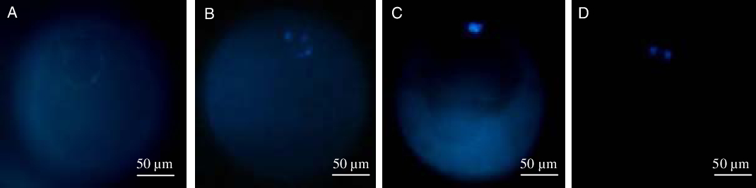
Oocytes were transferred to a 5-µl drop of Hoechst 33342 (0.01 mg/ml) on a slide and covered with a coverslip. After 20 min, nuclear morphology was observed using an inverted fluorescence microscope (Leica DMIRB). The nucleus was classified as follows: GV, germinal vesicle breakdown (GVB), metaphase I (MI), metaphase II (MII), or degenerate or unidentified (D/U) (excitation wavelength: 350 nm and emission wavelength: 461 nm) (Fig. 1)
Photographic presentation of the evaluation of nuclear stages of oocytes of experimental groups in inverted microscope with ultraviolet fluorescent light. After staining with Hoechst 33342 oocytes were classified into: (A) germinal vesicle (GV); (B) germinal vesicle breakdown (GVB); (C) metaphase I (MI); and (D) metaphase II (MII). ×400 magnification.

In vitro fertilization
Mature oocytes from all groups underwent IVF = day 0) with frozen Nelore bull (Bos taurus indicus) semen. The semen was prepared using the Percoll method (Maziero et al., Reference Maziero, Guaitolini, Paschoal, Kievitsbosch, Guastali, Moraes and Landim-Alvarenga2016; Paschoal et al., Reference Paschoal, Sudano, Schwarz, Maziero, Guastali, Crocomo and Landim-Alvarenga2017), with final concentration to 2 × 10⁶ sperm cells/ml. Oocytes were incubated for 24 h in an incubator with 5% CO2 in air at 38.5°C and 100% humidity.
Oocytes were fertilized in human tubal fluid (HTF®; Irvine Scientific, Santa Ana, California, USA) supplemented with 5 mg/ml BSA (A-8806), 11 mg/ml sodium pyruvate, 0.5 mg/ml caffeine, 3 mg/ml heparin, 0.3 mg/ml penicillamine, 0.11 mg/ml hypotaurine, 0.18 g/ml epinephrine, and 100 IU/µl amikacin (Paschoal et al., Reference Paschoal, Sudano, Schwarz, Maziero, Guastali, Crocomo and Landim-Alvarenga2017).
In vitro culture (IVC) and vitrification
Presumptive zygotes (n = 584) were cultured in synthetic oviductal fluid (SOFaa) (Paschoal et al., Reference Paschoal, Sudano, Schwarz, Maziero, Guastali, Crocomo and Landim-Alvarenga2017) supplemented with 2.7 mM myo-inositol, 0.2 mM sodium pyruvate, 5 mg/ml BSA (A-8806), 2.5% (v/v) FCS and 100 UI /ml amikacin. The embryos were kept in an incubator in 5% CO2, 5% O2 and 90% N2 at 38.5°C and 100% humidity. Blastocyst formation rate was observed on day 7 (day 0 = IVF). After that, embryos were vitrified.
Embryo vitrification was performed on days 6 (D6) and 7 (D7) of culture (15 replicas) according to the protocol of Maziero et al. (Reference Maziero, Guaitolini, Paschoal, Kievitsbosch, Guastali, Moraes and Landim-Alvarenga2016). Embryos produced in vitro were exposed to the vitrification solution 1 (SV1), composed of 5 M of ethylene glycol (EG) in base medium for 3 min, and transferred to a drop of 15 µl of the vitrification solution 2 (SV2) composed of 7 M of EG, 0.5 M galactose and 18% (w/v) Ficoll 70 in base medium. Embryos were then packed in 0.25-ml sterile straws (Nutricell), which were set up as follows: 1 cm of galactose (0.5 M galactose solution), 0.5 cm of air, 7 cm of galactose solution followed by an additional 0.5 cm of air. Embryos contained on SV2 were transferred to the straws (drop of 15 µl of SV2), followed by 0.5 cm of air, and the remaining space until the end of the straw was completed with galactose solution. The straws were sealed with polyvinyl alcohol.
The time necessary for loading and sealing a straw and transferring it into liquid nitrogen steam was not longer than 45 s. The straws were then held in liquid nitrogen steam for 1 min, and then dipped in the liquid. The embryos were stored for 1–6 months in liquid nitrogen (−196°C).
To warm up the straws, they were kept in air for 8 s and then in water at 37°C for 15 s. The embryos were then washed and cultured in SOFaa medium containing 10% FBS, for 12 h in a 5% CO2 incubator for further evaluation of re-expansion of the embryo.
Evaluation of apoptotic index
Rate of apoptosis was assessed after 7 days of IVC (day 0 = IVF) as previously described by Sudano et al. (Reference Sudano, Paschoal, Rascado, Magalhães, Crocomo, Lima-Neto and Landim-Alvarenga2011).
In total, 10–15 embryos were used in each experimental group for terminal deoxynucleotide transferase uridine nick-end labelling (TUNEL) analysis for identification of in situ internucleosomal DNA fragmentation, a result of apoptosis. Analysis was performed using the In Situ Cell Death Detection Kit, Fluorescein® (Roche 11 684 795 910, Mannheim, Germany).
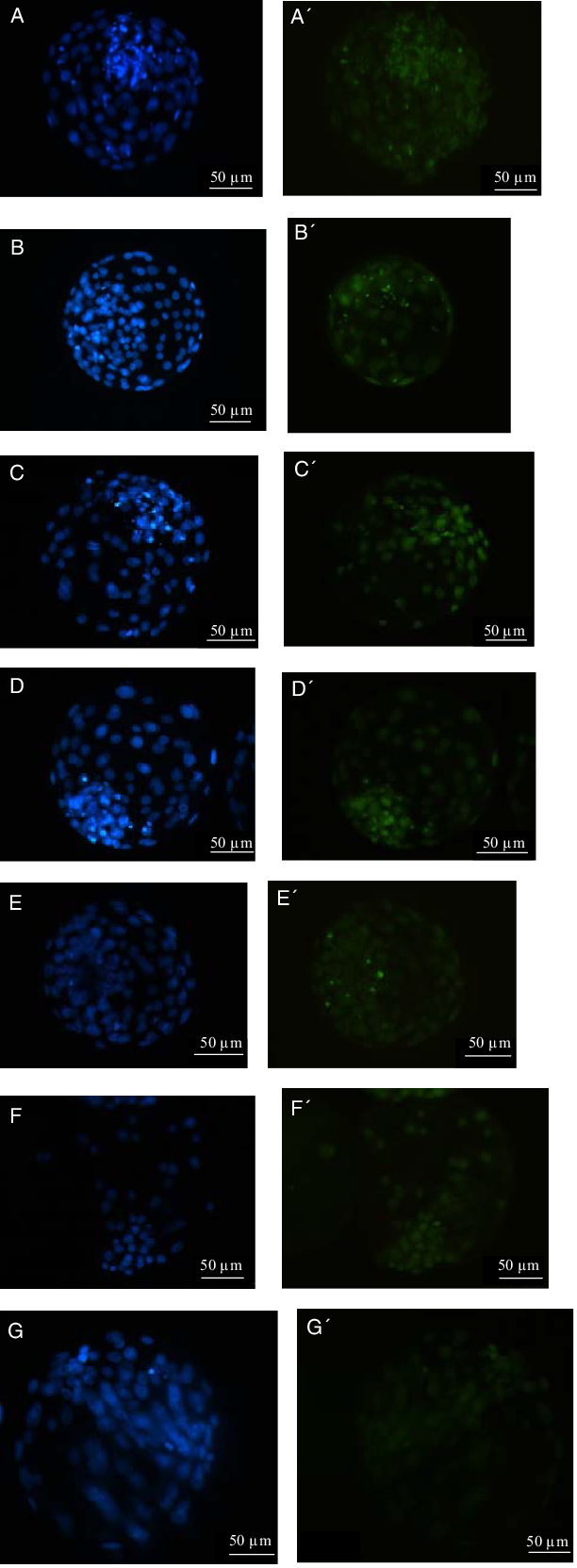
Embryos were washed in 0.1% (vol/vol) polyvinylpyrrolidone (PVP)/phosphate-buffered saline (PBS) and fixed in a solution of 4% (vol/vol) paraformaldehyde for 1 h at room temperature. Subsequently, embryos were transferred to a permeability solution [0.1% (vol/vol) Triton X-100 and 0.1% (vol/vol) sodium citrate in PBS] for 1 h. Embryos were incubated in positive and negative control solutions with DNase (Promega, M 6101, Madison, USA) for 1 h at 37°C in the dark. Then, the positive control and samples were incubated in 10 µl drops of TUNEL solution on slides for 1 h at 37°C in a dark and humid chamber (Fig. 2).
In vitro produced bovine embryos from different groups submitted to the TUNEL technique evaluated with an inverted fluorescence microscope. At the left side are the embryos with number of intact cells and the right side are the apoptotic embryos. (A, A′) Control; (B, B′) BL-I 50 6 h; (C, C′) ROS 6 h; (D, D′) RB 6 h; (E, E′) BL-I 12 h; (F, F′) R 12 h; (G, G′) RB 12 h (×400 magnification; bar: 100 mM). B, butyrolactone I; R: roscovitine; RB: association roscovitine + butyrolactone I; TUNEL, terminal deoxynucleotide transferase uracil nick-end labelling.

The negative control was incubated only with tracer solution, without the enzyme solution, to verify the signal. After washing, the samples and controls were subjected to a contrast step to visualize DNA with Hoechst 33342 solution (1 mg/ml) and were analyzed using a fluorescence inverted microscope. Using 450–490 nm excitation and 515 nm emission, green fluorescent nuclei [fluorescein isothiocyanate (FITC)] were considered TUNEL-positive cells with fragmented DNA. Using 365 nm excitation and 420 nm emission, the blue signal (Hoechst 333342 stain) indicated the presence and location of cell nuclei.
The number of apoptotic (green-stained) cells and the total (blue-stained) cells were counted using ImageJ 1:41 software (Wayne Rasband National Institutes of Health, Bethesda, USA). Based on these data, the percentage of apoptotic occurrences (the number of apoptotic cells compared with the total number of cells in the embryo) was calculated.
Statistical analysis
Data were analyzed using analysis of variance (ANOVA) and the SAS PROC GLM program (SAS Inst. Inc., Cary, NC, USA). Sources of variation in the model including treatment and replication were considered fixed and random effects respectively. If ANOVA results were significant, means were compared using the least-squares difference. Data were presented as mean and standard errors. A 5% significance level was used for all analyses.
Results
In total, 25 routines (50 oocytes/treatment) were performed between the nuclear maturation evaluation and the blastocyst production rate evaluation.
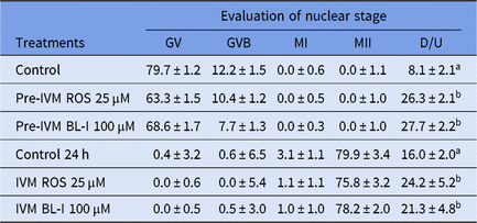
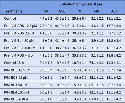
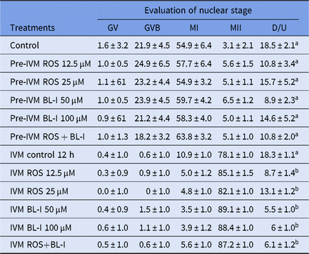
Regardless of drug used, the 24 h treatment resulted in a larger number of degenerated oocytes at the end of the IVM period (Table 1). Oocyte degeneration decreased with reduction of the meiotic blocking period to 6 or 12 h (Tables 2 and 3).
Nuclear maturation stage of oocytes treated for 24 h with roscovitine or butyrolactone I. In the top of the table are presented the results obtained just after the treatment and in the bottom are presented the results obtained after 24 h of in vitro maturation

D/U, Degenerate or unidentified; BL-I, butyrolactone; GV, germinal vesicle; GVB, germinal vesicle breakdown; MI, metaphase I; MII, metaphase II; ROS, roscovitine. Mean ± standard error.
a,b Values indicated by different superscript letters in the same column differ (P < 0.05).
Nuclear maturation stage of oocytes treated for 6 h with roscovitine or butyrolactone I. In the top of the table are presented the results obtained just after the treatment and in the bottom are presented the results obtained after 18 h of in vitro maturation. Total of five replicas with 350 oocytes

D/U, degenerate or unidentified; GV, germinal vesicle; GVB, germinal vesicle breakdown; MI, metaphase I; MII, metaphase II; ROS, roscovitine. Mean ± standard error. There was no statistical difference (P > 0.05).
Nuclear maturation stage of oocytes treated for 12 h with roscovitine or butyrolactone I. In the top of the table are the results obtained just after the treatment and in the bottom are the results obtained after 12 h of in vitro maturation. Total of five replicas with 350 oocytes

D/U, Degenerate or unidentified; GV, germinal vesicle; GVB, germinal vesicle breakdown; MI, metaphase I; MII, metaphase II; ROS, roscovitine. Mean ± standard error.
a,b Values indicated by different superscript letters in the same column differ (P < 0.05).
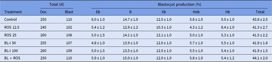
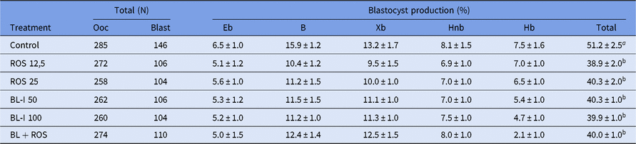
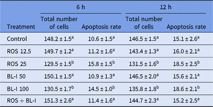
No significant differences were observed in blastocyst production rate in oocytes inhibited for 6 h compared with those in the control group (Table 4; P > 0.05). However, with inhibition of oocytes for 12 h (pre-IVM), a decrease in embryo production was observed compared with that in the control group (Table 5; P < 0.05). The total number of intact embryo cells decreased and the rate of apoptosis increased in ROS and BL-I groups with concentrations of 25 and 100 µM, respectively, following 6 h and 12 h of pre-IVM (Table 6; P < 0.05).
Evaluation of blastocyst rate on day 7 (day 0 = IVF) from oocytes inhibitors for 6 h (pre-IVM) with ROS, BLI and ROS + BL-I and control 18 h. Total of 10 (n = 10) replicas with 1520 oocytes

Embryo development stage evaluated on day 7: Eb, early blastocyst; B, blastocyst; Xb, expanded blastocyst; Hnb, blastocyst hatching; Hb, hatched blastocyst. Ooc, oocytes; Blast, blastocysts. ROS, roscovitine. BL-I, butyrolactone I. Mean ± standard error. There was no statistical difference (P > 0.05).
Evaluation of blastocyst rate on day 7 (day 0 = IVF) from oocytes inhibitors for 12 h (pre-IVM) with ROS, BL-I and ROS + BL-I and control for 12 h. Total of 10 (n = 10) replicas with 1611 oocytes

Embryo development stage evaluated on day 7: Eb, early blastocyst; B, blastocyst; Xb, expanded blastocyst; Hnb, blastocyst hatching; Hb, hatched blastocyst. Ooc, oocytes; Blast, blastocysts. ROS, roscovitine; BL-I, butyrolactone I. Mean ± standard error.
a,b Values indicated by different superscript letters in the same column differ (P < 0.05).
Evaluation of the total number of cells and apoptosis rate on day 7 blastocysts (day 0 = IVF) from oocytes inhibitors for 6 or 12 h (pre-IVM) with different concentrations of ROS, BL-I and ROS+BL-I and control for 18 h (IVM). Total of 10 replicas with 20 embryos per group

ROS, roscovitine; BL-I, butyrolactone I. Mean ± standard error mean.
a,b Values indicated by different superscript letters in the same column differ (P < 0.05).
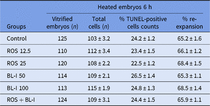
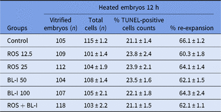
No differences in re-expansion after embryonic cryopreservation between groups and the number of apoptotic cells of devitrificated embryos (P > 0.05; Table 7).
Total number of cell and apoptotic cells of bovine heated blastocysts, total number of vitrified embryos and percentage of embryonic re-expansion from oocytes inhibitors for 6 h (pre-IVM) with different concentrations of ROS, BL-I and ROS + BL-I and control for 18 h (IVM). Total of 15 replicas with 15 embryos per group

ROS, roscovitine. BL-I, butyrolactone I. Mean ± standard error.
Discussion
The results showed that the use of ROS and BL-I at high concentrations for 24 h resulted in a higher oocyte degeneration rate than that in the control group (Table 1). Previous studies using the same concentrations and durations of ROS and BL-I treatment were effective in keeping bovine oocytes in germinal vesicles without cellular degeneration (Mermillod et al., Reference Mermillod, Tomanek, Marchal and Meijer2000; Lagutina et al., Reference Lagutina, Ponderato, Lazzari and Galli2002; Barreto et al., Reference Barreto, Castro, Garcia and Mingoti2011).
The reasons for these differences were not clear. However, high degeneration rates were observed across several repeated tests performed in our laboratory. Therefore, ROS concentration was reduced to 12.5 µM, with 6 h (Table 2) and 12 h (Table 3) of meiotic inhibition (pre-IVM), and corresponding 18 h and 12 h reversion (IVM) to evaluate embryo production rates.
Use of 25 µM or 12.5 µM ROS for 6, 12, or 24 h inhibited nuclear maturation (Tables 1–3). The mechanisms involved in meiosis inhibition control and progression are not fully understood. Some studies have reported that culture medium composition is important in maintaining meiotic arrest of bovine oocytes resulting from inhibition (Bilodeau-Goeseels, Reference Bilodeau-Goeseels2006). In mouse oocytes, culture medium composition affects in vitro maturation. According to Downs and Verhoeven (Reference Downs and Verhoeven2003), differences in combinations and concentrations of energy sources influence spontaneous nuclear maturation rates and efficiency of IVM-inhibiting drugs. However, some molecules that are capable of inhibiting nuclear maturation in mouse oocytes are not effective in bovine oocytes.
In this study, meiotic inhibition with ROS for 12 h (ROS 12.5 µM = 38.9 ± 2.0%; ROS 25 µM = 40.3 ± 2.0%) reduced embryonic production rate compared with that in the control group (C = 51.2 ± 2.5%). These results were higher than those obtained by Adona and Leal (Reference Adona and Leal2004). They observed 35% blastocysts (D8) in the control group, while the group treated with ROS (25 µM/24 h) was comprised of 24.2% blastocysts.
In contrast, Barreto et al. (Reference Barreto, Castro, Garcia and Mingoti2011) reported similar embryonic production rates between the control group (29.6 ± 6.6%) and the ROS treatment group (25 µM) across the following treatment durations: 16, 20, and 24 h in IVM (35.2 ± 6.8%, 31.7 ± 2.0% and 33 ± 4.7%, respectively). Mermillod et al. (Reference Mermillod, Tomanek, Marchal and Meijer2000) also observed similar embryonic production rates between oocytes in the control group and in the group treated with 25 µM ROS for 24 h during IVM. These authors reported blastocyst formation rates of 23 ± 6% for the control group (D7) and of 18 ± 5% for the group treated with ROS.
Adona and Leal (Reference Adona and Leal2004) reported results similar to our study (Tables 1–3). In this study, 100 µM BL-I for 24 h was effective in blocking meiosis (Table 1).
High oocyte degeneration rate was observed at pre-IVM with 100 µM BL-I treatment for 24 h (18.1 ± 4.2%; Table 1). These results contrasted with those reported by Ferreira et al. (Reference Ferreira, Vireque, Adona, Meirelles, Ferriani and Navarro2009) in which the oocyte degeneration rate was only 4.8% after 18 h of incubation, and 2.6% after 24 h of inhibition.
When BL-I concentration was decreased to 50 µM for 6 h and 12 h pre-IVM, the number of degenerated oocytes decreased to 15.8 ± 2.4% (Table 2) and 8.9 ± 2.3% (Table 3), respectively. These results were superior to the data obtained by Ferreira et al. (Reference Ferreira, Vireque, Adona, Meirelles, Ferriani and Navarro2009).
According to previous studies, the GVB takes place between 6 and 9 h after onset of IVM (Sirard and First, Reference Sirard and First1988; Sirard et al., Reference Sirard, Parrish, Ware, Leibfried-Rutledge and First1988; Bilodeau-Goeseels, Reference Bilodeau-Goeseels2006). In addition, immediately following follicular aspiration, a large number of oocytes can be found in the GVB (Sirard et al., Reference Sirard, Parrish, Ware, Leibfried-Rutledge and First1988). Use of BL-I at a concentration of 50 µM for 6 h (Table 4) resulted in an embryonic production rate (41.9 ± 1.8% blastocysts – D7) similar to the control group (43.8 ± 2.5%).
These data are similar to previous reports showing that BL-I treatment resulted in the same embryonic production rate as the control group (Kubelka et al., Reference Kubelka, Motlík, Schultz and Pavlok2000; Lonergan et al., Reference Lonergan, Dinnyes, Fair, Yang and Boland2000; Adona and Leal, Reference Adona and Leal2004; Adona et al., Reference Adona, Pires, Quetglas, Schwarz and Leal2008). However, Maziero et al. (Reference Maziero, Guaitolini, Paschoal, Kievitsbosch, Guastali, Moraes and Landim-Alvarenga2016) observed improvements in production rates and post-vitrification survival of embryos using commercial medium – In Vitro Brasil®, with 50 µM BL-I treatment for 6 h.
Adona et al. (Reference Adona, Pires, Quetglas, Schwarz and Leal2008) obtained 31.3% blastocysts (D7) in the group treated with 100-µM BL-I for 24 h and 36.5% blastocysts (D7) in the control group. Similarly, Lonergan et al. (Reference Lonergan, Dinnyes, Fair, Yang and Boland2000) reported similar blastocyst rates between the control group and the group treated with BL-I, with 28.4% blastocysts (D8) in the control group and 27% in the group treated with 100-µM BL-I for 24 h. Such data differ from those obtained in this study when using a meiotic inhibition time of 12 h, in which the blastocyst formation rate was much lower than that of the control group (BL-I = 39.3 ± 1.0% × C = 51.2 ± 2.5%; Table 6).
Total embryonic cells and number of apoptotic cells did not differ (Table 6) when treated with BL-I at the 50 µM concentration for 6 or 12 h. However, at 100 µM, less embryos were present and rate of apoptosis was higher with both meiotic inhibition times (6 and 12 h). According to Byrne et al. (Reference Byrne, Southgate, Brison and Leese1999), the best way to evaluate embryonic quality was through the cellular apoptosis index, which associates high incidence of apoptosis with reduction of embryonic viability.
The combination of BL-I and ROS has been used to block GVB of bovine oocytes at lower concentrations than with individual use. In this study, these drugs were used for two inhibition times (6 or 12 h), with reversion of blocking for 18 and 12 h, respectively. The concentrations used were 25 µM BL-I and 6.25 µM ROS, half of the concentrations used in previous studies.
Previous reports have shown that administration of these drugs at the concentration used in this study was able to keep 100% of oocytes in VG without compromising later embryonic development (Ponderato et al., Reference Ponderato, Lagutina, Crotti, Turini, Galli and Lazzari2001). However, these results were not observed in this study, as high índices of oocytes in GVB were observed for both 6 (C= 43.6 ± 6.5 × ROS + BL-I = 38.2 ± 4.4; Table 2) and 12 h pre-IVM (C = 21.9 ± 4.5 × ROS + BL-I= 18.2 ± 3.2; Table 3). Despite this, the combination of BL-I and ROS was efficient in reverting meiosis, as evidenced by the high number of oocytes in MII, after inhibition. Previous studies have demonstrated that BL-I and ROS were efficient in keeping oocytes in GV and then oocytes were capable of resuming normal development when no longer exposed to drugs that inhibit meiosis (Ponderato et al., Reference Ponderato, Lagutina, Crotti, Turini, Galli and Lazzari2001; Lonergan et al., Reference Lonergan, Faerge, Hyttel, Boland and Fair2003; Adona et al., Reference Adona, Pires, Quetglas, Schwarz and Leal2008).
The embryonic rates obtained using combined BL-I and ROS for 6 h were similar to that of the control group (ROS + BL-I = 44.11 ± 2.0% × C= 43.8 ± 2.5%; Table 4). Such data show that use of these drugs in combination did not affect embryonic development. Adona et al. (Reference Adona, Pires, Quetglas, Schwarz and Leal2008) observed similar results between control and treated groups when using BL-I + ROS in IVM medium for 24 h. Other authors have also reported similarities between treated and control groups (Ponderato et al., Reference Ponderato, Lagutina, Crotti, Turini, Galli and Lazzari2001). However, in this study, when oocytes were treated with BL-I and ROS for 12 h, there was a decrease in embryo production (C= 51.2 ± 2.5% × ROS+BL-I = 40.0 ± 1.0%; Table 5).
Embryos from the control group and the group treated with ROS+BL-I in combination for 6 and 12 h were similar in number of cells. Similarly, the groups had apoptosis rates similar to the control group (Table 6) demonstrating embryonic viability following removal of BL-I and ROS. Adona et al. (Reference Adona, Pires, Quetglas, Schwarz and Leal2008) observed similarities in number of blastocyst cells between the control group and the group treated with ROS and BL-I in combination.
Meiotic arrest has been effective in vitro when using drugs that inhibit the activity of MPF (Mermillod et al., Reference Mermillod, Tomanek, Marchal and Meijer2000; Maziero et al., Reference Maziero, Guaitolini, Paschoal, Kievitsbosch, Guastali, Moraes and Landim-Alvarenga2016). Additionally other drugs that block meiosis, such as cAMP (adenylate cyclase) and phosphodiesterase inhibitors, have been effective maintained in germ cell vesicle oocytes (Eppig, Reference Eppig1991; Downs and Verhoeven, Reference Downs and Verhoeven2003). However, bovine treatment with meiosis blockers resulted in only transient blockade (Sirard et al., Reference Sirard, Parrish, Ware, Leibfried-Rutledge and First1988) and development of post-treatment oocytes do not differ from the untreated group (Paschoal et al., Reference Paschoal, Sudano, Schwarz, Maziero, Guastali, Crocomo and Landim-Alvarenga2017; Razza et al., Reference Razza, Sudano, Fontes, Franchi, Belaz, Santos, Castilho, Rocha, Eberlin, Machado and Nogueira2018).
No differences in re-expansion after embryonic cryopreservation between groups and the number of apoptotic cells of devitrificated embryos were observed (Tables 7 and 8), also Cuello et al. (Reference Cuello, Gomis, Alminana, Maside, Sanchez-Osorio, Gil, Sanchez, Parrilla, Vázquez, Roca and Martinez2013), when comparing fresh embryos to vitrified and heated embryos, observed a similarity in the total of cells. Maziero et al. (Reference Maziero, Guaitolini, Paschoal, Kievitsbosch, Guastali, Moraes and Landim-Alvarenga2016) observed that meiosis arrest using BL-I or this association with ROS increased the rate of blastocyst formation and the association of ROS + BL-I resulted in further resistance to the embryo cryopreservation process.
Total number of cell and apoptotic cells of bovine heated blastocysts, total number of vitrified embryos and percentage of embryonic re-expansion from oocytes inhibitors for 12 h (pre-IVM) with different concentrations of ROS, BL-I and ROS + BL-I and control for 12 h (IVM). Total of 15 replicas with 15 embryos per group

ROS, roscovitine. BL-I, butyrolactone I. Mean ± standard error.
Regarding treatment not improving the results obtained, many authors reported the same situation, one answer would be that blocking oocyte meiosis does not completely inhibit protein synthesis and phosphorylation or transcription. Wu et al. (Reference Wu, Sun, Mao, Lai, Park, Prather, Didion and Day2002) also observed that mitogen activated protein kinase (MAPK), although inhibited during blockade, is rapidly activated after inhibitor removal. This indicates that during blockage there may be accumulation of factors necessary for meiotic progression.
The likely causes of poor efficiency in embryo production may be correlated with inhibitor concentrations. Lonergan et al. (Reference Lonergan, Dinnyes, Fair, Yang and Boland2000) demonstrated that meiotic blockade with high BL-I concentration (150 μM) had adverse effects on embryonic development of bovine oocytes. Among the changes observed by electron microscopy in the study by Lonergan et al. (Reference Lonergan, Faerge, Hyttel, Boland and Fair2003) in bovine oocytes treated with BL-I (100 μM) or roscovitine (25 μM), the cumulus cells were longer, with a wavy and invaginated oocyte nucleus, mitochondria with the very modified shape (pleomorphic), enlarged endoplasmic reticulum and some degenerate-looking cortical granules. In agreement with this observation, it can be assumed that a correlation between inhibition time and higher toxicity would occur, as lower exposure to drugs (6 h) did not impair embryo production when compared with the control. Conversely, most studies using 100 μM BL-I do or do not reduce (Lonergan et al., Reference Lonergan, Dinnyes, Fair, Yang and Boland2000; Ponderato et al., Reference Ponderato, Lagutina, Crotti, Turini, Galli and Lazzari2001; Imai et al., Reference Imai, Kobayashi, Kaneyama, Kojima and Nagai2002) or may even increase development to the blastocyst stage (Hashimoto et al., Reference Hashimoto, Minami, Takaura and Imai2002; Coy et al., Reference Coy, Romar, Ruiz, Cánovas, Gadea, Vázquez and Matás2005b).
In addition, there are reports of fetal development (Ponderato et al., Reference Ponderato, Crotti, Turini, Duchi, Galli and Lazzari2002) and birth (Coy et al., Reference Coy, Romar, Ruiz, Cánovas, Gadea, Vázquez and Matás2005b) with similar treatments. The reasons for this discrepancy are unclear, but could be caused by seasonal variation in oocyte quality that could be more sensitive to drug use for an extended time (12 or 24 h). Although there was a reduction in blastocyst formation in inhibition for 12h, the hatching rate and cell number were not negatively affected by any treatment, indicating that although it may affect the proportion of blastocysts formed, their quality was not. This may indicate an effect of treatment on the quality of oocytes but not the embryos generated. Rizos et al. (Reference Rizos, Ward, Duffy, Boland and Lonergan2002) observed that the proportion of IVF-produced embryos is affected by intrinsic oocyte quality, but that embryo quality is affected by IVC.
The lack of negative effect on the number of embryo cells obtained from blocked oocytes was also observed by Mermillod et al. (Reference Mermillod, Tomanek, Marchal and Meijer2000). Hashimoto et al. (Reference Hashimoto, Minami, Takaura and Imai2002), however, obtained an increase in the number of cells. Ponderato et al. (Reference Ponderato, Lagutina, Crotti, Turini, Galli and Lazzari2001), evaluating the quality of embryos by hatching rate after vitrification of blastocysts also did not observe differences between blocked and unblocked oocyte embryos, as in our study. The results of embryonic development presented here are in accordance with the literature (Mermillod et al., Reference Mermillod, Tomanek, Marchal and Meijer2000; Ponderato et al., Reference Ponderato, Lagutina, Crotti, Turini, Galli and Lazzari2001; Ponderato et al., Reference Ponderato, Crotti, Turini, Duchi, Galli and Lazzari2002; Adona and Leal, Reference Adona and Leal2004).
Ponderato et al. (Reference Ponderato, Lagutina, Crotti, Turini, Galli and Lazzari2001) in their study mixed the inhibitors ROS (12.5 μM) and BL-I (6.25 μM), reducing their concentration, having good embryonic development results, but similar to untreated oocytes, as well as our study, with 6 h of inhibition. However, the use of low concentrations of inhibitor in the absence of macromolecules did not negatively affect embryonic development. Further studies are needed to better use inhibitors (BL-I and ROS) to improve the efficiency of bovine oocyte maturation in vitro by adding additives during the blocking period (antioxidants, hormones, growth factors or factors that are involved with oocyte competence) to study the mechanisms involved in cytoplasmic maturation.
Conclusion
Although the results observed did not support the initial hypothesis that meiotic blocking would improve embryonic quality, 6 h of inhibition might facilitate handling of oocytes in commercial programmes for in vitro embryonic production. Additionally, 6 h of inhibition might allow transportation and synchronization of the fertilization timing for oocytes of different animal species.
Further studies are needed to better use inhibitors (BL-I and ROS) to improve the efficiency of bovine oocyte maturation in vitro by adding additives during the blocking period (antioxidants, hormones, growth factors or factors that are involved with oocyte competence) to study the mechanisms involved in cytoplasmic maturation.
Financial support
This work was supported by the Fundação de Amparo à Pesquisa do Estado de São Paulo, Brazil. Grant numbers: 2010/18994-4 and 2011/08926-4. We thank the University Paranaense – UNIPAR for financial support for the article translation.
Conflicts of interest
Authors have no conflict of interest with this study.
Ethical standards
The ovaries were collected at a commercial slaughterhouse, therefore it was not necessary to obtain approval of the Ethics Committee.