Background
Cochlear implantation is an established technique used to enhance auditory perception in individuals with profound hearing loss. The cochlear implant stimulates the auditory nerve directly. However, the auditory signal delivered by the implant is both temporally and spectrally degraded.Reference Massida, Belin, James, Rouger, Fraysse and Barone1 Despite this degraded signal, patients with cochlear implants have the potential to hear and discriminate speech. Many patients with cochlear implants develop good open-set speech perception, resulting in marked improvements in their quality of life.Reference Peterson, Pisoni and Miyamoto2, Reference Orabi, Mawman, Al-Zoubi, Saeed and Ramsden3 Nevertheless, a significant proportion of patients with an implant have poor speech perception.
Before we can determine the mechanisms responsible for the poor outcomes following cochlear implants, it is necessary to understand how individuals learn to process speech using the implant. However, the exact central processes responsible for speech perception in implantees remain unknown. This review aimed to provide a summary of the present knowledge regarding how patients process speech using a cochlear implant.
Recipients of cochlear implants may be broadly divided into two groups. The pre-lingual group refers to patients who develop hearing loss prior to the acquisition of speech and language (i.e. children), and the post-lingual group refers to patients who become deaf after the acquisition of speech. The ages and aetiology for hearing loss differ significantly between pre- and post-lingual hearing loss groups. This review focused on the adult post-lingual group.
To understand the cortical processes involved in speech processing, it is necessary to observe the cortical changes in the brain when an auditory stimulus is presented. As cochlear implants possess an internal magnet, the only brain imaging technique that can be used on implantees is positron emission tomography (PET), which involves radiation.
Current literature suggests that in normal-hearing individuals, language processing occurs in the temporal lobes. Specifically, basic acoustic analysis occurs in the primary auditory cortex, which is located in the superior temporal gyrus of the temporal lobe. Higher processing of language takes place in additional areas of the temporal gyri, in particular, the auditory association area and prefrontal cortices bilaterally.Reference Miyamoto and Wong4–Reference Giraud and Truy9 Higher processing involves the processing of phonetic and sublexical speech sounds, and the accessing of the lexicon and semantics to retrieve the meaning of the presented word.
The studies by Belin et al.Reference Belin, Zilbovicius, Crozier, Thivard, Fontaine and Masure10–Reference Zatorre, Belin and Penhune12 have revealed that in right-handed, normal-hearing individuals, the left hemisphere is predominantly involved in the rapid temporal processing of sounds such as speech. In contrast, the right hemisphere favours spectral processing as in music. This hemispheric preference has been attributed to the anatomical differences between the two hemispheres. The neural networks in the left hemisphere have greater myelination, allowing more rapid conduction of electrical impulses, and are therefore more sensitive to rapidly changing acoustic stimuli. Thus, there is left hemispheric dominance for language in right-handed, normal-hearing individuals.Reference Okazawa, Naito, Yonekura, Sadato, Hirano and Nishizawa13
In studies of cochlear implant patients, to our knowledge there has only been one report to date in which the patients had bilateral implants and were subjected to simultaneous binaural auditory stimuli.Reference Strelnikov, Rouger, Eter, Lagleyre, Fraysse and Demonet14 All previous studies have focused on monaural stimulation in patients with unilateral cochlear implants whose hearing was compared to normal-hearing individuals for whom one of the ears was blocked off.
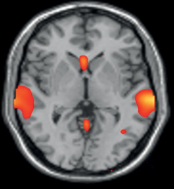
For the monaural group, speech perception was associated with activations in similar regions of the superior temporal gyrus as in normal-hearing individuals.Reference Naito, Hirano, Honjo, Okazawa, Ishizu and Takahashi6, Reference Naito, Hirano, Okazawa, Takahashi, Ishizu and Fujiki15 However, these activations were more diffuse and intense compared with those observed in normal-hearing individuals (Figure 1).Reference Limb, Molloy, Jiradejvong and Braun16 Furthermore, additional activations have been observed in the middle temporal gyri, Broca's area and its right homologue, cingulate gyrus, prefrontal cortex, visual cortices, and the cerebellum.Reference Strelnikov, Rouger, Eter, Lagleyre, Fraysse and Demonet14, Reference Naito, Hirano, Okazawa, Takahashi, Ishizu and Fujiki15, Reference Naito, Okazawa, Honjo, Hirano, Takahashi and Shiomi17–Reference Mortensen, Mirz and Gjedde23 In the bilateral implantee group, speech perception was associated with recruitment of the same regions of the temporal lobes as in their normal-hearing controls.Reference Strelnikov, Rouger, Eter, Lagleyre, Fraysse and Demonet14
Axial positron emission tomography image showing activations of a unilateral cochlear implantee during speech perception.

The activation of additional cortical regions in the unilateral implantee group suggests that these areas were recruited to compensate for their incoming degraded signal in order to process speech. The more diffuse and intense activations are likely to represent the recruitment of a larger neuronal network to perform the task, or the development of new supplementary neuronal pathways that enable speech processing using an implant. In contrast, the bilateral implantees’ cortical activations simulated those of normal-hearing individuals.Reference Strelnikov, Rouger, Eter, Lagleyre, Fraysse and Demonet14 Several studies have documented improved speech outcomes in patients with bilateral cochlear implants, which may account for this finding.Reference Tyler, Gantz, Rubinstein, Wilson, Parkinson and Wolaver24–Reference Balkany, Boggess and Dinner26 Hence, the bilateral implant group have the additional advantage of binaural hearing and may be more efficient at processing language.
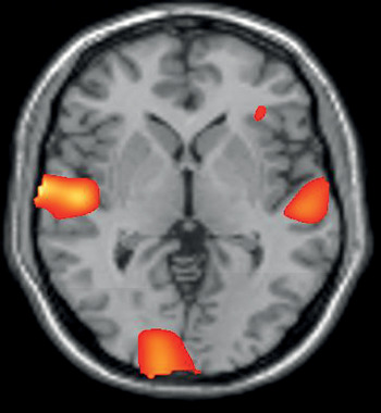
Interestingly, the recruitment of additional cortical regions in unilateral implantees has been both variable and inconsistent across the studies.Reference Strelnikov, Rouger, Eter, Lagleyre, Fraysse and Demonet14, Reference Naito, Hirano, Okazawa, Takahashi, Ishizu and Fujiki15, Reference Naito, Okazawa, Honjo, Hirano, Takahashi and Shiomi17–Reference Mortensen, Mirz and Gjedde23 The visual cortices seem to be the only extra-auditory region that is more consistently involved with language processing in unilateral implantee groups, despite the absence of any visual stimulation (Figure 2).Reference Giraud and Truy9 This region has been noted to be recruited both in new implant recipients and those who are experienced implant users.Reference Green, Ramsden, Julyan and Hastings21, Reference Giraud, Price, Graham, Truy and Frackowiak22 However, in the study by Strelnikov et al.,Reference Strelnikov, Rouger, Eter, Lagleyre, Fraysse and Demonet14 activation in the visual cortices was not apparent in patients with bilateral cochlear implants. It seems that unilateral implantees may rely on visual cues gained via lip reading. This audio-visual integration may improve speech perception performance by compensating for the degraded incoming auditory signal from the cochlear implant. It is likely that the visual cortices and the other cortical regions thought to contribute to language processing in unilateral implantees are not essential for speech processing. Instead, these regions may be supplementary and recruited by individuals as necessary.
Axial positron emission tomography image showing extra-auditory posterior activations of the visual cortices in a unilateral cochlear implantee during speech perception.

Previous work suggests that individuals with normal hearing demonstrate minimal activity in the auditory regions of the brain when the auditory stimulus consists of multi-talker babble.Reference Miyamoto and Wong4–Reference Miyamoto, Wong, Pisoni, Hutchins, Sehgal and Fain7 In contrast, the unilateral implantees exhibit strong activations in the language processing areas.Reference Wong, Miyamoto, Pisoni, Sehgal and Hutchins5, Reference Miyamoto, Wong, Pisoni, Hutchins, Sehgal and Fain7 A possible explanation for this contrast is that individuals with cochlear implants receive speech as degraded signals and therefore cannot easily differentiate between multi-talker babble and meaningful speech. Hence, those with cochlear implants process babble as speech, unlike normal-hearing individuals who are able to ignore it.
The effect of multi-talker babble in bilateral implantees has yet to be determined. It would be interesting to study this effect as research suggests that bilateral implantees have better speech perception, especially in noise, compared with unilateral implantees.Reference Tyler, Gantz, Rubinstein, Wilson, Parkinson and Wolaver24, Reference Mosnier, Sterkers, Bebear, Godey, Robier and Dequine27 Hence, one would expect less activity in the language processing regions compared with the unilateral implantee group.
As mentioned above, in right-handed, normal-hearing individuals, the left auditory region is specialised for processing language. This hemispheric lateralisation for language is suggested to exist in post-lingually deafened adults with cochlear implants too.Reference Limb, Molloy, Jiradejvong and Braun16 Fujiki et al.Reference Fujiki, Naito, Hirano, Kojima, Shiomi and Nishizawa18 demonstrated greater activations in the left hemisphere of unilaterally implanted patients exposed to speech. Enhanced activations in the left auditory hemisphere in response to speech were also documented by Naito et al;Reference Naito, Okazawa, Honjo, Hirano, Takahashi and Shiomi17 however, these observations did not reach statistical significance. Other studiesReference Okazawa, Naito, Yonekura, Sadato, Hirano and Nishizawa13, Reference Green, Ramsden, Julyan and Hastings21 did not find any hemispheric lateralisation for language. In the study by Strelnikov et al., which investigated bilateral implantees, the authors commented on the possibility of left hemispheric dominance, however, the results failed to reach statistical significance.Reference Strelnikov, Rouger, Eter, Lagleyre, Fraysse and Demonet14 The above evidence suggests that the issue of left hemispheric lateralisation in patients with cochlear implants remains unresolved.
The study by Fujiki et al.Reference Fujiki, Naito, Hirano, Kojima, Shiomi and Nishizawa18 had the largest sample size; these authors investigated hemispheric lateralisation in 14 patients with cochlear implants. The other studies involved no more than six patients. In two studies by Belin and colleagues,Reference Belin, Zilbovicius, Crozier, Thivard, Fontaine and Masure10, Reference Zatorre and Belin11 hemispheric lateralisation was investigated in normal-hearing individuals using 10 and 12 subjects respectively. To increase the power of the study analyses and arrive at more definitive conclusions, it may be necessary to conduct further studies with larger numbers of patients.
Good versus poor speech perception
Several studies have compared cortical activity in patients with unilateral implants who have good speech perception with patients who perform poorly with their implant. Positron emission tomography studies reveal a significant increase in blood flow through the auditory regions in those with good speech perception.Reference Fujiki, Naito, Hirano, Kojima, Shiomi and Nishizawa18, Reference Green, Julyan, Hastings and Ramsden28 A recent study by Green et al.Reference Green, Julyan, Hastings and Ramsden29 reported increased cortical activity in the primary auditory and auditory association areas of patients with good speech perception. However, there was only a weak enhancement of the primary auditory area in patients with poor speech perception. Interestingly, when cochlear implant patients have their speech coding strategy upgraded their speech perception improves, and a parallel increase in activation is observed in the auditory association areas.Reference Fujiki, Naito, Hirano, Kojima, Kamoto and Nishizawa30
The auditory association area seems to play a pivotal role in language processing in implantees. Literature suggests that unless appropriate neural networks are established between the auditory association area and the incoming cochlear implant signal, then the speech perception outcome is likely to be poor.Reference Fujiki, Naito, Hirano, Kojima, Shiomi and Nishizawa18, Reference Green, Julyan, Hastings and Ramsden28, Reference Green, Julyan, Hastings and Ramsden29 Further investigation is required to establish the exact mechanisms responsible for reduced activity in the auditory association area. In addition, it would be beneficial to determine if activity in the auditory association area pre-implant can be used as a predictive tool for post-implantation outcomes.
There is increasing interest in the temporal voice areas located in the superior temporal gyrus. This area is believed to be important in voice discrimination, and hence, speech perception. The temporal voice area is dominantly activated when speech is used as a stimulus in both normal-hearing individuals and cochlear implant users with good speech perception. However, these dominant activations are not present in implant users with poor speech perception.
Studies reveal that many implantees reach high levels of speech perception over time if the voice they hear is familiar. However, they fail to achieve equivalent standards when they are challenged to discriminate voices.Reference Massida, Belin, James, Rouger, Fraysse and Barone1, Reference Coez, Zilbovicius, Ferrary, Bouccara, Mosnier and Dahan31 Cochlear implants are poorly adapted for voice discrimination, music and environmental sounds.Reference Massida, Belin, James, Rouger, Fraysse and Barone1 Improving implant technology in these fields and incorporating voice discrimination training may improve future outcomes for implant users with poor speech perception.
Conclusion
Speech processing in cochlear implantees is a complex process, and the cortical networks that develop in each individual to enable speech comprehension are heterogeneous. Based on the literature, we can conclude that users of unilateral implants seem to recruit additional cortical regions to process speech, partly to compensate for the degraded signal received from the implant. Bilateral implantees seem to process speech in a similar manner to normal-hearing individuals. However, we are only aware of one published study on patients with bilateral cochlear implants. Further work is necessary in this cohort before we can draw definitive conclusions.
To appreciate neuroplasticity it is necessary to follow patients over a longer period of time and to closely monitor the cortical changes that take place from pre-implant to post-implant. Only when we develop a better understanding of neuroplasticity can we specifically address individual difficulties experienced by cochlear implant recipients. Rehabilitation programmes can offer specific management early on to improve the outcome of implantation. It is likely that functional neuroimaging will play a crucial role in the selection process for potential cochlear implant candidates, as well as helping clinicians to predict outcomes.
Current implants are known to greatly improve speech discrimination. It is essential that we excel in this complex task, and consider the need to adapt these implants in order for the patient to discriminate voices and environmental sounds, and to appreciate music.
To conclude, individuals with cochlear implants develop individual strategies to process speech, thus exhibiting varying degrees of neuroplasticity.