Introduction
In recent decades, preantral follicles have been considered as sources of numerous oocytes available for in vitro embryo manipulation and production and are applied in animal reproduction (Gupta and Nandi Reference Gupta and Nandi2012; Silva et al. Reference Silva, Van Den Hurk and Figueiredo2016). The use of preantral follicles has become a key strategy for human-assisted reproduction, especially for patients with impaired fertility, patients at risk of premature ovarian failure, patients with non-malignant diseases and even patients with genetic abnormalities or prepubertal girls suffering from blood-borne malignancies. Isolated preantral follicles can mature in vitro without the risk of reintroducing malignant cells or requiring surgical intervention, as could be the case with the culture and reintroduction of ovarian tissue fragments (Frydman and Grynberg Reference Frydman and Grynberg2016; Telfer Reference Telfer2019; Dolmans et al. Reference Dolmans, Donnez and Cacciottola2020; Stejskalová et al. Reference Stejskalová, Vankelecom, Sourouni, Ho, Götte and Almquist2021; Candelaria et al. Reference Candelaria, Botigelli, Guiltinan, Shikanov and Denicol2025).
Detailed investigations into the mechanisms of follicular development are limited by the absence of precise culture systems that support adequate follicular growth. Furthermore, understanding preantral folliculogenesis in non-rodent species is hampered by the scarcity of appropriate in vitro models and limited knowledge of ovarian development and function, especially regarding preantral folliculogenesis in large mammals (i.e. cows and humans), which exhibit prolonged duration and considerable volumetric changes between the primordial and pre-ovulatory follicle stages (Telfer and Andersen Reference Telfer and Andersen2021; Candelaria et al. Reference Candelaria, Botigelli, Guiltinan, Shikanov and Denicol2025). Folliculogenesis in vivo is a complex process involving a series of sequential steps in which preantral follicles are activated to initiate growth and undergo ovulation or atresia (99.9%). This has spurred interest in the development of an in vitro culture system that can maintain follicular growth at all stages and prevent follicle loss in vivo (Xu et al. Reference Xu, Hua, Zhang, Jiang, Zhang, Ma, Zheng, Sun, Shen, Sha, Cooke and Shi2011; Haag et al. Reference Haag, Magalhães-Padilha, Fonseca, Wischral, Gastal, King, Jones, Figueiredo and Gastal2013; Araújo et al. Reference Araújo, M., Figueiredo and Gastal2014; Dolmans et al. Reference Dolmans, Donnez and Cacciottola2020).
In vitro follicle cultures can be performed in two-dimensional (2D) and three-dimensional (3D) culture systems, where the follicle is placed on top of the substrate in 2D and immersed in the substrate in 3D (Rossetto et al. Reference Rossetto, Lima, Saraiva, Lima-Verde and Figueiredo2011; Figueiredo et al. Reference Figueiredo, Lima, Silva and Santos2018). Two-dimensional systems have been used for several years as traditional methods of follicular culture (Gore-Langton and Daniel Reference Gore-Langton and Daniel1990; Cortvrindt et al. Reference Cortvrindt, Smitz and Van Steirteghem1996; West et al. Reference West, Shea and Woodruff2007; Bizarro-Silva et al. Reference Bizarro-Silva, Santos, Gerez, González, Lisboa and Seneda2018) and have demonstrated promise in supporting bovine preantral follicle growth (Araújo et al. Reference Araújo, Gastal, Wischral, Figueiredo and Gastal2015; Rossetto et al. Reference Rossetto, Saraiva, Bernuci, Silva, Brito, Alves, Magalhães-Padilha, Báo, Campello, Rodrigues and Figueiredo2016), whereas 3D culture systems are relatively new. Three-dimensional culture systems mimic the ovarian tissue stroma and thus resemble the conditions under which follicles develop in vivo. The use of bioengineered 3D hydrogel culture systems permits timely follicle degradation while retaining the mechanical support necessary to maintain its spherical structure (Ihm et al. Reference Ihm, Lee, Han, Lim and Hubbell2015; Mendez et al. Reference Mendez, Zhou and Shikanov2018; Correia et al. Reference Correia, Lima, Sousa, Ferreira, Cadenas, Paes, Alves, Shikanov and Figueiredo2020). Although previous studies have explored hydrogel-based matrices, including alginate and polyethylene glycol (PEG) systems, for isolated follicle culture (Correia et al. Reference Correia, Lima, Sousa, Ferreira, Cadenas, Paes, Alves, Shikanov and Figueiredo2020; Candelaria et al. Reference Candelaria, Botigelli, Guiltinan, Shikanov and Denicol2025), comparatively few investigations have addressed the use of agarose as a support matrix for in situ culture of ovarian cortex fragments containing multiple preantral follicles. All initial attempts to grow in situ tissue fragments resulted in no cell maintenance owing to the inefficiency of culture methods and media (Sadr et al. Reference Sadr, Ebrahimi, Shahhoseini, Fatehi and Favaedi2015; Green and Shikanov Reference Green and Shikanov2016). Thus, we developed a 2D culture method with agar gel support that would provide an interface between gas-media cells and deliver the same conditions as a 3D system (Sato et al. Reference Sato, Katagiri, Gohbara, Inoue, Ogonuki, Ogura, Kubota and Ogawa2011), allowing timely degradation by the follicle while retaining the mechanical support necessary to maintain its spherical structure (Mendez et al. Reference Mendez, Zhou and Shikanov2018). Unlike alginate-based encapsulation systems, which are primarily designed for isolated follicle culture and require enzymatic or chemical dissolution for follicle retrieval, the agarose gel support system employed herein provides a non-degradable, inert scaffold that maintains the native tissue architecture of the ovarian cortex while permitting nutrient and gas exchange at the tissue–medium interface (Dillon et al. Reference Dillon, Yu, Sridharan, Ranieri and Bellamkonda1998; Diekjürgen and Grainger Reference Diekjürgen and Grainger2017).
In this context, although several animal models have been established and accepted as alternatives to research on human reproduction, restricted access to human gametes and the ethical implications of using human cells have delayed progress in this field (Kim et al. Reference Kim, Tingen and Woodruff2010; Telfer Reference Telfer2019). The use of bovine specimens should be considered because of these limitations. Several physiological similarities exist between cattle and humans that favour cattle as models over other species. For example, humans and cattle share the duration of folliculogenesis and single ovulation, as well as oocyte lipid content and embryonic metabolism (Leese Reference Leese2012). Working with in vitro cow models is simplified by easy access to ovaries from slaughterhouses and well-established protocols for reproductive biotechnology. The use of isolated oocytes from post-mortem dissected ovaries of cows slaughtered in abattoirs complies with the 3Rs principle (reduction, replacement and reuse) (Langbeen et al. Reference Langbeen, De Porte, Bartholomeus, Leroy and Bols2015; Asimaki et al. Reference Asimaki, Vazakidou, van Tol, Oei, Modder, van Duursen and Gadella2022).
In this study, we compared 2D and 3D in vitro culture systems to evaluate their relative efficacy in supporting the development of preantral follicles in ovarian tissue. We evaluated follicular integrity, follicular growth and development and oxidative stress responses.
Materials and methods
All chemicals used were purchased from Sigma-Aldrich (St. Louis, MO, USA) unless otherwise noted.
Collection and transport of ovaries
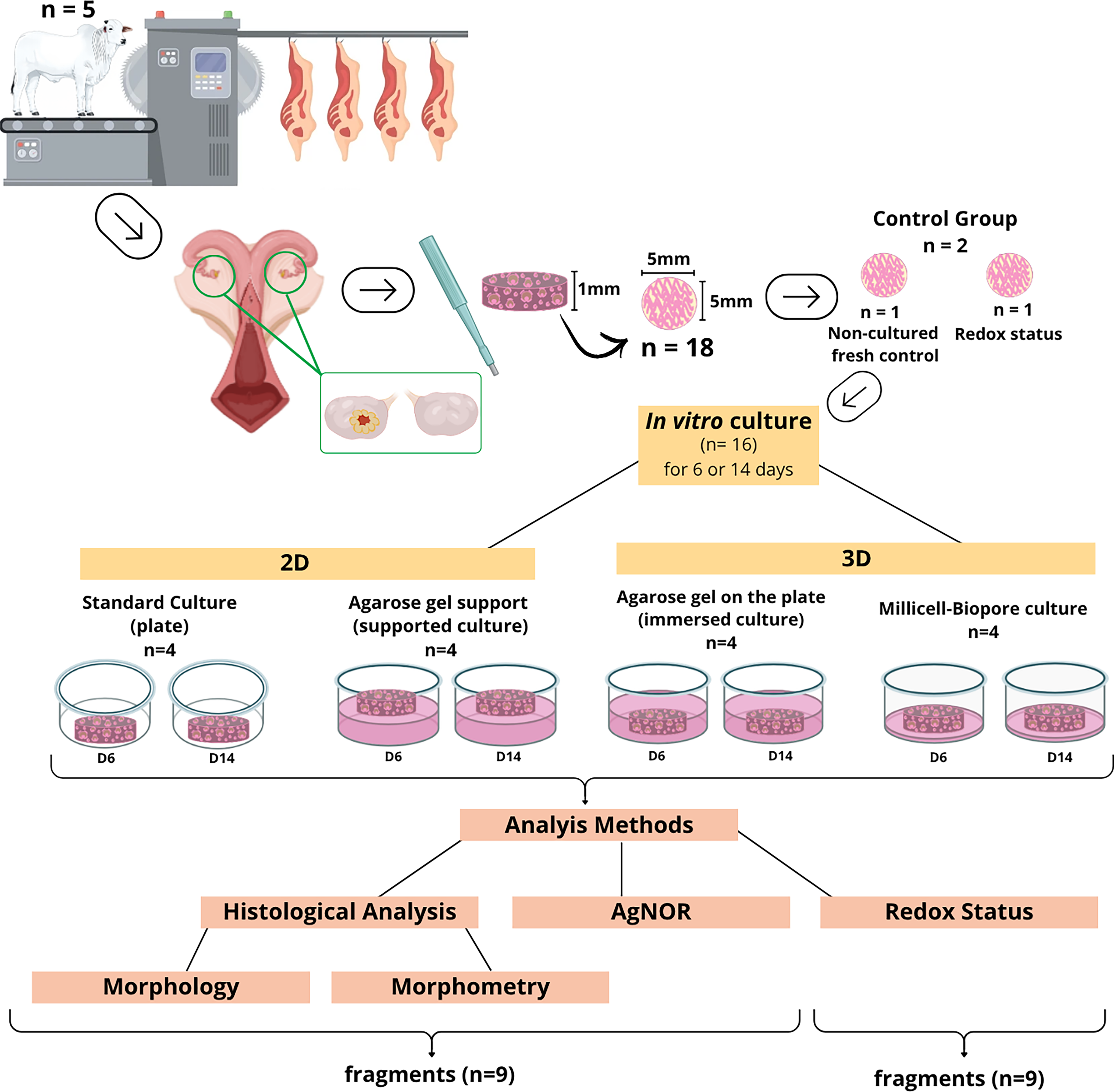
Ovaries (n = 10) were collected from a slaughterhouse of Bos taurus indicus heifers (Nellore Breed), aged approximately 24–26 months (±1.2 months) that exhibited ovarian activity (judging by the presence of the corpus luteum and small antral follicles) and had good body condition (body score of 3–4 on a scale of 0–5) (Figure 1). After isolation, the ovaries were washed in 70% ethanol for 10 s and phosphate-buffered saline (PBS; Embriolife®, Vitrocell, Brazil). Next, they were shredded and transported at a controlled temperature to the laboratory (4°C for 45 min). Bovine ovaries were transported in the minimal essential medium (MEM; Gibco BRL, Rockville, MD, USA; osmolarity 300 mOsm/L, pH 7.2) supplemented with 100 IU/mL penicillin and 100 mg/mL streptomycin.
Experimental design. In vitro culture of control bovine preantral follicles (MEM) with different culture methods (standard culture, agarose gel-supported culture, medium-immersed agarose gel culture and Millicell–Biopore culture).

In vitro culture of preantral ovarian follicles
Both ovaries from each animal (n = 5) were carefully processed. Initially, the surrounding tissue and ligaments were removed, and subsequently the ovarian cortex was divided into fragments (n = 18), each approximately 5 × 5 × 1 mm, using a disposable sterile dermatological punch (5 mm ⌀; Kolplast, São Paulo, Brazil). From each animal, a fragment was randomly selected and immediately fixed in Bouin’s solution (control non-cultured or fresh control; D0). The remaining fragments from the ovarian cortex were cultured individually for 6 (D6) or 14 (D14) days in 1 mL aliquots of MEM (Figure 1). Fragments were in 24-well culture plates in an incubator at 38.5°C with an atmosphere of 5% CO2 in air and saturated humidity. The control culture consisted of MEM supplemented with ITS (6.25 mg/mL of insulin + 6.25 mg/mL of transferrin + 6.25 mg/mL of selenium), 0.23 mM of pyruvate, 2 mM of glutamine, 2 mM of hypoxanthine, 1.25 mg/mL of bovine serum albumin (BSA; Gibco BRL, Rockville, MD, USA), 20 UI/mL of penicillin and 200 mg/mL of streptomycin. For the experimental conditions, the fragments were divided into four groups: i) standard culture, directly on a culture plate (2D), ii) supported culture on a gel monolayer (2D), iii) immersed culture on an agarose gel (plating coated) covered in medium (3D) and iv) Millicell–Biopore culture (Merck, Darmstadt, Germany; 3D) (Figure 1). For in vitro culture in methods ii and iii, the position of the ovarian cortex faced the agarose matrix. Every 2 days, the culture medium was replaced with fresh medium at a controlled temperature (38.5°C) and under light-controlled conditions to avoid direct exposure of the fragments. For this purpose, light sources were reduced in all treatments. The culture period used was based on published studies on the time course of primordial follicle activation in other species (Braw-Tal and Yossefi Reference Braw-Tal and Yossefi1997; Fortune et al. Reference Fortune, Kito, Wandji and Srsen1998; Figueiredo et al. Reference Figueiredo, Lima, Silva and Santos2018).
Agarose gel preparation
Gel formation solution was prepared by dissolving agarose (Agarose Molecular Biology Grade; Kasvi, Brazil) in sterile deionised water (MilliQ) to obtain a 1.5% agarose solution (p/v). At this concentration, the agarose gel exhibits a gel strength of approximately 1200 g/cm2 and a pore size ranging from 100 to 300 nm, which permits adequate diffusion of nutrients and gases while providing structural support (Dillon et al. Reference Dillon, Yu, Sridharan, Ranieri and Bellamkonda1998; Diekjürgen and Grainger Reference Diekjürgen and Grainger2017). After obtaining the gel, 1 cm3 of support was prepared (adapted from Sato et al. Reference Sato, Katagiri, Gohbara, Inoue, Ogonuki, Ogura, Kubota and Ogawa2011), and 1 mL (per well) was added to coat the plate wells, creating a uniform structure for the supported and immersed treatments (Silva et al. Reference Silva, Bergamo, Búfalo, Bonato and Seneda2017). The gel was completely replaced on the eighth day (D8) of culture.
Histological processing
For histological analysis of ovarian morphology and ovarian follicles, control fragments (non-cultured) and fragments cultured for 6 or 14 days were fixed by immersion in Bouin’s solution for 24 h and subsequently stored in 70% alcohol (Figure 1). After fixation, the tissues were dehydrated by incubation in an increasing concentration gradient of ethanol solution, clarified, xylol-diaphanised, paraffin-embedded and sectioned into blocks for histological analysis. Subsequently, each piece was sectioned at 5 µm with 5 µm intervals between the rotating tissue sections (Leica®, Wetzlar, Germany), and sections were mounted on slides for microscopy. The slides were stained with periodic acid–Schiff (PAS) and haematoxylin. For PAS and haematoxylin staining, briefly, the tissue slides were initially de-paraffinised with xylene and then rehydrated using 70% ethanol before staining with periodic acid (Sigma; St. Louis, MO, USA) for 5 min and Schiff’s reagent for 10 min. Finally, the slides were counterstained with haematoxylin (Andrade et al. Reference Andrade, Seneda, Alfieri, Bracarense and Figueiredo2012).
Subsequently, 150 (30 per animal) follicles per treatment were monitored throughout the culture period. To prevent recounting, preantral follicles were counted only in sections where the oocyte nucleus was visible (Candelaria and Denicol Reference Candelaria and Denicol2020).
Follicular integrity and development analysis
All sections were examined under a light microscope (10× and 40×; Nikon, Tokyo, Japan). Preantral follicles were classified according to the stage of follicular development as (1) primordial (containing a layer of somatic cells known as flat or flat granulosa cells around the oocyte) and (2) primary (a single layer of cuboid granulosa cells around the oocyte) or (3) secondary (two or more cubic granulosa cell layers). Follicles were quantified at different stages of development (primordial, primary and secondary) in the control group and after in vitro culture under different conditions to evaluate follicle activation and growth (Andrade et al. Reference Andrade, Seneda, Alfieri, Bracarense and Figueiredo2012).
Classification according to integrity was performed based on the following morphological variables: (I) integrity of oocytes and granulosa cells, (II) presence or absence of pycnotic bodies, (III) observable ooplasmic retraction, (IV) granulosa cell organisation and (V) presence of an unrippled basement membrane firmly adhered to the granulosa cells (Bizarro-Silva et al. Reference Bizarro-Silva, Bergamo, Costa, González, Yokomizo, Rossaneis, Verri Junior, Sudano, Andrade, Alfieri and Seneda2024). Based on these criteria, follicles were considered morphologically intact when the oocyte was intact, non-pycnotic and surrounded by granulosa cells organised in discrete layers and degenerated when the oocyte was shrunken with a pycnotic nucleus and/or surrounded by disorganised granulosa cells away from the basement membrane.
Only intact follicles were considered when evaluating follicular activation and growth rates. The percentages of primordial and developing follicles (primary and secondary) were calculated after 0 (control treatment), 6 or 14 days of culture in different groups. As mentioned above, the preantral follicles were counted only in the section where the oocyte nucleus was observed.
Morphometry of in vitro cultured preantral follicles
Five morphologically intact preantral follicles were observed per treatment to analyse the cultured follicle and oocyte diameters, and images were then captured. In total, 270 preantral follicles were evaluated in non-cultured treatments (n = 30), 6-day cultures (n = 120) and 14-day cultures (n = 120). The slides were examined under a Leica® microscope (DMSL, Leica Microsystems, Germany), and images were captured by a MOTICAM 2500 digital camera (5.0 MPixel) and were analysed using Motic Images Plus 2.0 ML. Follicle and oocyte measurements were performed as described by Silva-Buttkus et al. (Reference Silva-Buttkus, Jayasooriya, Mora, Mobberley, Ryder, Baithun, Stark, Franks and Hardy2008), and oocyte and follicle diameters were calculated from the arithmetic means of two perpendicular measurements.
Evaluation of granulosa cell proliferation
Argyrophilic nucleolar organiser regions (AgNOR) staining was performed to estimate the granulosa cell proliferation index of preantral follicles present in non-cultured controls and follicles cultured in vitro for 6–14 days. AgNOR staining enables quantification of the number of argyrophilic nucleolar organising regions (NOR), which show affinity for silver (Ag), and were calculated per granulosa cell nucleus. Briefly, AgNOR-stained slides were prepared using the same procedure as that used for histological processing. Subsequently, samples were de-paraffinised, dehydrated, incubated for 15 min in Triton X 0.5%, and then incubated for 10 min in AgNOR solution at 37°C. The solutions used were (A) AgNOR 0.5 or 1 g/mL in deionised water and (B) 1 or 2% gelatin in deionised water containing 1% formic acid. The staining solution consisted of a mixture of A (2 volumes) and B (1 volume). In each case, the reactions were stopped by rinsing several times with deionised water, as described by Castro et al. (Reference Castro, Carvalho, Silva, Santos, Campello, Figueiredo and Rodrigues2014).
At least five follicles were evaluated in each treatment group for quantification. The features of all visualised follicular granulosa cells were considered. For positive staining (brown staining), we considered follicles in which granulosa cells had at least one NOR signal. The slides were visualised under a light microscope (40× and 1000×), and the NOR of all visible granulosa cell nuclei in the different treatment groups was counted.
Redox status
Redox status was determined in non-cultured ovarian tissue samples and in samples cultured in vitro for 6 or 14 days by colourimetric assessment of lipid peroxidation, superoxide anion production and antioxidant capacity as detailed below. The bovine ovarian tissue samples cultured in the different methods were collected and kept at −80°C for at least 48 h before processing for the aforementioned assays.
Antioxidant capacity
The ability of the sample to resist oxidative damage was determined by ferric reducing antioxidant power (FRAP), its ability to quench the 2,2-azinobis-3-ethylbenzothiazoline 6-sulfonic acid (ABTS) radical cation and by measuring reduced glutathione (GSH) levels, as described and adapted according to Pinho-Ribeiro et al. (Reference Pinho-Ribeiro, Zarpelon, Mizokami, Borghi, Bordignon, Silva, Cunha, Alves-Filho, Cunha, Casagrande and Verri2016).
FRAP and ABTS assay
The FRAP and ABTS assays were performed as previously described by Katalinic et al. (Reference Katalinic, Modun, Music and Boban2005) and adapted to a 96-well microplate format (Pinho-Ribeiro et al. Reference Pinho-Ribeiro, Zarpelon, Mizokami, Borghi, Bordignon, Silva, Cunha, Alves-Filho, Cunha, Casagrande and Verri2016). Ovarian tissue samples were homogenised in 1.15% (500 μL) ice-cold KCl buffer, centrifuged (200 g for 10 min at 4°C), and the supernatant collected for assays. For the FRAP assay, the supernatants (20 μL) were mixed with the freshly prepared FRAP reagent (150 μL), and the reaction mixture was incubated at 37°C for 30 min. The absorbance was measured at 595 nm (Multiskan GO; Thermo Scientific). For the ABTS assay, 10 µL of the supernatant was mixed with diluted ABTS solution (200 μL) as described in Hohmann et al. Absorbance was measured at 730 nm after 6 min of incubation (25°C). The results were normalised against a standard Trolox curve (0.02–20 nmol) and expressed as Trolox equivalents per mg of protein.
Reduced glutathione quantification
The samples were homogenised in 200 μL of 0.02 M EDTA. Each homogenate was mixed with 25 μL of 50% trichloroacetic acid and was homogenised three times for 15 min. The mixture was centrifuged (400 g for 15 min at 4°C). The supernatant was added to 200 μL of 0.2 M TRIS buffer, pH 8.2 and to 10 μL of 0.01 M DTNB. After 5 min, absorbance was measured at 412 nm, followed by balancing with a blank reagent containing no supernatant. A standard curve was generated using GSH. The results are expressed as nanomoles of GSH per mg of protein.
Lipid peroxidation
Lipid peroxidation was assessed by measuring thiobarbituric acid reactive substance (TBARS). Ovarian tissue samples were homogenised in 1.15% (500 μL) of KCl buffer, and the homogenate was used for this assay. The homogenates (50 µL) were transferred into microtubes, and 1 M FeCl3 (5 µL), ascorbic acid (5 µL), 2.8% TCA (50 µL), and 1% TBA (50 µL) were added, and the mixture was incubated at 100°C for 15 min. The supernatants were transferred into 96-well microplates, and malondialdehyde (MDA), an intermediate product of lipid peroxidation, was determined by the difference in its absorbance at 535 and 572 nm (Multiskan GO). The results are reported as optical density (OD) at 535–572 nm per milligram of protein.
Superoxide anion production
The production of superoxide anions was determined by reduction of the redox dye nitroblue tetrazolium (NBT). Tissue homogenates in 1.15% KCl buffer (500 µL) were transferred to a 96-well plate, followed by the addition of 100 µL of NBT solution (1 mg/mL) and incubated for 1 h at 37°C. The supernatant was carefully removed, and the precipitated formazan was solubilised in 120 µL of KOH and 120 µL of DMSO. OD was measured at 600 nm (Multiskan GO), and results are presented as OD per mg of protein.
Statistical analysis
All analyses were performed using the Action 3.1 version of R 3.0.2 (Campinas, SP, Brazil). Data were initially subjected to residual normality (Shapiro–Wilk) and variance homogeneity (Bartlett) tests. The average number of morphologically intact and degenerate preantral follicles, primordial follicles and developing follicles obtained in the control and the different in vitro culture methods for 6 or 14 days (thereafter termed D6 and D14) was analysed using analysis of variance (ANOVA) and Tukey’s test (P ≤ 0.05). For comparisons within the same culture method for 6 or 14 days, data were analysed using Student’s t-test. Follicle morphometry (mean follicle diameter and oocytes) was subjected to Kruskal–Wallis analysis, followed by Dunn’s post hoc test when significant differences were detected. For the oxidative stress tests, data were subjected to ANOVA, followed by Tukey’s test for each method and culture time. All data are presented as mean ± standard error of the mean (SEM). Values were considered statistically significant at P < 0.05. The experimental unit for all analyses was the individual animal (n = 5). For each animal, 30 follicles per treatment were evaluated, yielding 150 follicles per treatment across the five biological replicates. Statistical analyses were performed on the mean values obtained per animal within each treatment, thereby avoiding pseudo-replication. The hierarchical structure of the data (follicles nested within animals) was thus accounted for by using the animal-level mean as the unit of analysis.
Results
Analysis of follicular morphology and growth
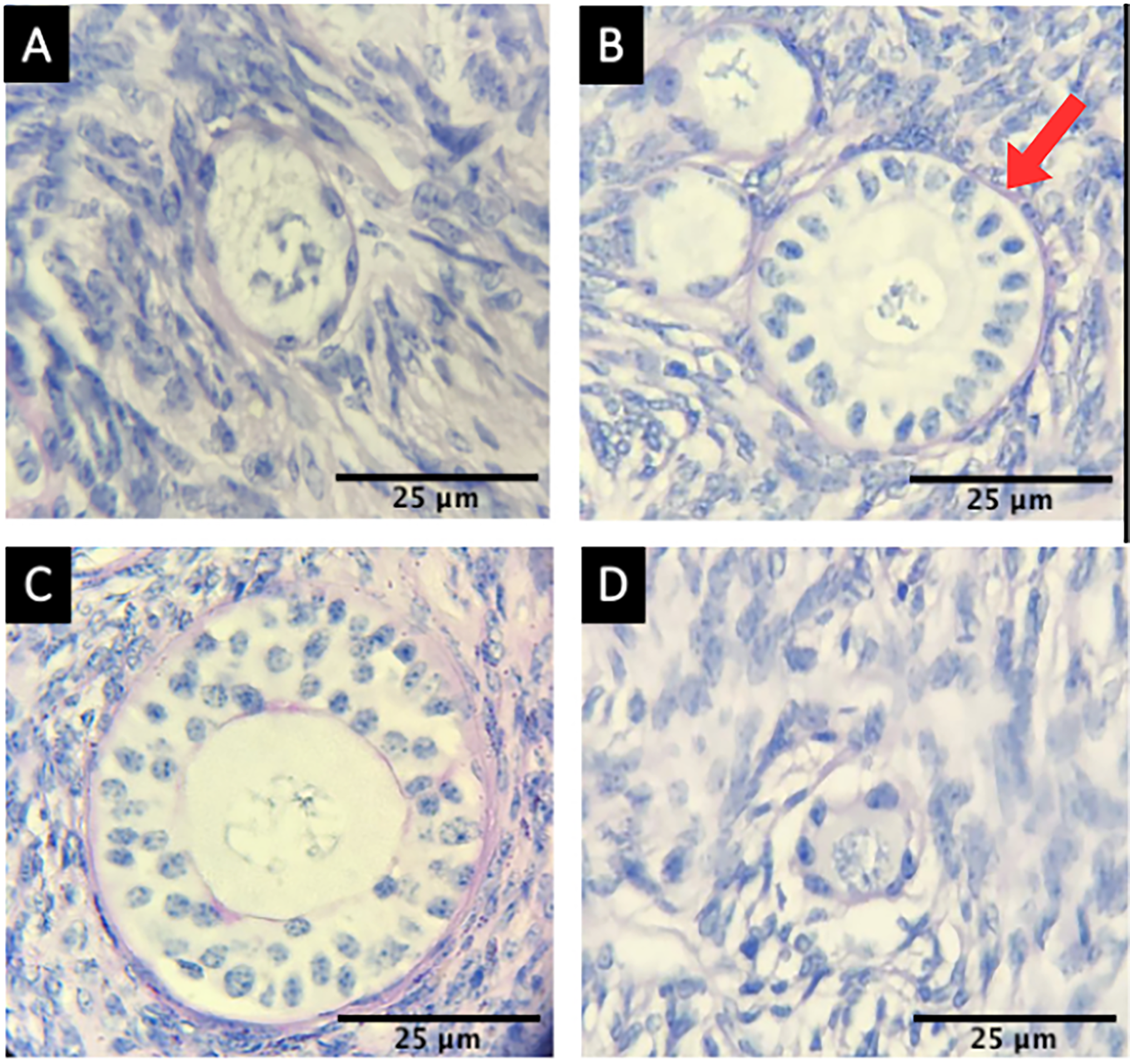
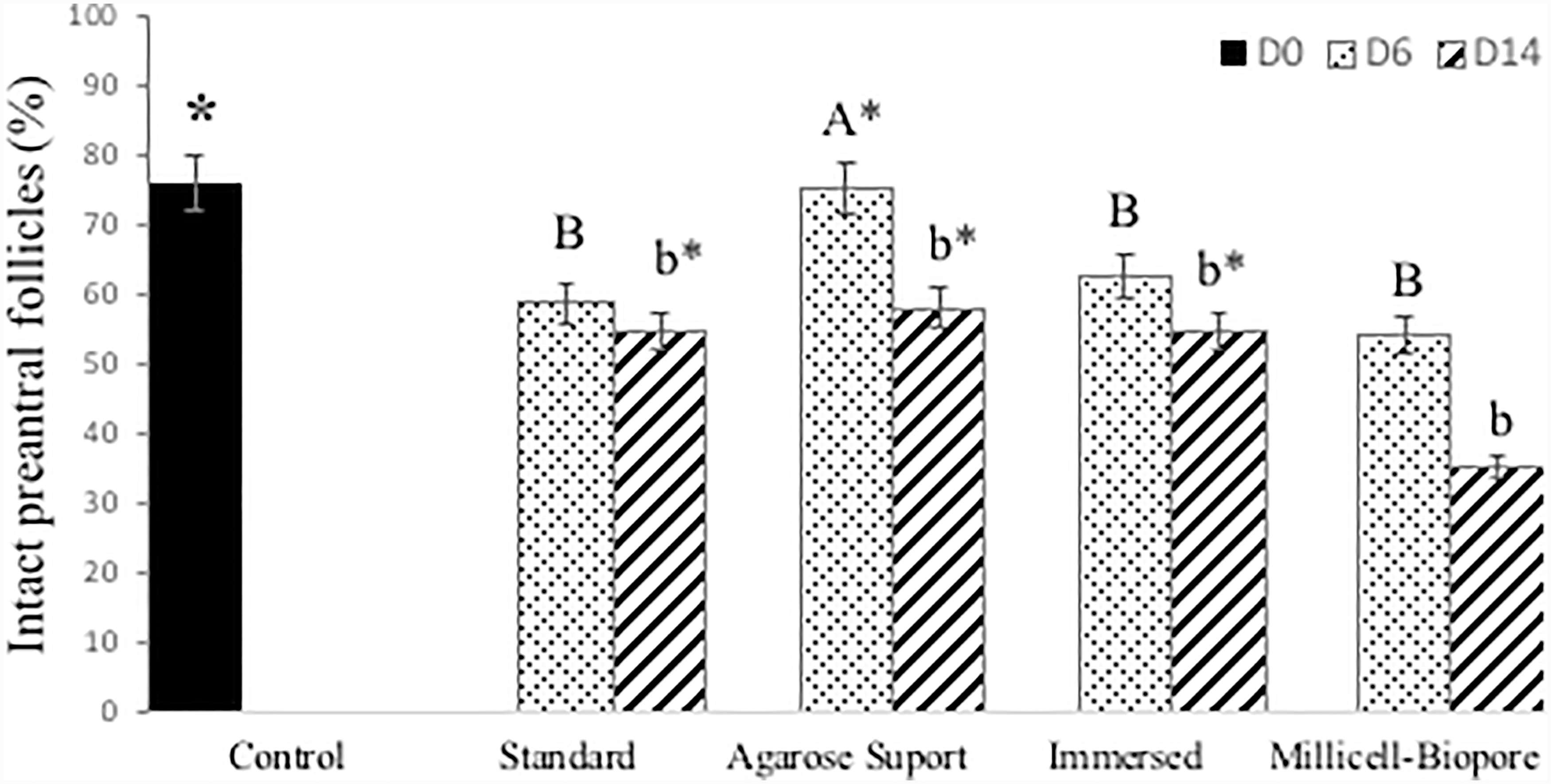
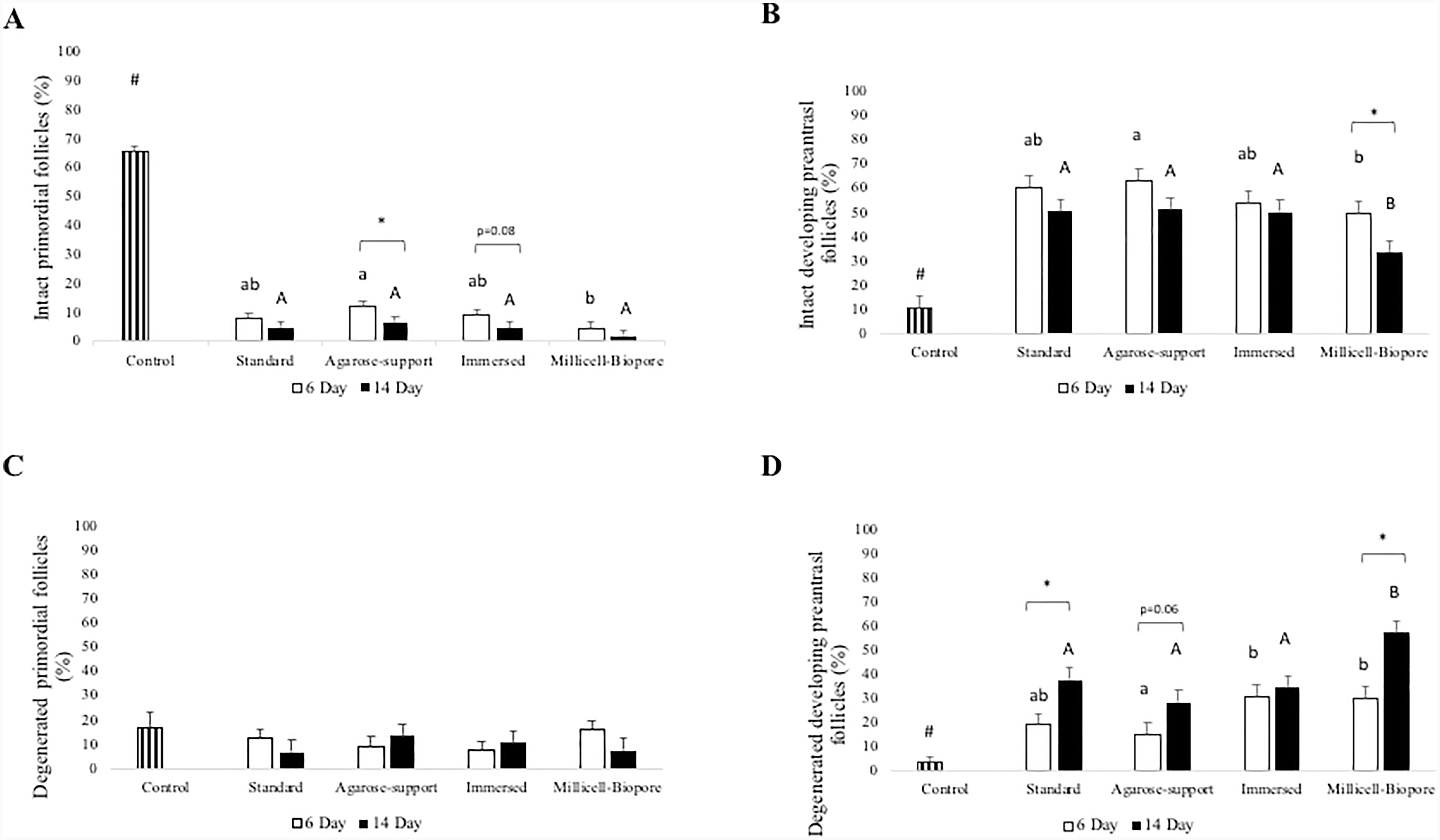
In total, 1330 preantral follicles were evaluated, of which 326 (24.5%) were primordial and 1004 (75.5%) were developing (Figure 2). The morphological characteristics were evaluated for integrity (intact: Figure 2A–2C; degenerated: 2D) and development (primordial: 2A and D; development: 2B and C). In general, after the beginning of the culture, the proportion of primordial follicles decreased among the different treatments, leading to an increase in the percentage of developing follicles (P < 0.05). Figure 3 summarises the percentage of follicles with intact morphology on day 0 before culture (non-cultured or fresh control) and on days 6 and 14 of culture. The percentages of intact and degenerate follicles according to the stage of development are shown in Figure 4 (intact: 4A and B; degenerate: 4C and D).
Histological photomicrograph of the morphology of bovine preantral follicles (Bos taurus indicus). Whole primordial follicle (A), whole primary follicle (arrow) (B), whole secondary follicle (C) and degenerated primordial follicle (D). Periodic acid Schiff (PAS) and haematoxylin stains, 40× and 100×.

Percentage of intact preantral (primordial, primary + secondary) follicles in non-cultured ovarian tissue (control, day 0) and after in vitro culture using different culture methods (standard culture, agarose gel-supported culture, culture agarose gel immersed in medium and cultivated Millicell–Biopore) for 6 or 14 days. *Compared with the control group. A, B Indicate differences among treatments within 6 days. a, b Indicate differences among treatments within 14 days (P < 0.05).

Percentages of intact and degenerate follicles according to the stage of development. Intact primordial (A), intact developing (B), degenerated primordial (C) and degenerated developing (D) preantral follicles in non-cultured ovarian tissue (Control, day 0) and after in vitro culture with different follicular culture methods (standard culture, culture on agarose gel support, medium-immersed agarose gel culture and Millicell–Biopore culture) for 6 or 14 days. *Indicates differences between 6 days and 14 days. # Indicates differences between control and other treatments (except in comparison with agarose support at B and D). a, b Indicate differences among treatments within 6 days. A, B Indicate differences among treatments within 14 days.

Figure 4 shows the follicles analysed according to the effect of different culture methods, day of culture and a comparison of the culture method with the day. A significant transition (follicular activation) from primordial follicles (Figure 4A) to developing follicles was observed (Figure 4B) compared with the control and culture days (D6 and D14). In addition, the methods of culture with agarose support and agarose gel immersion in the medium demonstrated reduced number of primordial follicles on D6 and D14 (P < 0.05 and P = 0.08, respectively; Figure 4A). Furthermore, for the intact culture method of developing follicles, Millicell–Biopore showed a significantly reduced percentage of follicles at D6 and D14 (P < 0.05, Figure 4B).
Although normal (intact) and atretic (degenerate) follicles were observed in all treatments, after 6 days of culture, the fragments grown on the agarose gel support displayed a higher proportion of intact follicles (75.3%; 113/150) compared to the other groups: 58.7% (88/150) for the standard culture, 62.6% (94/150) for culture with agarose gel immersed in the medium and 54% (81/150) for Millicell–Biopore culture (P = 0.0034, Figure 3). After 14 days of culture, the culture methods showed no significant differences in follicle integrity rates (P = 0.6195, Figure 3).
No differences were observed in the percentages of developing follicles on D6 between the culture methods tested (P = 0.6871, Figure 4). After 14 days of growth, standard cultures, cultures on agarose gel support and cultures on agarose gel immersed in the medium exhibited a higher percentage of morphologically intact developing follicles: 54.7% (82/150), 58% (87/150) and 54.7% (82/150), respectively, when compared to the Millicell culture (35.3%; 53/150). The treatments did not differ in other samples for the rate of development in D14 (P = 0.5879, Figure 4).
Morphometry of in vitro cultured preantral follicles
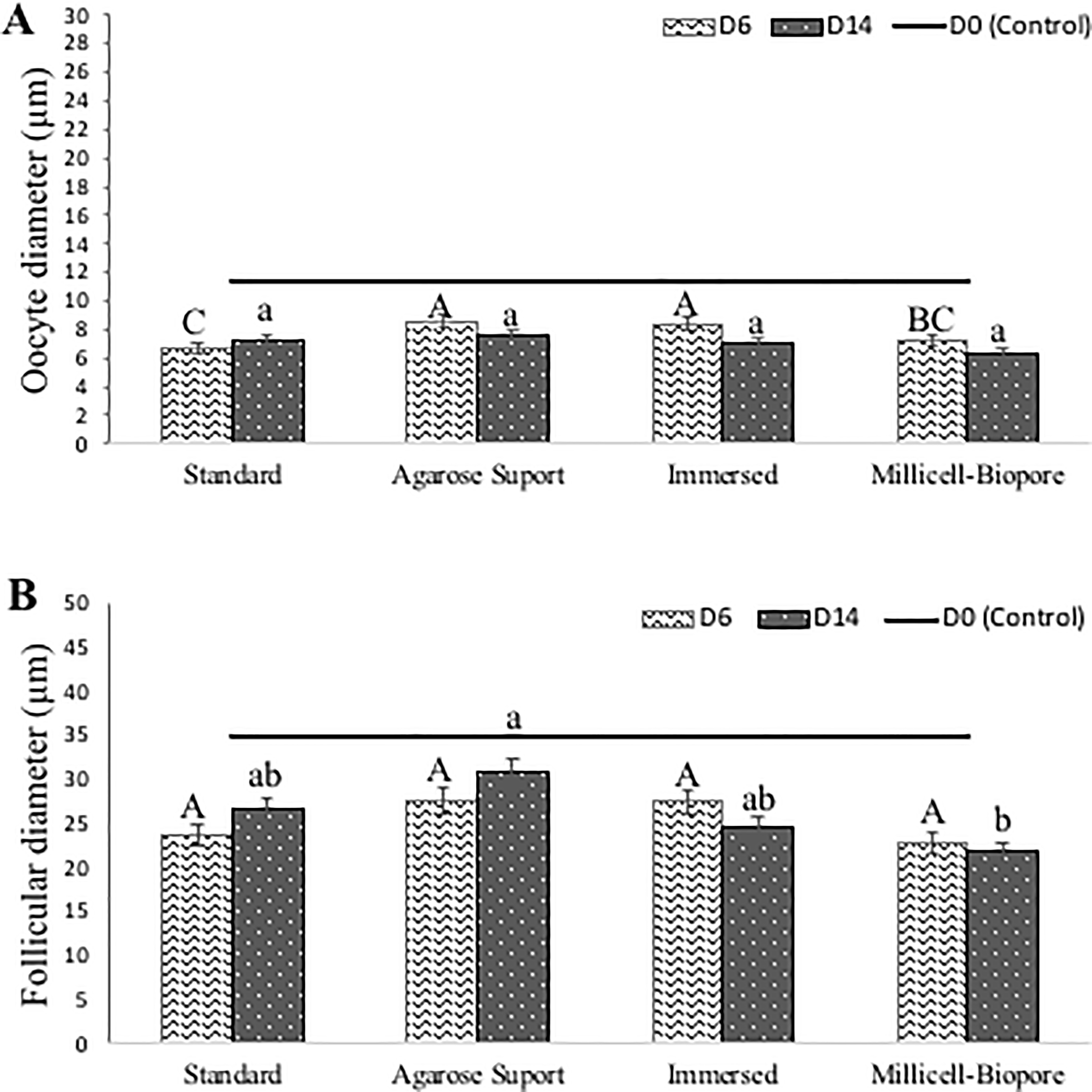
For morphometric analysis of preantral follicles, oocyte and follicle dimensions were measured in tissues cultured using different in vitro culture methods. Morphometric data were analysed for primordial and developing (primary + secondary) follicles separately. The average diameters of oocytes and follicles observed on days 6 and 14 of culture are shown in Figure 5A and 5B, respectively.
Average oocyte (A) and follicle (B) diameter in micrometers (μm) of bovine preantral follicles grown for 6 or 14 days under different in vitro culture methods (standard culture, agarose gel support culture, culture with agarose gel immersed in the medium and Millicell–Biopore culture). A, B Indicate differences among treatments within 6 days. a, b Indicate differences among treatments within 14 days (P < 0.05).

No difference was noted in the oocyte diameter in the follicles at any stage of development (P > 0.05). In addition, oocyte diameter in the follicles showed no difference between any of the development methods and controls (P > 0.05). On D6, the methods on the gel support culture (8.6 ± 1.6 μm) and immersed culture (8.4 ± 1.1 μm) showed significant differences compared to the other culture systems (standard culture: 6.7 ± 1.1 μm and Millicell culture: 7.2 ± 1.1 μm; P < 0.05). Regarding the oocyte diameter in follicles cultured for 14 days, no significant difference was observed between the culture methods (P > 0.05).
On day 0 (non-cultured control), preantral follicle diameter was 34.9 ± 5.5 μm. After 6 days of in vitro culture, the mean diameter of preantral follicles (primordial, primary and secondary) showed similar results between the four groups (standard culture: 23.7 ± 4.0 μm, gel support culture: 27.6 ± 5.9 μm, immersed culture: 27.5 ± 4.9 μm and Millicell culture: 22.9 ± 4.4 μm; P > 0.05). However, the follicles grown on the agarose support had a larger mean diameter after 14 days than those grown on the Millicell (gel support culture: 30.8 ± 10.4 μm, standard culture: 26.6 ± 7.4 μm, immersed culture: 24.7 ± 5.1 μm and Millicell culture: 21.8 ± 2.7 μm; P < 0.05).
Evaluation of granulosa cell proliferation
Based on NOR labelling, a qualitative analysis of granulosa cells from control preantral follicles or those cultured using different methods (standard culture, agarose gel support culture, immersed agarose gel culture and Millicell-Biopore culture; Table 1) was performed for 6 or 14 days.
Qualitative analysis of granulosa cells of preantral follicles marked (stained-brown) with AgNOR (nucleophilic organising regions related to argyrophilic proteins) present in the control cells or in those cultured using different methods (standard culture, culture on agarose gel support, medium-immersed agarose gel culture and Millicell-Biopore culture) – % (n/t)

Table 1. Long description
A table comparing granulosa cell culture methods and their effects on preantral follicles. The table has 3 rows and 5 columns. The columns are labeled Control D0, Cultivation period, Standard % (n/t), Support % (n/t), Immersed % (n/t), and Millicell-Biopore % (n/t). The rows are labeled with different cultivation periods and methods. Row 1: Control D0, 35.6 (46/129). Row 2: D6, Standard 3.52 (3/85), Support 66.0 (66/100), Immersed 26.4 (37/140), Millicell-Biopore 35.0 (7/20). Row 3: D14, Standard 10.7 (3/28), Support 52.3 (11/21), Immersed 0, Millicell-Biopore 20.0 (6/30). The table shows the percentage and number of granulosa cells marked with AgNOR in different culture methods over 6 and 14 days.
* Denotes a statistically significant difference compared to the other culture methods within the same evaluation period (P < 0.05).
AgNOR = Argyrophilic Nucleolar Organizer Regions.
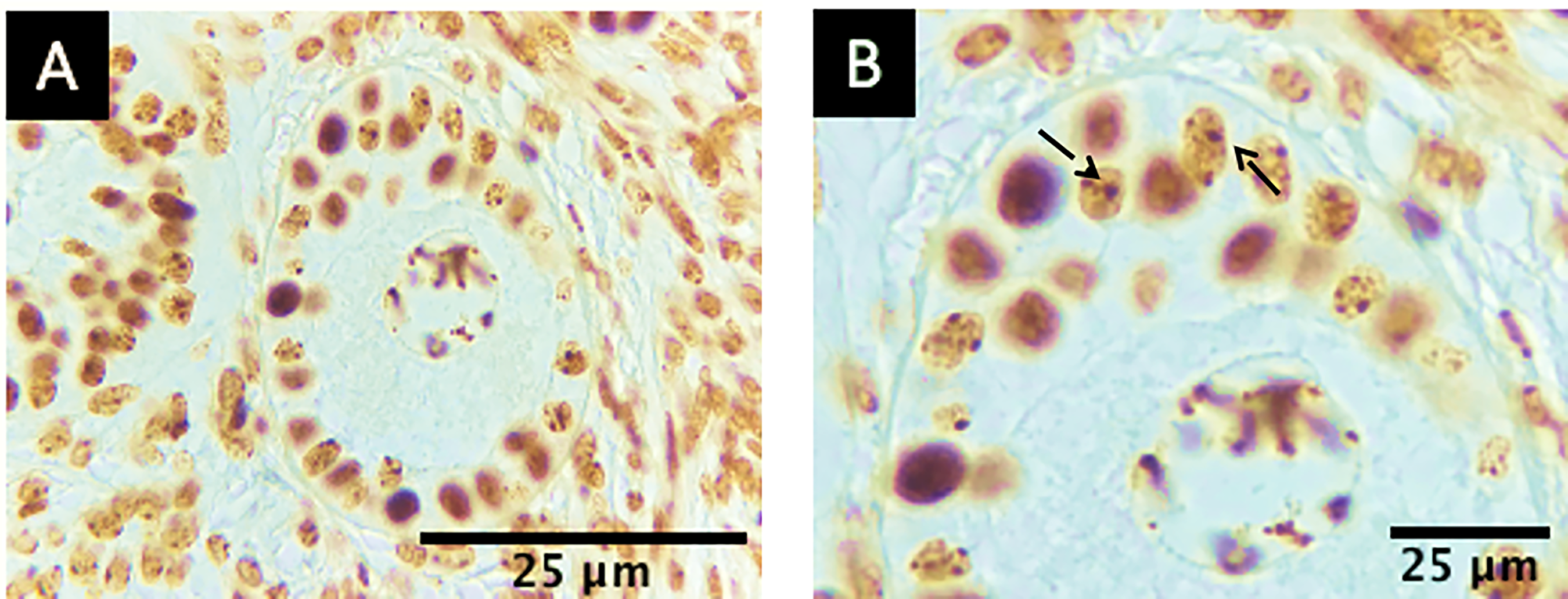
A total of 72 preantral follicles were analysed by AgNOR staining. Cell proliferation was evaluated by quantifying NOR (Figure 6A and 6B), which revealed more NOR-stained granulosa cells than total follicle granulosa cells on the agarose gel support at days 6 and 14. In contrast, the agarose gel culture method immersed in the medium did not show any marked granulosa cells after 14 days of culture.
Photomicrograph of cell proliferation for NOR quantification of bovine preantral follicles (Bos taurus indicus). (A) NOR-labelled primordial follicle and (B) positive granulosa cells (black arrow) AgNOR. 40× and 100×.

Redox status
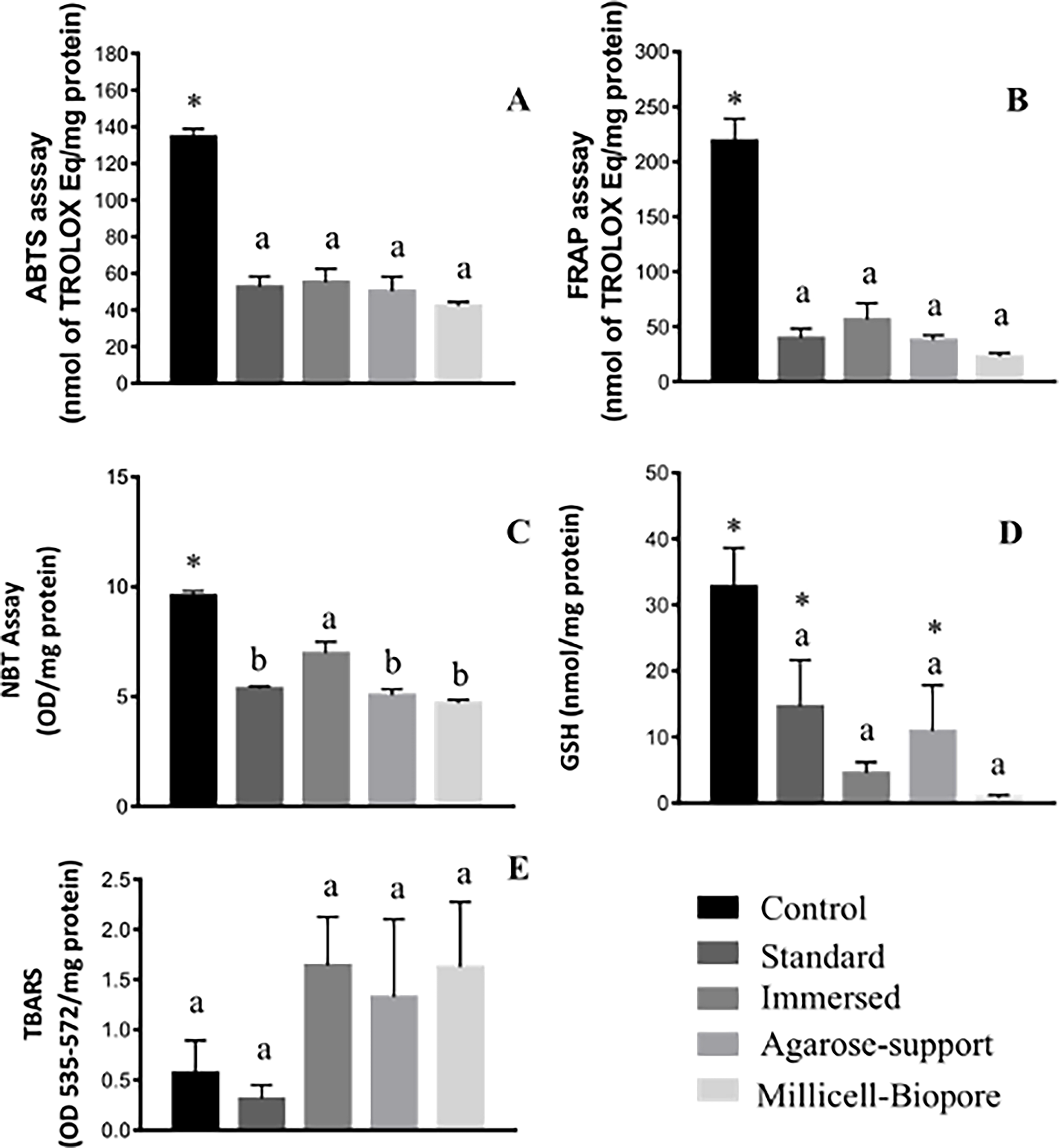
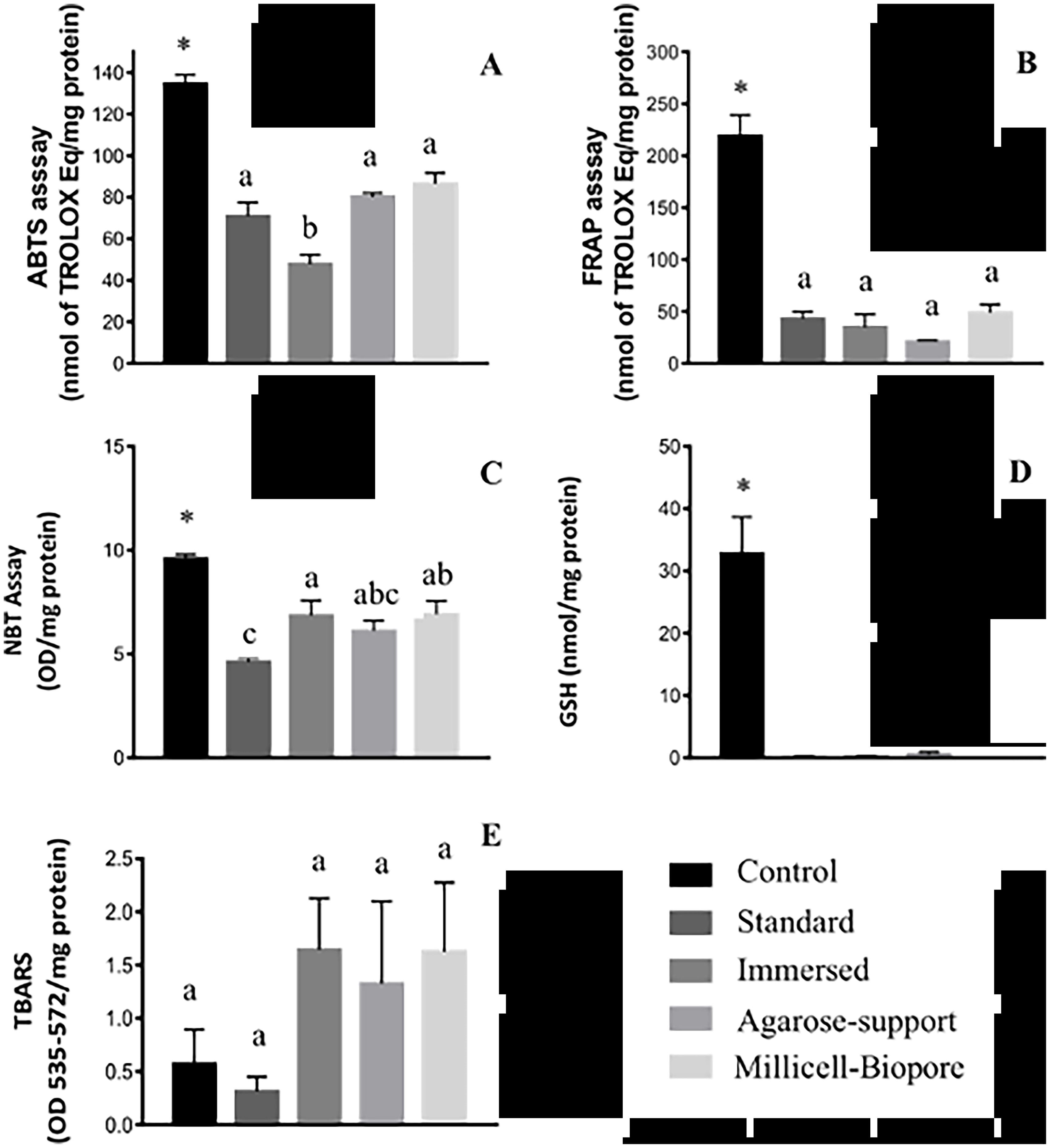
Assessment of the redox status of ovarian samples revealed that all samples cultured using the standard culture method, agarose gel support method, immersed agarose gel culture and Millicell culture showed a reduction in superoxide anion levels, antioxidant capacity and lipid peroxidation compared with the control group. The test results are presented in Figure 7 (D6) and Figure 8 (D14).
Effects of different culture methods (standard culture, agarose gel-supported culture, medium-immersed agarose gel culture and Millicell–Biopore culture) on oxidative stress during the culture of preantral follicles in ovarian tissue at D6. (A) Free radical scavenging capacity (ABTS) and (B) ferric reducing antioxidant power (FRAP) were compared with a Trolox curve. Superoxide anion production (C), reduced glutathione quantification (D) and lipid peroxidation (E) were determined by NBT, GSH and TBARS tests, respectively. Samples were collected after 6 days of in vitro culture. *Compared with the control group. A, B Indicate differences among treatments (P < 0.05).

Effects of different culture methods (standard culture, agarose gel-supported culture, medium-immersed agarose gel culture and Millicell–Biopore culture) on oxidative stress during the culture of preantral follicles in ovarian tissue at D14. (A) Free radical scavenging capacity (ABTS) and (B) ferric reducing antioxidant power (FRAP) were compared with a Trolox curve. Superoxide anion production (C), reduced glutathione quantification (D) and lipid peroxidation (E) were determined by NBT, GSH and TBARS tests, respectively. Samples were collected after fourteen days of in vitro culture. *Compared with the control group. A, B Indicate differences among treatments (P < 0.05).

At D6, it was observed that the agarose gel culture method on the plate presented higher superoxide anion levels compared to other culture methods (NBT; mean ± SEM = 5.37 ± 0.1 in standard culture, 5.06 ± 0.2 in gel-supported culture, 6.64 ± 0.5 in immersed culture and 4.69 ± 0.1 OD/mg protein in Millicell culture). At D14, the same was observed for the immersed culture method and Millicell culture, which had higher superoxide anion levels than the standard method (6.42 ± 0.7, 6.91 ± 0.6 and 4.61 ± 0.2 OD/mg protein, respectively). On D6, no significant differences in the antioxidant capacity (FRAP and ABTS assays) of ovarian tissues subjected to different culture methods were observed. In contrast, the culture in the medium-immersed agarose gel conditions presented lower antioxidant capacity when compared to the standard, supportive and Millicell methods at D14 (70.85, 85.17, 47.78 and 86.43 Trolox equivalent/mg protein, respectively), as shown by the ABTS test. No differences were present in the FRAP test between the culture methods.
Quantification of GSH levels revealed that at D6, standard and agarose-supported cultures exhibited GSH levels similar to those of the control (P > 0.05) and reduced GSH levels in the agarose gel-supported culture immersed in medium and the Millicell culture methods (P < 0.05). On D14, none of the culture methods maintained GSH levels comparable to those of the control (P > 0.05). Lastly, no significant difference was observed in the TBARS levels between the control and ovarian tissues subjected to different culture methods on D6 or D14 (P > 0.05).
Discussion
This is among the first studies comparing 2D and 3D in situ cultures with an agarose-based extracellular matrix for morphological preservation and development of bovine preantral follicles over 14 days. While previous investigations have employed alginate, polyethylene glycol and hyaluronan hydrogels for isolated follicle culture in rodent and caprine models (Desai et al. Reference Desai, Abdelhafez, Calabro and Falcone2012; Asgari et al. Reference Asgari, Valojerdi, Ebrahimi and Fatehi2015; Correia et al. Reference Correia, Lima, Sousa, Ferreira, Cadenas, Paes, Alves, Shikanov and Figueiredo2020), the present study specifically addresses the use of agarose as a support matrix for in situ culture of bovine ovarian cortex fragments, a configuration that preserves the native tissue architecture and paracrine signalling environment of preantral follicles.
We used 2D and 3D systems for in situ follicle cultures (multiple follicle cultures) to evaluate the efficiency of the methods in the presence or absence of an extracellular matrix. Compared to other methods, in vitro culture on an agarose gel support (2D) presented better conditions for maintaining follicular viability and integrity, larger follicle and oocyte diameters, greater granulosa cell proliferation and antioxidant capacity during 14 days of preantral follicle culture. Although the results of the present study indicate that the 2D system provides more favourable conditions for follicular viability, previous studies have highlighted the important advantages of 3D support matrices. Kapałczyńska et al. (Reference Kapałczyńska, Kolenda, Przybyła, Zajączkowska, Teresiak, Filas, Ibbs, Bliźniak, Łuczewski and Lamperska2018) reported that 3D matrices help preserve the follicular architecture and better mimic in vivo conditions by maintaining adequate access to oxygen and nutrients. Consequently, 3D culture systems may contribute to follicular developmental outcomes closer to those observed in animal models (Jensen and Teng Reference Jensen and Teng2020). Nevertheless, the present culture system must positively affect survival, proliferation, follicular morphology, gene expression and steroidogenesis to mimic the natural environmental differences in in vitro developing follicles (Araújo et al. Reference Araújo, M., Figueiredo and Gastal2014; Sadr et al. Reference Sadr, Ebrahimi, Shahhoseini, Fatehi and Favaedi2015).
The 2D cell culture has been used for cell culture since the beginning of the 20th century (Ferreira et al. Reference Ferreira, Gaspar and Mano2018). It is important to emphasise that the culture of preantral follicles under this (standard) condition is associated with technical limitations, such as compromised paracrine communication between granulosa cells and oocytes (Andersen et al. Reference Andersen, Auk-Emblem and Dornish2015), resulting in the fixation of granulosa cells to the culture plate, with the growing cells attached to a plastic surface (Breslin and O’Driscoll Reference Breslin and O’Driscoll2013). The advantages of 2D cultures include simple, low-cost maintenance of cell cultures and the ability to perform functional tests (Kapałczyńska et al. Reference Kapałczyńska, Kolenda, Przybyła, Zajączkowska, Teresiak, Filas, Ibbs, Bliźniak, Łuczewski and Lamperska2018). It is well-established that any alteration in the morphology of follicular cells causes delayed growth and incomplete development of preantral follicles, similar to that observed in 2D culture systems (Jamalzaei et al. Reference Jamalzaei, Valojerdi, Montazeri and Baharvand2020).
In this context, this study improved the culture system using an agarose substrate, perhaps by improving the physiological requirements for follicular growth. A similar system has been described for culturing sperm from mice, yielding interesting results regarding spermatogenesis (Sato et al. Reference Sato, Katagiri, Gohbara, Inoue, Ogonuki, Ogura, Kubota and Ogawa2011). Furthermore, the use of agarose as support for the ovarian cortex provides an environment that supports survival and follicular growth during culture. A recent study suggested that the agarose gel culture system supports follicle growth and the formation of oocyte-granulosa complexes (Le et al. Reference Le, Nguyen, Lam, Lam, Nguyen, Nguyen and Bui2024). Our findings are consistent with the data described by Ghezelayagh et al. (Reference Ghezelayagh, Abtahi, Khodaverdi, Valojerdi, Mehdizadeh and Ebrahimib2021), obtained from human follicle cultures.
Compared to that of the uncultured control, the morphological integrity of preantral follicles was preserved only in 2D culture on agarose support (73% and 58% of intact follicles after 6 or 14 days of culture, respectively). This is likely due to the presence of mechanisms that drive the formation of an extracellular gel matrix that protects the 3D structures from adhering to the plate wall. In addition, the agarose concentration directly reflects the gel mesh size (pores), which controls the gas-medium-cell interface, allowing for greater or lesser perfusion of the medium (Diekjürgen and Grainger Reference Diekjürgen and Grainger2017). A recent study observed a continuous increase in antrum formation and the oocyte-cumulus-granulosa intact after 14 days of in vitro growth (antrum formation 2D culture system vs. 3D culture system, 7.64 ± 2.74 vs 67.29 ± 3.98, respectively) in a 3D-agarose culture system for oocytes from preantral follicles in porcine. The agarose matrix used to culture oocyte–cumulus–granulosa complexes isolated from preantral follicles maintained the spherical structure of the complexes, with granulosa cells proliferating around the oocytes (Le et al. Reference Le, Nguyen, Lam, Lam, Nguyen, Nguyen and Bui2024). In addition, the culture of vitrified bovine ovarian tissue on agarose gel inserts has been demonstrated to preserve follicular integrity, supporting the suitability of this matrix for maintaining the ovarian tissue architecture during in vitro culture (Alkali et al. Reference Alkali, Colombo, Luciano, Nizanski, Hassan, Dziegiel and Luvoni2023).
Studies with isolated goat follicles demonstrated that when comparing 2D and 3D alginate gel systems, the viability of follicles was 45.5% and 67.5%, respectively (Pessoa et al. Reference Pessoa, Rocha, Brito, Silva, Chaves, Magalhães-Padilha, Campello, Rodrigues, Nunes-Pinheiro and Figueiredo2014). The physical characteristics of the alginate hydrogel pore network enable efficient diffusion of nutrients and metabolic waste, including the bidirectional transport of hormones and other proteins essential for tissue viability and follicular development. Furthermore, alginate is not of animal origin, contributing to its high biocompatibility (Andersen et al. Reference Andersen, Auk-Emblem and Dornish2015). In addition, certain studies on in vitro 3D culturing of follicles isolated from rodents have identified follicular integrity and survival percentages similar to those found in the present study (70.6% at 8 days and 63.3% at 12 days of alginate hydrogel culture (Asgari et al. Reference Asgari, Valojerdi, Ebrahimi and Fatehi2015) and 80% at 12 days culture with hyaluronan hydrogel (Desai et al. Reference Desai, Abdelhafez, Calabro and Falcone2012). The best results were obtained using a 2D culture with an extracellular matrix and in situ follicles. Goat and cattle 2D systems of follicle culture over 18 days showed 80.8% and 77.7% viability, respectively (Rossetto et al. Reference Rossetto, Santos, Silva, Duarte, Silva, Campello and Figueiredo2013). These discrepancies between the survival and follicular integrity data, extracellular matrix data and duration data may also be related to differences between species and specific systems and methods used in the culture of preantral follicles.
Another important factor to consider during in vitro culture of ovarian follicles is the components of the culture medium, such as minerals, vitamins, nucleosides and amino acids. Owing to certain peculiarities of in vitro development, the culture medium is the first source of nutrients in contact with the follicles. Therefore, among the available media, MEM is considered the medium of choice for the culture of preantral follicles due to its ability to maintain follicle integrity and in vitro development (Araújo et al. Reference Araújo, M., Figueiredo and Gastal2014; Jimenez et al. Reference Jimenez, Araújo, Penitente-Filho, De Azevedo, Silveira and Torres2016). Thus, in this study, follicular development data were similar (over 50% of follicles were developing) for all culture methods for the first 6 days. The same phenomenon occurred after 14 days of culture; however, the Millicell–Biopore (3D) culture method resulted in follicles that resembled those of the uncultured control. This suggests that this method was not as efficient as the other methods in maintaining follicular development under the specified conditions.
Morphometric responses demonstrated a reduction in follicle and oocyte sizes after culture for 6 or 14 days. At 6 days of culture, the average dimensions of follicles cultured using the different methods were similar; however, at 14 days, the culture using the insert (Millicell–Biopore) yielded smaller follicles than those in the other treatments. In addition, the average oocyte and follicle diameters cultured for 6 days were larger in the 2D and 3D methods in the presence of the extracellular matrix. Further, at 14 days of culture, the 2D and 3D methods maintained the oocyte diameter. Follicle growth depends primarily on morphological changes in somatic and granulosa cells. Thus, any alteration in the morphology of follicular cells causes delayed growth and incomplete development of the preantral follicle (Jamalzaei et al. Reference Jamalzaei, Valojerdi, Montazeri and Baharvand2020). The dispersion of granulosa cells in 2D culture systems impairs their functional interactions with oocytes and disrupts the signalling mechanisms necessary for follicular development. Cellular communication plays a significant role in the transmission of autocrine and paracrine signals (Andersen et al. Reference Andersen, Auk-Emblem and Dornish2015). Contrary to the present results, another research group observed in a study with fresh and vitrified preantral mouse follicles that the gene expression of Bmp4 (paracrine growth factor secreted by theca and granulosa cells) was higher in 3D culture systems than in 2D systems at all concentrations evaluated Jalili et al. (Reference Jalili, Khani Hemmatabadi, Bakhtiyari, Abdolmaleki and Moradi2021). Bmp4, along with Bmp7, is responsible for regulating follicular growth and the transition of primordial follicles to their primary form (West-Farrell et al. Reference West-Farrell, Xu, Gomberg, Chow, Woodruff and Shea2009).
Different results were found for follicular growth in other species, showing that follicles from goats, canines and horses can be maintained with a larger diameter in culture than what can be seen in controls (Matos et al. Reference Matos, Silva, Rodrigues and Figueiredo2007; Serafim et al. Reference Serafim, Araújo, Silva, Duarte, Almeida, Chaves, Campello, Lopes, Figueiredo and Silva2010; Aguiar et al. Reference Aguiar, Lunardi, Lima, Rocha, Bruno, Magalhães-Padilha and Figueiredo2016). The discrepancies between previous results and those obtained in the present study may be due to the absence of a specific growth factor or hormone in the culture medium necessary for follicular development.
Cell proliferation capacity was assessed using nucleolus organising regions (NORs) as parameters for in vitro cultured antral follicles, and the results demonstrated that proliferation was significantly higher in the culture with agarose gel support than in cultures maintained with other methods. Alkali et al. (Reference Alkali, Colombo, Luciano, Nizanski, Hassan, Dziegiel and Luvoni2023) evaluated the follicular integrity of vitrified ovarian tissue in agarose gel culture and demonstrated high proliferation of follicular cells, attributing this to the immediate availability of nutrients to the tissue fragments, which were partially covered by a thin film of medium with simultaneous lower gas exchange. The agarose gel support approach has been successfully used to evaluate vitrified and subsequently cultured follicles (Brito et al. Reference Brito, Domingues, Rodrigues, Maside, Lunardi, Wu, Figueiredo, Pieczarka and Santos2018) and the developmental competence of the oocyte-granulosa complex of preantral follicles in pigs (Le et al. Reference Le, Nguyen, Lam, Lam, Nguyen, Nguyen and Bui2024). Furthermore, this technique has been widely used to analyse the activity, isolation and analysis of cancer cell lines (Klopfleisch et al. Reference Klopfleisch, Klose and Gruber2010; Preciado et al. Reference Preciado, Lam, Azarin, Lou and Aksan2021). We believe that the decrease in the number of NORs is related to the culture method itself, which may slow granulosa cell proliferation, likely due to the influence or interruption of the cell cycle (Choi et al. Reference Choi, Lee, Lee, Yoon, Bae and Choi2008).
Follicular growth begins with follicular cell development and differentiation following stimulation of this follicle (Araújo et al. Reference Araújo, M., Figueiredo and Gastal2014). Nucleolar organiser regions (NORs) are loops of DNA actively associated with the nucleolus, which code for ribosomal RNA consisting of argyrophilic non-histone proteins. They appear as black and brown dots on staining with silver stains and are called argyrophilic nucleolar organiser regions (Ramraje et al. Reference Ramraje, Kosale and Dahake2025). Therefore, active chromosomes are marked during the cell cycle, and the number of NORs indicates the proliferative activity of these follicles (Silva et al. Reference Silva, Serakides, Nascimento, Nunes, Ribeiro and Ocarino2003). Consequently, cells with more intense proliferative activity exhibit a greater number of NOR (Derenzini Reference Derenzini2000), which are considered reliable markers of proliferation (McLaughlin et al. Reference McLaughlin, Kinnell, Anderson and Telfer2014; Brito et al. Reference Brito, Domingues, Rodrigues, Maside, Lunardi, Wu, Figueiredo, Pieczarka and Santos2018).
Reactive oxygen species (ROS) are reactive molecules that play several physiological roles, such as intracellular cell signalling, cell death, immune defence and induction of mitogenic responses. However, an imbalance between ROS generation and detoxification of these reactive metabolites results in oxidative stress and damage to DNA, proteins and lipids, compromising cell and tissue health (Pizzino et al. Reference Pizzino, Irrera, Cucinotta, Pallio, Mannino, Arcoraci, Squadrito, Altavilla and Bitto2017). Thus, the quantification of ROS and overall antioxidant capacity is commonly used to assess tissue health. Herein, we analysed several parameters to determine the effects of different culture methods on the redox status of the ovarian tissue. Culture methods using an agarose support maintained superoxide anion levels throughout the culture period. In contrast, the medium immersion agarose gel method yielded the highest levels of superoxide anions. The concurrent reduction in both ROS levels and antioxidant capacity observed across all cultured groups relative to the non-cultured control is noteworthy. This pattern likely reflects a general down-regulation of metabolic activity in the cultured tissue, rather than a selective impairment of antioxidant defences. In the non-cultured (fresh) control, the tissue retains its full in vivo metabolic rate, which is associated with higher baseline ROS production and correspondingly higher antioxidant enzyme activity. Upon transfer to in vitro conditions, the reduced oxygen tension, absence of vascular perfusion and limited nutrient renewal may collectively attenuate cellular metabolism, leading to proportionally lower ROS generation and a concomitant decrease in antioxidant capacity (Soto-Heras and Paramio Reference Soto-Heras and Paramio2020). Prolonged exposure of the culture system may interfere with the antioxidant activity of the sample. This was verified by GSH analysis after 14 days of culture. This may be due to the inefficiency of cellular or tissue metabolism, and it is necessary to add substances to the culture medium to assist these cells. However, it was not possible to verify the factors involved in these circumstances, and further studies are required.
Interestingly, the increase in ROS levels may be associated with a reduced number of NOR-labelled cells per follicle, as histological analysis revealed a decrease in the number of intact follicles in the medium-immersed agarose gel group. This observation suggests a potential link between oxidative stress and impaired granulosa cell proliferation, wherein elevated superoxide anion levels may trigger cell cycle arrest or apoptosis, thereby reducing the number of actively dividing cells detectable by AgNOR staining (Choi et al. Reference Choi, Lee, Lee, Yoon, Bae and Choi2008). Oxidative stress, which results from an imbalance in the production and elimination of reactive intracellular oxygen species (Soto-Heras and Paramio Reference Soto-Heras and Paramio2020), as well as thermal stress (Aguiar et al. Reference Aguiar, Hyde, Pedroza and Denicol2020), is considered one of the main factors affecting the results of assisted reproduction techniques. In this experiment, we minimised external interference during in vitro culture to the utmost. For this, luminous and temperature controls were performed during manipulation.
To contextualise these findings within reproductive biotechnology, a few considerations regarding the study’s current scope can guide future investigations. Although the biological replication (n = 5 animals) matches standard protocols in the field (Araújo et al. Reference Araújo, Gastal, Wischral, Figueiredo and Gastal2015; Bizarro-Silva et al. Reference Bizarro-Silva, Santos, Gerez, González, Lisboa and Seneda2018), expanding the sample size in future long-term studies could further refine the detection of subtle physiological variations. Additionally, because preserving structural and biochemical integrity is an absolute prerequisite for oocyte quality, our characterisation of these parameters establishes a vital benchmark, setting the stage for future validation through functional endpoints such as in vitro fertilisation. The prominent differences detected at D6 underscore the system’s capacity to optimizse the critical early window of tissue culture, before outcomes stabilise by D14. Finally, the biochemical redox data presented here provide a valuable overview of the oxidative environment, establishing a clear trajectory for future transcriptomic and mechanistic characterisation.
In conclusion, these findings demonstrate that the agarose gel support system offers a platform for maximising the recovery of viable preantral follicles, while providing fundamental insights into the initial events of bovine folliculogenesis. By establishing this structural and biochemical baseline, our study provides a framework that naturally facilitates future downstream milestones in reproductive biotechnology, such as in vitro oocyte maturation and embryo development, while supporting further expanded scale-up investigations.
Author contributions
Camila Bizarro da Silva: Conceptualisation-Lead, Data curation-Lead, Investigation-Lead, Methodology-Equal, Project administration-Lead, Writing – original draft-Equal; Larissa Zamparone Bergamo: Data curation-Equal, Investigation-Equal, Methodology-Equal; Marcela Bortoletto Cerezzetti: Conceptualisation-Equal, Data curation-Equal, Investigation-Equal; Rafaela Schoma Cardoso: Investigation-Equal, Writing – original draft-Equal; Miriam Nagashima Hohmann: Data curation-Equal, Investigation-Equal; Waldiceu A. Verri Jr.: Conceptualisation-Equal, Data curation-Equal; Mateus José Sudano: Conceptualisation-Equal, Investigation-Equal; Marcelo Marcondes Seneda: Conceptualisation-Equal, Formal analysis-Equal, Funding acquisition-Equal, Project administration-Equal, Resources-Equal, Supervision-Equal, Validation-Equal, Writing – review & editing-Equal.
Funding information
This study was partially funded by the Coordenação de Aperfeiçoamento de Pessoal de Nível Superior, Brazil (CAPES; Finance Code 405495/2023-4) and INCT LEITE - 465725/2014.
Competing interests
Authors declare no conflicts of interest.
Ethical Standards
The authors state that all the experimental procedures were conducted in accordance with the guidelines of the Animal Experimentation Ethics Committee of the State University of Londrina (identification number: 031.2024).