Implications
The supplementation of a suitable quantity of all-trans retinol to oocyte maturation medium or embryo culture medium followed by culture under appropriate gaseous environments protects oocytes and embryos from oxidative damage, thereby greatly enhancing the yields of viable embryos. This could be applied in Animal Biotechnology to increase lamb crops from high-yielding breeds of farm animals, which are seasonal breeders or which have reproduction problems and also to propagate endangered animal species.
Introduction
Assisted reproduction technology (ART) in farm animals has revolutionized farming practices around the globe. In vitro embryo production (IVEP) technologies have enabled the production in large numbers of embryos of superior breeds in various domestic animals. They have also enabled the production of large numbers of embryos for scientific research purposes from slaughtered and/or live animals. In vitro fertilisation (IVF) and in vitro embryo culture (IVEC) constitute technology for embryo production. Although several decades of research have gone into IVF, the culture conditions that promote the maximal embryo yield are yet to be standardised. Several factors affect the IVF–IVEC process (Camargo et al., Reference Camargo, Viana, Sá, Ferreira, Ramos and Vale Filho2006). One of the most critical among them is oxidative stress (Guérin et al., Reference Guérin, El Mouatassim and Ménézo2001).
The fact that oxidative stress beyond a certain threshold leads to defective embryo development has been well documented. The deleterious effects of pro-oxidants and reactive oxygen species (ROS) on gamete and embryo development in vitro have been studied in bovine (Fujitani et al., Reference Fujitani, Kasai, Ohtani, Nishimura, Yamada and Utsumi1997), humans (Bedaiwy et al., Reference Bedaiwy, Falcone, Mohamed, Aleem, Sharma, Worley, Thornton and Agarwal2004), mice (Nasr-Esfahani and Johnson, Reference Nasr-Esfahani and Johnson1991), porcine (Kitagawa et al., Reference Kitagawa, Suzuki, Yoneda and Watanabe2004) and many other vertebrates. Although gametes and embryos possess some innate antioxidant capacity, it is not sufficient to counter the oxidative stress during in vitro culture. Supplementation of the culture media with antioxidants to counter oxidative stress has been in practice for quite some time now, with various potential molecules being tested for their antioxidant abilities in in vitro oocyte maturation and embryo-culture media. Some of these antioxidant supplements include metal chelators, antioxidant enzymes, thiol compounds, proteins and vitamins (Guérin et al., Reference Guérin, El Mouatassim and Ménézo2001).
Vitamin A and its metabolites are known to regulate cell growth, differentiation and vertebrate embryogenesis (Gudas, Reference Gudas1994). Retinoid supplementation to chemically defined maturation media has been shown to improve the blastocyst formation rate in bovine (Livingston et al., Reference Livingston, Eberhardt, Edwards and Godkin2004), while in mice it is found to hinder blastocyst development (Huang et al., Reference Huang, Shen, Chang, Wu and Hsuuw2003). The significance of endogenous retinoids in the development of porcine (Parrow et al., Reference Parrow, Horton, Maden, Laurie and Notarianni1998) and murine oocytes (Romert et al., Reference Romert, Tuvendal, Simon, Dencker and Eriksson1998) of exogenous retinoids in the development of sheep (Eberhardt et al., Reference Eberhardt, Will and Godkin1999) and gilt (Whaley et al., Reference Whaley, Hedgpeth, Farin, Martus, Jayes and Britt2000) oocytes and of dietary vitamin A in the maturation and development of bovine oocytes in vivo (Ikeda et al., Reference Ikeda, Kitagawa, Imai and Yamada2005) has been well documented. Thus, retinol and its metabolites have a key role in vertebrate gamete and embryo formation and development both in vitro and in vivo.
In an attempt to determine the optimal in vitro culture environment for gametes and embryos, different gaseous environments have been used. Thompson et al. (Reference Thompson, Simpson, Pugh, Donnelly and Tervit1990) reported that culturing sheep and cattle embryos at 5% to 10% O2 levels was optimal for embryo development in vitro. Similar observations were also made by Bernardi et al. (Reference Bernardi, Flechon and Delouis1996) and Leoni et al. (Reference Leoni, Rosati, Succu, Bogliolo, Bebbere, Berlinguer, Ledda and Naitana2007). In this context, this study was carried out to determine the antioxidant potential of vitamin A (all-trans retinol) in sheep in vitro oocyte and embryo culture under conditions of low and high O2 concentrations, respectively.
Material and methods
Unless otherwise stated, all chemicals used in this experiment were purchased from Sigma-Aldrich Chemicals Private Ltd, Bangalore, India.
Collection of oocytes
Sheep ovaries were obtained from a local abattoir and transported to the laboratory suspended in 0.9% saline supplemented with 50 μg/ml gentamycin in insulated containers within an hour of slaughter. Upon arrival, the ovaries were washed repeatedly in normal saline, trimmed free of extraneous tissue and rinsed in normal saline. The cumulus–oocyte complexes (COCs) were isolated from follicles by the slicing method (Pawshe et al., Reference Pawshe, Totey and Jain1994) and subsequently washed thrice in Tyrode’s lactate – N – [2-hydroxyethyl] piperazine – N′ – [2-ethanesulphonic acid] (TL-HEPES) medium. The COCs were assessed morphologically and only those that had a compact non-atretic cumulus oophorus–corona radiata and a homogenous ooplasm were selected for in vitro maturation.
Maturation of oocytes in vitro
The COCs were washed thrice – first in TL-HEPES medium and subsequently in maturation medium composed of TCM 199 (Invitrogen Corporation, USA) supplemented with 10% (v/v) foetal calf serum (FCS; Gibco Laboratories, Grand Island, USA), 5.5 mg/ml sodium pyruvate, 25 μg/ml gentamycin sulphate, 5.0 μg/ml LH, 0.5 μg/ml FSH and 1 μg/ml estradiol (E2). Twenty COCs were placed in 100 μl droplets of maturation medium, covered with sterile mineral oil and matured for 24 h at 39°C either at 5% CO2 in air (which is approximately 20% O2) or at 5% CO2, 5% O2 and 90% N2, based on the design of the experiment.
Fertilisation of oocytes in vitro
The sheep testis obtained from the local abattoir was transported to the laboratory suspended in 0.9% saline supplemented with 50 μg/ml gentamycin in an insulated container within an hour of slaughter. The sheep testis was washed in saline and trimmed free of covering tissues. The tail of the epididymis, presumed to contain mature sperms, was cut using a sterile blade and the sperm-rich fluid that oozed out was directly laid on bovine serum albumin-free brackett and oliphant (BSA-free BO) medium (Brackett and Oliphant, Reference Brackett and Oliphant1975; containing 10 mM caffeine sodium benzoate and 10 μg/ml heparin) in a petri dish. Sperm selection was carried out in a Percoll (Pharmacia, Uppsala) density gradient (45%/90%) placed in a CO2 incubator at 39°C for 2 h. Approximately 2 to 3 ml of the semen sample in the sperm TL medium was layered over the pre-incubated gradient solution in sterile centrifuge tubes, and centrifuged at 1400 r.p.m. for 10 min at room temperature. The supernatant was discarded and the sperm sediment was rewashed thrice by centrifugation at 1400 r.p.m. for 10 min in the BSA-free BO medium described above. The final pellet was resuspended in 1 ml of the BSA-free BO medium diluted with 1 ml BO medium containing 20 mg/ml BSA supplemented with 10 μg/ml heparin. A final sperm concentration of approximately 1 to 2 × 10⁶ per ml BO medium was used for fertilisation. Mature sheep COCs were then washed in the BO medium and distributed at a rate of 20 per 100 μl drops of fertilisation medium under mineral oil. Capacitated spermatozoa (2 μl) were added to these fertilisation drops and incubated for 18 h at 39°C at 5% CO2, 5% O2 and 90% N2.
Embryo culture in vitro
On completion of the incubation period, the oocytes were washed to remove the cumulus cells by repeated pipetting through a small-bore pipette. They were then cultured in a modified synthetic oviductal fluid medium (Tervit et al., Reference Tervit, Whittingham and Rowson1972) containing 2% (v/v) basal medium eagle essential amino acids and 1% (v/v) minimum essential medium non-essential amino acids, 3 mg/ml BSA, 0.6 mM sodium pyruvate, 10 μg/ml gentamycin at a rate of 20 embryos per 100 μl droplets for 8 days at 39°C under 5% CO2 in air (which is approximately 20% O2) or under 5% CO2, 5% O2 and 90% N2 based on the design of the experiment. The medium was changed once every 48 h to avoid toxic accumulation of ammonia and to replenish the nutrients.
Blastocyst cell number analysis
Blastocyst cell number analysis was performed to assess the morphological quality of the embryos. Expanded day eight blastocysts from each treatment group were fixed and stained as per the method described by Mermillod et al. (Reference Mermillod, Vansteenbrugge, Wils, Mourmeaux, Massip and Dessy1993). These expanded blastocysts were then individually transferred onto glass microscopic slides and dried at room temperature. They were then fixed with 70% ethanol for 24 h. The fixed blastocysts were then stained with 10 μg/ml bisbenzamide (Hoechst 33342, Sigma-Aldrich Chemicals Private Ltd, Bangalore, India) and 2.3% sodium citrate. The slides were observed under an epifluorescence microscope, fitted with excitation filter (330 to 380 nm) and barrier filter (420 nm). The total numbers of nuclei in each blastocyst were counted.
Comet assay
DNA damage in individual embryos cultured for 3 days under 20% O2, in the presence or absence of vitamin A supplementation was assessed by comet assay (Takahashi et al., Reference Takahashi, Keicho, Takahashi, Ogawa, Schultz and Okano2000). About 10 to 20 embryos were washed twice in a mixture of phosphate-buffered saline (PBS) and polyvinylpyrrolidone (4 mg/ml). Then, the embryos from each experimental group were transferred to a 200 μl drop of 1% low-melting temperature agarose (Genei, Bangalore, India) in PBS at 39°C; the agarose drop was placed on a 35 mm plastic petri dish. Using a stereo-dissection microscope to visualise the embryos, the embryos were gently mixed with the 1% low-melting temperature agarose and then captured in a total volume of about 10 μl using a mouth-operated glass pipette. The embryos were then quickly transferred onto a glass microscopic slide pre-coated with 1% high melting temperature agarose (Genei, Bangalore, India). Then, the slides were placed for 5 min on ice to solidify the agarose. The embryos were then lysed by incubating the slides for 3 h at ambient temperature in a lysing buffer composed of 10 mM Tris, pH 10, containing 100 mM sodium EDTA, 2.5 mM NaCl, 10 μg/ml proteinase K, 1% sodium sarcosinate and 1% Triton X-100. Then, the slides were removed from the lysing solution and placed on a horizontal gel electrophoresis unit. The unit was filled with fresh electrophoresis buffer (1 mM sodium EDTA, 300 mM NaOH) to a level 0.25 cm above the slides, followed by equilibration of the slides in the electrophoresis buffer for 20 min. Electrophoresis was then carried out at 25 V for 20 min. The slides were then neutralised by immersing in 0.4 M Tris--HCl (pH 7.5) for 5 min at ambient temperature. Staining of DNA was carried out by adding a 20 μl drop of acridine orange (5 μg/ml) to the slide for 2 min followed by 1 min of washing in sterile distilled water. Stained DNA was observed under a fluorescence microscope. Quantification of DNA damage was done by measuring the length of the streak of the DNA comet tail. The length was calculated by comparing with a photograph of a micrometre of the same magnification as that of the embryos.
Experimental design
Vitamin A (all-trans retinol) was dissolved in 100% ethyl alcohol; appropriate dilutions were made and stored at −80°C until use. The ethanol concentration during oocyte or embryo culture was <0.05%. In experiment I, varying concentrations of all-trans retinol (0, 2, 4, 6, 8 and 10 μM) were added to the oocyte maturation medium followed by culture in a 5% O2 environment, whereas, in experiment II, the aforesaid concentrations of all-trans retinol were added to the oocyte maturation medium followed by culture in a 20% O2 environment. Experiment III was carried out in a 5% O2 environment wherein varying concentrations of all-trans retinol (0, 2, 4, 6, 8 and 10 μM) were added to the embryo culture medium and in experiment IV, varying concentrations of all-trans retinol (0, 2, 4, 6, 8 and 10 μM) were added to the embryo culture medium followed by culture in a 20% O2 environment.
Thus the experimental design can be summarised as follows:
– Experiment I: Oocyte maturation with all-trans retinol at a 5% O2 environment and embryo culture without all-trans retinol at a 5% O2 environment.
– Experiment II: Oocyte maturation with all-trans retinol at a 20% O2 environment and embryo culture without all-trans retinol at a 5% O2 environment.
– Experiment III: Oocyte maturation without all-trans retinol at a 5% O2 environment and embryo culture with all-trans retinol at a 5% O2 environment.
– Experiment IV: Oocyte maturation without all-trans retinol at a 5% O2 environment and embryo culture with all-trans retinol at a 20% O2 environment.
Statistical analysis
The Statistical Package for Social Sciences (SPSS 11.O, Chicago, IL, USA) software was used for statistical analysis. In each experimental group, oocytes were randomly distributed. The percentages of oocytes that were fertilised and the embryos that had developed to morula and blastocyst were subjected to arcsine transformation before analysis. The total cell count data (of blastocysts) were directly subjected to analysis. All data were subjected to one-way ANOVA followed by Tukey’s test to determine the differences among the experimental groups. Differences at a P < 0.05 were considered statistically significant.
Results
Experiment I
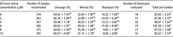
The results of experiment I are shown in Table 1. The addition of all-trans retinol to an in vitro oocyte maturation medium and subsequent culture under a 5% O2 environment did not result in significant increases in the percentage of cleavages, when compared with the controls. However, the percentage of embryos that underwent cleavage, was significantly reduced in the 10 μM supplemented group (P < 0.01) when compared with the 4 and 6 μM supplemented groups. The percentage of compact morulae was significantly higher in the 6 μM supplemented group compared with the 10 μM supplemented group (P < 0.05). Supplementation of 6 μM of all-trans retinol resulted in a significant increase in the total percentage of blastocysts compared with the control, 2 and 8 μM supplemented groups (P < 0.05) and 10 μM supplemented group (P < 0.001). The percentage of blastocysts from the 4 μM supplemented group was significantly greater than that of the 10 μM supplemented group (P < 0.05). The total cell number was also significantly higher in the 6 μM supplemented group when compared with the control, 2 and 8 μM supplemented groups (P < 0.05) and 10 μM supplemented group (P < 0.01).
Effect of all-trans retinol supplementation to oocyte maturation medium on development of pre-implantation sheep embryos cultured in 5% O2 environment

Values are listed as mean ± s.e.m.; means in the same column with different superscripts were significantly different. A total of six replicates were used per all-trans retinol supplement concentration/control.
Experiment II
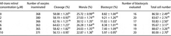
Data from Experiment II are shown in Table 2. The supplementation of all-trans retinol to the in vitro oocyte maturation medium and subsequent culture under a 20% O2 environment did not yield any significant results with respect to the percentages of embryos that developed to morulae and blastocyst compared with the control group. However, the percentage of embryos cleaved was significantly higher in the 6 μM supplemented group compared with the control, 2 and 8 μM supplemented groups (P < 0.05) and 10 μM supplemented group (P < 0.01). The 10 μM supplemented group showed significantly lesser percentage of cleavage, morulae and blastocysts compared with the 4 μM supplemented group (P < 0.05). The total cell number was significantly higher in the 4 (P < 0.05) and 6 μM (P < 0.01) supplemented groups compared with the 10 μM supplemented group (P < 0.01).
Effect of all-trans retinol supplementation to oocyte maturation medium on development of pre-implantation sheep embryos cultured in 20% O2 environment

Values are listed as mean ± s.e.m.; means in the same column with different superscripts were significantly different. A total of six replicates were used per all-trans retinol supplement concentration/control.
Experiment III
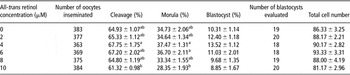
The effects of all-trans retinol in the embryo culture medium at 5% O2 levels are shown in Table 3. The rates of cleavage, morula and blastocyst formation were not significantly different between the supplemented and the control groups. The 10 μM supplemented group showed a significantly lesser percentage of cleavage and morulae compared with the 4 μM supplemented group (P < 0.05). The percentage of morulae from the 10 μM supplemented group was also significantly less than that observed in the 6 μM supplemented group (P < 0.05). No significant difference was observed in the percentage of blastocysts and total cell number among the control and supplemented groups.
Effect of all-trans retinol supplementation to embryo culture medium on development of pre-implantation sheep embryos cultured in 5% O2 environment

Values are listed as mean ± s.e.m.; means in the same column with different superscripts were significantly different. No significant difference in rates of blastocyst formation or blastocyst total cell number could be observed between control and supplemented groups or among the supplemented groups. A total of six replicates were used per all-trans retinol supplement concentration/control.
Experiment IV
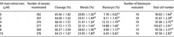
The results of Experiment IV are shown in Table 4. No significant difference was observed in the rate of cleavage among the control and supplemented groups. With respect to the formation of compact morulae, the rates were significantly higher in the 4 (P < 0.05) and 6 μM (P < 0.01) supplemented groups compared with the 10 μM supplemented group. The rate of blastocyst formation was significantly higher in the 6 μM supplemented group compared with the control (P < 0.01), 2 (P < 0.01), 8 (P < 0.05) and 10 μM (P < 0.001) supplemented groups. The rate of blastocyst formation was significantly higher in the 4 μM supplemented group (P < 0.05) compared with the 10 μM supplemented group. The total cell count was significantly higher (P < 0.01) in the 6 μM supplemented group than the control (P < 0.05), 2 (P < 0.05) and 10 μM (P < 0.01) supplemented groups.
Effect of all-trans retinol supplementation to embryo culture medium on development of pre-implantation sheep embryos cultured in 20% O2 environment

Values are listed as mean ± s.e.m.; means in the same column with different superscripts were significantly different. No significant difference in cleavage rates could be observed between control and supplemented groups or among the supplemented groups. A total of six replicates were used per all-trans retinol supplement concentration/control.
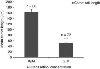
Figure 1 shows the extent of DNA damage in individual embryos on day 3 of the culture at 20% O2 levels, in the absence or presence, respectively, of all-trans retinol supplementation. Under the fluorescence microscope, the DNA from individual embryos that migrated in the gel was visualised as a comet tail-like streak. Supplementation of 6 μM all-trans retinol resulted in a significant reduction (P < 0.001) in the comet tail length compared with that of the control.
Effect of all-trans retinol supplementation to embryo culture medium at 20% O2 levels on DNA damage in individual sheep embryos determined via comet assay. Values are expressed as mean ± s.e.m.; ***significant difference (P < 0.001) was observed in 6 μM supplemented group compared with the control group.

Discussion
This study shows that supplementation of all-trans retinol at the 6 μM concentration in the oocyte maturation medium and culture in a 5% O2 environment enhanced the blastocyst formation rate and the blastocyst total cell count compared to the control. Lower or higher concentrations of all-trans retinol did not significantly affect the blastocyst formation rate compared with the control. Our results are consistent with studies involving all-trans retinol supplementation to the oocyte maturation media, resulting in the enhanced blastocyst formation in bovine (Duque et al., Reference Duque, Diez, Royo, Lorenzo, Carneiro, Hidalgo, Facal and Gomez2002; Livingston et al., Reference Livingston, Eberhardt, Edwards and Godkin2004). However, our results are not consistent with those obtained in porcine, wherein, all-trans retinol supplementation to the oocyte maturation media had yielded better rates of cleavage, but not of blastocyst formation (Almiñana et al., Reference Almiñana, Gil, Cuello, Caballero, Roca, Vazquez, Gomez and Martinez2008), which could possibly be due to the difference in culture conditions used by these authors.
All-trans retinol supplementation to the oocyte maturation medium at 6 μM concentration and subsequent culture in a 20% O2 environment enhanced the percentage of oocytes that underwent cleavage compared with control group, but not those of morulae, blastocysts or the blastocyst total cell number. This probably indicates, as suggested by Livingston et al. (Reference Livingston, Eberhardt, Edwards and Godkin2004), that optimal retinol supplementation during in vitro maturation improves the viability of oocytes under greater oxidative stress, thereby leading to greater rates of fertilisation and cleavage. The mechanism by which retinoids aid embryo development in various vertebrates during IVF has not yet been fully deciphered. Mohan et al. (Reference Mohan, Malayer, Geisert and Morgan2002) suggested that retinoids could act either directly on the bovine oocytes or via cumulus cells that surround the oocyte or via both these mechanisms in an autocrine or paracrine manner. Livingston et al. (Reference Livingston, Eberhardt, Edwards and Godkin2004) stated that retinoids possibly act by exerting effects directly or indirectly on the antioxidant defence mechanisms, by modulating the growth factor gene expression and by improving the quality of mRNAs. Ikeda et al. (Reference Ikeda, Kitagawa, Imai and Yamada2005) stated that retinoids enhance cytoplasmic maturation of cattle oocytes possibly by the following five mechanisms: functioning as an antioxidant, regulating the expression of the gonadotrophin receptor, inducing midkine, suppressing cyclooxygenase synthesis and nitric oxide synthesis in follicle cells, cumulus-granulosa cells and oocytes.
All-trans retinol supplementation to the embryo culture medium in a 5% O2 environment does not enhance the rate of cleavage, morula or blastocyst formation compared with the control group. This could be attributed to the varying antioxidant requirements for the oocytes matured, fertilised and cultured in vitro (Ali et al., Reference Ali, Bilodeau and Sirard2003). The supplementation of all-trans retinol in the embryo culture medium in a 20% O2 concentration resulted in enhanced blastocyst formation and total cell number in the 6 μM supplemented group compared with the control or most other supplemented groups. This effect possibly provides an indication that all-trans retinol possibly acts as an effective antioxidant under conditions of greater oxidative stress. This was also deciphered from our comet assay results wherein the embryos cultured in a 20% O2 environment, in the absence of all-trans retinol, exhibited greater DNA damage than did embryos with retinol supplementation.
Metabolism of molecular O2 is the key to embryo development as evidenced in mice by Houghton et al. (Reference Houghton, Thompson, Kennedy and Leese1996). Reactive oxygen species (ROS) are produced due to aerobic metabolism (Harvey et al., Reference Harvey, Kind and Thompson2002). The level of ROS production is important, especially during in vitro oocyte/embryo culture (Guérin et al., Reference Guérin, El Mouatassim and Ménézo2001). Production of ROS and susceptibility to ROS by early embryos vary with the stage of development of the embryo (Morales et al., Reference Morales, Tilquin, Rees, Massip, Dessy and Van Langedonckt1999). This has been attributed to the metabolic changes undergone by the embryos during their development. At the onset of development to the blastocyst stage, the rate of adenosine triphosphate (ATP) synthesis increases to support rapid protein synthesis and also to support the elevated activity of the ion-transport systems such as Na+/K+-dependent ATPase (Leese, Reference Leese1995). Increased ATP synthesis coupled with increased uptake of O2 and energy substrates such as glucose and pyruvate indicates increased oxidative metabolism (Thompson et al., Reference Thompson, Partridge, Houghton, Cox and Leese1996). This could lead to increased ROS production during the development of embryos, which therefore necessitates the supplementation of antioxidants at appropriate levels to the culture media to attain optimal embryo development.
Thus, maintaining an optimal level of ROS by adding antioxidants to the culture media seems to be absolutely essential for the proper production of gametes, successful interaction of gametes, fertilisation and development of embryos in vitro (Miesel et al., Reference Miesel, Drzejczak and Kurpisz1993; Sanocka and Kurpisz, Reference Sanocka and Kurpisz2004). Some authors have reported improvements in the overall outcome of IVF–IVEC – embryo transfer procedure (Ptak et al., Reference Ptak, Clinton, Tischner, Barboni, Mattioli and Loi2002). Others have attempted to supplement the culture media with different sets of nutrients or co-culture the oocytes and embryos to obtain better results (Rexroad and Powell, Reference Rexroad and Powell1993; Urdaneta et al., Reference Urdaneta, Jiménez-Macedo, Izquierdo and Paramio2003; Karja et al., Reference Karja, Kikuchi, Fahrudin, Ozawa, Somfai, Ohnuma, Noguchi, Kaneko and Nagai2006). Several studies have reported the incidence of developmental arrest or in vitro developmental block of embryos at specific stages. These include developmental blocks at the two-cell stage in mice (Nasr-Esfahani and Johnson, Reference Nasr-Esfahani and Johnson1991), four- to eight-cell stage in bovine (Camous et al., Reference Camous, Heyman, Meziou and Ménézo1984), and sixteen-cell stage in goat (Sakkas et al., Reference Sakkas, Batt and Cameron1989), etc. Although the molecular mechanisms underlying such developmental blocks are yet to be deciphered, ROSs are also presumed to have a role in the same (Noda et al., Reference Noda, Matsumoto, Umaoka, Tatsumi, Kishi and Mori1991). Hajializadeh et al. (Reference Hajializadeh, Babaei, Nematollahi-Mahani and Azizollahi2008) observed that retinoic acid supplementation to culture media helps murine embryos overcome the two-cell block.
Retinoids are touted to be key regulators of the redox signalling pathways (Olson, Reference Olson1993). In neuronal cells, retinoic acid protects against oxidative damage by inhibiting staurosporine-induced glutathione depletion, thereby helping to maintain adequate glutathione levels (Ahlemeyer and Krieglstein, Reference Ahlemeyer and Krieglstein2000). Retinoids also act by reducing the ROS content and increasing the protein levels of Mn-SOD (superoxide dismutase) and Cu-Zn SOD in staurosporine-treated neuronal cells. Such maintenance of appropriate glutathione levels is also essential for oocyte maturation, fertilisation and development (Guérin et al., Reference Guérin, El Mouatassim and Ménézo2001). Livingston et al. (Reference Livingston, Rich, MacKenzie and Godkin2009) observed that the glutathione content and the antioxidant enzyme expression did not differ significantly between the in vivo matured oocytes from retinol-treated ewes and control ewes, thereby indicating the necessity for further research to decipher the exact mechanism by which retinol confers antioxidant protection to the ovine oocyte.
Although a significant amount of research has done in IVF – embryo production technology – the desired output, in terms of large numbers of the healthy progeny, is yet to be obtained. Future research directed towards deciphering the molecular mechanisms by which antioxidants such as all-trans retinol aid in the IVF–IVEC process and imbibe developmental competence on oocytes and embryos would help better ART.
Conclusions
The results help to better understand the culture conditions in which all-trans retinol supplementation would improve ovine oocyte maturation and embryo development in vitro. Our results show that supplementation of 6 μM all-trans retinol to the culture media in an appropriate O2 environment may enhance the developmental competence of ovine oocytes and embryos in vitro.
Acknowledgements
The technical expertise provided by Dr Palanisamy and technical assistance provided by Mr Kartheeswaran, Mr Shankar and Ms Nithya, Department of Animal Biotechnology, Tamil Nadu Veterinary and Animal Sciences University, Chennai, are gratefully acknowledged.