INTRODUCTION
Two basic modes of feeding that correlate with their epifaunal or infaunal lifestyles have been adopted by sea urchins (Echinodermata: Echinoidea). ‘Regular’ sea urchins use Aristotle's lantern to rasp or bite algae, seagrasses, encrusting organisms, sessile animals, or carrion. Some ‘regular’ sea urchin species are exclusively herbivorous, while others are generalists (De Ridder & Lawrence, Reference De Ridder, Lawrence, Jangoux and Lawrence1982). In contrast, irregular sea urchins are deposit feeders that feed on organic matter contained within the underlying or surrounding sediment. Those irregular sea urchins still possessing Aristotle's lantern use it to rasp or crush biofilm-coated sediment grains, whereas those lacking it collect sediment grains using mechanisms such as ‘podial particle picking’ (Telford & Mooi, Reference Telford and Mooi1996).
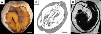
In Spatangoida, an abundant and distinctive taxon among echinoids, sediment is carried to the mouth in a number of ways through the action of specialized spines and tube feet, usually following particular pathways along the body surface (Chesher, Reference Chesher1963; De Ridder et al., Reference De Ridder, Jangoux and De Vos1987). In these animals, sediment occupies most of the digestive tract lumen but is presumably never present in the siphon and the gastric caecum, structures that remain fluid-filled. The siphon is a slender tube that bypasses the stomach by opening at the distal part of the oesophagus and by rejoining the digestive tract at the proximal part of the intestine. The siphon actively pumps seawater from the oesophagus to the intestine, most likely in order to provide it with oxygenated water (Stott, Reference Stott1955) and to avoid dilution of the enzyme-rich contents of the stomach (Buchanan, Reference Buchanan1969), that is the main site of digestive and absorptive processes (De Ridder et al., Reference De Ridder, Jangoux, Van Impe, Keegan and O'Connor1985). It has also been suggested that the siphon could facilitate advancement of the digestive tract content (Ferguson, Reference Ferguson, Florkin and Scheer1969). In contrast to the siphon, the gastric caecum, a large, non-muscular pouch (Figure 1), has only a single, slit-like opening which branches off the anterior part of the stomach (De Ridder & Jangoux, Reference De Ridder and Jangoux1993; Ziegler et al., Reference Ziegler, Mooi, Rolet and De Ridder2010).
Anatomy of the digestive tract of the spatangoid sea urchin Echinocardium cordatum. (A) Aboral view of the internal anatomy of a dissected specimen. Note the semi-transparent gastric caecum between the opaque coils of the intestine (scale bar = 1 cm); (B) schematic drawing of the course of the digestive tract (modified from De Ridder & Jangoux, Reference De Ridder and Jangoux1993) (scale bar = 1 cm); (C) virtual horizontal section through a 3D magnetic resonance imaging dataset of a formalin-fixed specimen at the level of the gastric caecum. Note the absence of sediment from the caecal lumen (scale bar = 1 cm). a, anus; as, anterior stomach; e, oesophagus; gc, gastric caecum; I, intestine; ic, intestinal caecum; is, intermediate stomach; m, mouth; ps, posterior stomach; r, rectum; s, siphon.

Despite its prominent aspect, the gastric caecum has never been the focus of physiological research and its precise function remains unclear. It has been considered to be a glandular structure (Agassiz, Reference Agassiz1872–1874), an absorptive organ (Cuénot, Reference Cuénot and Grassé1948; De Ridder & Jangoux, Reference De Ridder and Jangoux1993), or a fermentation chamber filled with symbiotic microorganisms (Thorsen, Reference Thorsen1998, Reference Thorsen1999). Three macroscopically visible features of the gastric caecum are particularly remarkable: its relatively large size, its thin, almost transparent wall, and finally, its permanently swollen aspect in vivo.
Differences in shape among irregular sea urchins (Ziegler et al., Reference Ziegler, Faber, Mueller and Bartolomaeus2008) as well as the evolutionary origin (Ziegler et al., Reference Ziegler, Mooi, Rolet and De Ridder2010) of this enigmatic structure have recently been described. According to these results, a gastric caecum is found in most sea urchin species. However, in ‘regular’ sea urchins its macroscopic aspect does not differ from the rest of the digestive tract and its size relative to the rest of the digestive tract is significantly smaller than in most irregular species. Therefore, a presumably more specialized gastric caecum occurs exclusively in irregular sea urchins, and might appear to be an adaptation to the infaunal lifestyle predominant in the Irregularia.
Echinocardium cordatum (Pennant, 1777) is the best-studied spatangoid species in terms of physiology and ecology (see e.g. Nichols, Reference Nichols1959a–Reference Nichols and Nicholsc; Buchanan, Reference Buchanan1966; Péquignat, Reference Péquignat1970; Higgins, Reference Higgins1974; Bromley & Asgaard, Reference Bromley and Asgaard1975; Buchanan et al., Reference Buchanan, Brown, Coombs, Pirie and Allen1980; Smith, Reference Smith1980; De Ridder et al., Reference De Ridder, Jangoux, Van Impe, Keegan and O'Connor1985, 1987; De Ridder, Reference De Ridder1987; Cramer, Reference Cramer1991; Kanazawa, Reference Kanazawa1992; De Ridder & Jangoux, Reference De Ridder and Jangoux1993; Osinga et al., Reference Osinga, Kop, Malschaert and Van Duyl1997; Thorsen, Reference Thorsen1998, Reference Thorsen1999; Warnau et al., Reference Warnau, Temara, Ameye and Jangoux1998; De Amaral et al., Reference De Amaral, Nunes and Jangoux2007). Specimens of this species live burrowed in fine- to medium-grained sand (Nichols, Reference Nichols1959a; Buchanan, Reference Buchanan1966), and the burrow is connected to the seawater through a vertical chimney with a funnelled opening. This chimney serves as an ‘umbilical cord’ towards the open seawater (De Ridder et al., Reference De Ridder, Jangoux and De Vos1987), and allows both seawater circulation and selective trapping of detritus (De Ridder et al., Reference De Ridder, Jangoux, Van Impe, Keegan and O'Connor1985; Cramer, Reference Cramer1991). By focusing on the spatangoid species E. cordatum, the present study aims to shed light on the role of the gastric caecum.
MATERIALS AND METHODS
Fifty specimens of Echinocardium cordatum (Pennant, 1777) (Echinodermata: Echinoidea: Spatangoida: Loveniidae) were collected in Wimereux (Pas-de-Calais, France) by digging on a sandy beach at low tide in March 2006, July 2009 and May 2011. Sediment and seawater from the immediate surrounding area were simultaneously sampled. To measure the osmolarity, the caecal and the coelomic fluids were taken from 15 individuals kept briefly in a recirculating open-water circuit aquarium. Seawater samples from the aquaria were taken at the same time using sterile syringes. Each aquarium (25 cm height, 25 cm width and 45 cm length) contained a 15 cm layer of the collected sediment as well as five sea urchins.
In order to visually trace seawater inside the digestive tract, five sea urchins were kept in separate aquaria containing a 10 cm thick layer of sediment from the sampling site and coloured seawater (1 M neutral red) for 30 and 60 minutes, respectively, before being dissected. As a control, five more sea urchins were kept in separate aquaria filled with unstained seawater before dissection. The dissections were made from the aboral side in order to access the gastric caecum directly and to collect caecal fluid (1 ml) with sterile syringes. The optical density (at 540 nm) of these samples was then analysed with a spectrophotometer (Thermo Scientific NanoDrop 1000) in order to measure the concentration of neutral red. Since the data were not normally distributed, non-parametric tests were used to identify significant differences in the dataset. Wilcoxon rank tests were used for comparison and analyses were performed with Systat 5.2.1 (Wilkinson, Reference Wilkinson1988).
To measure osmotic concentrations of body fluids, specimens were dissected as described above and the caecal and coelomic fluids were obtained with sterile syringes. In addition, seawater was sampled from the aquarium. Each sample (1 ml) was centrifuged for 10 minutes at 2000 G using a microcentrifuge (Tomy HF120). After centrifugation, the osmotic concentration of the supernatant was measured with an osmometer (Vapro 5520). After normality of the data was verified, analyses of variance (one-way ANOVA) were performed using Systat 5.2.1. Directions of significant differences were obtained with a Tukey–Kramer post-hoc test (α = 0.05).
In order to characterize the particles suspended in the caecal fluid and in seawater, contrast phase microscopy (CPM) and scanning electron microscopy (SEM) were employed. Using CPM, seawater sampled over the seafloor in the surroundings of the animals as well as the caecal fluid was observed. The caecal fluid (1 ml) was sampled with sterile syringes after aboral dissection. Both samples (i.e. seawater and caecal fluid) were put on sterile microscope slides covered by sterile coverglasses and observed with a contrast phase microscope (Leitz Diaplan). Diatoms found in these samples were identified, and four species were counted in order to compare their abundance. After normality of the data was verified, a Student's t-test was performed using Systat 5.2.1. For SEM, only the caecal fluid was sampled with sterile syringes and filtered through Durapore (Millipore) filters with a pore size of 0.22 µm and a filter diameter of 25 mm in order to capture suspended particles and microorganisms. Subsequently, filters were fixed in Bouin's fluid for 24 hours, rinsed with 70% ethanol until picric acid elimination, dehydrated in a graded ethanol series and finally coated with gold before SEM observation (JEOL-6100). For magnetic resonance imaging (MRI), fifteen E. cordatum were dredged at depths between 40 and 50 m in the North Sea off Helgoland in April 2006. Four individuals were fixed in 7% formaldehyde solution and transferred to 70% ethanol for storage. For MRI, these specimens were gradually immersed in distilled water and placed inside 50 ml Falcon tubes. Magnevist (Bayer-Schering) was added at a final concentration of 2 mM. Imaging was conducted on a 7 T Pharmascan 70/16 AS pre-clinical scanner with a 1H-resonance frequency of 300 MHz (Bruker Biospin). The maximum strength of the gradient system was 300 mT/m. A linear birdcage resonator with an inner diameter of 38 mm was used for excitation and signal detection. A 3D FLASH protocol with (81 µm)3 spatial resolution, (384)3 pixel matrix size, 6.7 ms echo time, 30 ms repetition time, 12 averages, and a scan time of ~15 hours was employed. Data acquisition was carried out with Paravision 4.0 (Bruker Biospin). Image processing was carried out using ImageJ 1.42q (NIH) and its Volume Viewer plugin.
RESULTS
In all the individuals studied by dissection (N = 50) and by MRI (N = 15), the gastric caecum was always completely free of sediment and was filled with a transparent fluid (Figure 1).
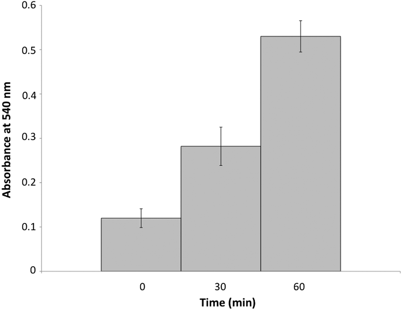
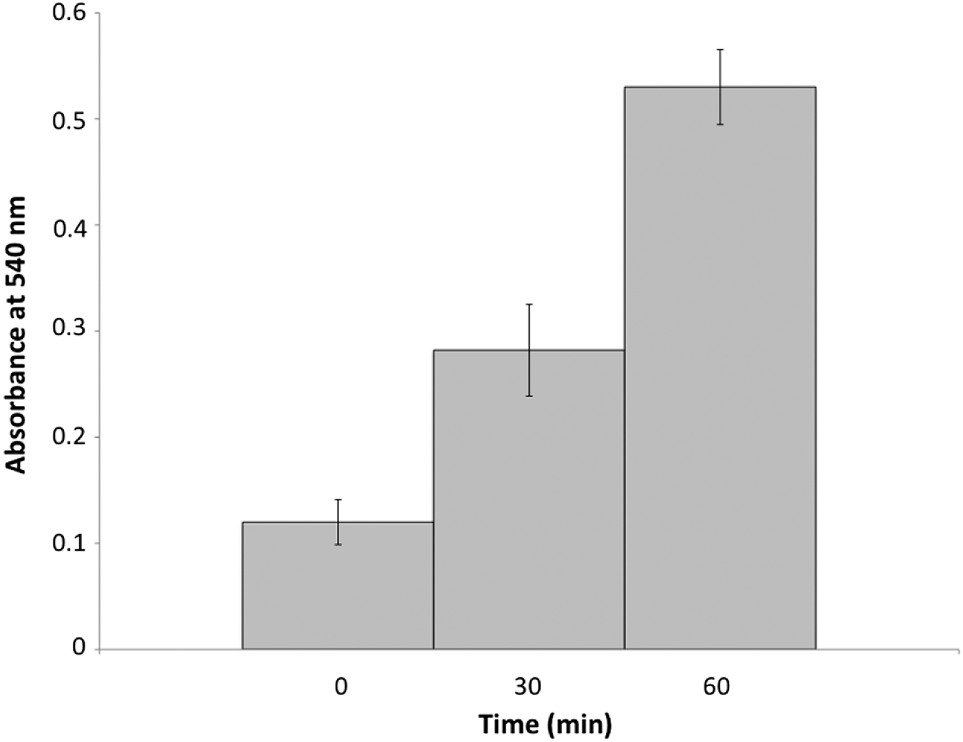
In sea urchins kept in dyed seawater (N = 5), the fluid of the gastric caecum turned red after approximately 30 minutes. The dyed seawater has been observed only inside the foregut and within the gastric caecum, but never in the hindgut. Absorbance measurements of the caecal fluid reveal a significant increase in colour over time (Figure 2). In the beginning (t = 0 minute), the absorbance reaches 0.12 ± 0.021, while 30 minutes later, it has been raised to 0.282 ± 0.043 (P = 0.0121). Finally, at the end of the experiment, it reaches 0.53 ± 0.035 (P = 0.0088).
Absorbance measurements at 540 nm of samples from the caecal fluid of Echinocardium cordatum for specimens placed in seawater (0 minute) and in seawater with neutral red (1 M) for 30 minutes and 60 minutes. According to the Wilcoxon rank test, each series is significantly different from the other series (N = 5, α = 0.05).
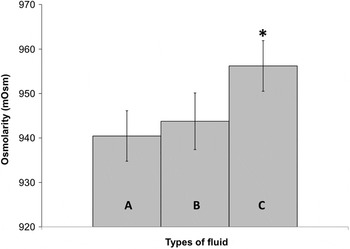
Osmolarity measurements of the coelomic and the caecal fluids (N = 15) indicate that the osmolarity of the caecal fluid (943.76 ± 6.37 mOsm, P = 0.6612) and that of the surrounding seawater (940.45 ± 5.68 mOsm, P = 0.6809) did not differ significantly and that both of them were significantly lower than the osmolarity of the coelomic fluid (956.21 ± 5.69 mOsm, P = 0.0038; Figure 3).
Osmolarity measurements in mOsm of seawater (A), the caecal fluid (B), and the coelomic fluid (C). The asterisk (*) signifies a significant difference according to a Tukey–Kramer post-hoc test (N = 15, α = 0.05).

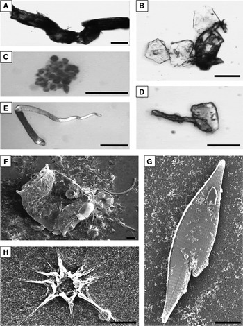
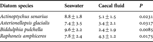
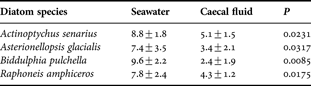
A variety of detrital particles do occur in the caecal fluid. These particles were identified as plant (Figure 4A) or animal (Figure 4B) fragments, flocculent organic aggregates of undetermined origin (Figure 4C), and pipe-like structures resembling fungal sporangia sensu Egglishaw (Reference Egglishaw, Melchiorri-Santolini and Hopton1972) (Figure 4D). Egg capsules of parasitic platyhelminthes with an undulating extremity were also regularly observed (Figure 4E). The most abundant particles were diatoms. Four species, namely Actinoptychus senarius (Ehrenberg) Ehrenberg, 1843, Biddulphia pulchella Gray, 1821 (Figure 4F), Raphoneis amphiceros (Ehrenberg) Ehrenberg, 1844 (Figure 4G) and Asterionellopsis glacialis (Castracane) Round, 1990 (Figure 4H) were systematically present in all the samples, but were significantly less abundant in the caecal fluid as compared to the seawater samples (N = 15; Table 1).
Contrast phase (A–E) and scanning electron (F–H) microscopic images of detrital particles from the caecal fluid of Echinocardium cordatum. (A) Plant fragment (scale bar = 25 µm); (B) remains of chitinous fragments from a crustacean carapace (scale bar = 25 µm); (C) organic aggregates of undetermined origin (scale bar = 25 µm); (D) pipe-like structures, presumably fungal sporangia (scale bar = 25 µm); (E) tapered capsules with an undulating extremity, possibly egg capsules of platyhelminthes (scale bar = 100 µm); (F–H) diatoms: Biddulphia pulchella on the left and Actinoptychus senarius on the right (F, scale bar = 10 µm); Raphoneis amphiceros (G, scale bar = 10 µm); and Asterionellopsis glacialis (H, scale bar = 10 µm).

Concentration of diatoms (/ml) within the caecal fluid as well as in the seawater originating from the area surrounding the specimen (N=15, mean±SE), (t-test, α=0.05).
DISCUSSION
Numerous burrowing deposit feeders have developed mechanisms to collect sediment from the seafloor surface. In Echinocardium cordatum, surface sediment reaches the animal by falling down the chimney of the burrow. Once located on top of the animal, it is carried to the mouth by specialized spines (De Ridder et al., Reference De Ridder, Jangoux and De Vos1987). Within the oesophagus, the sediment with its load of organic matter is moulded into a compact cylinder owing to intensive mucus secretion and to the peristalsis of the oesophagus and anterior stomach (Holland & Ghiselin, Reference Holland and Ghiselin1970; De Ridder, Reference De Ridder1987; De Ridder & Jangoux, Reference De Ridder and Jangoux1993).
In addition, E. cordatum, like most infaunal organisms, also generates a steady influx of seawater from the seafloor into its burrow (Foster-Smith, Reference Foster-Smith1978). This well-described circulation is due to currents created by ciliated spines. It conveys seawater first over the respiratory tube-feet and, later on, into the mouth and the sanitary drain (Nichols, Reference Nichols1959a, Reference Nicholsb; Buchanan, Reference Buchanan1966). Owing to the even macroscopically visible peristaltic movements of the oesophagus and of the siphon, sea urchins in general are able to ingest seawater (Stott, Reference Stott1955; Buchanan, Reference Buchanan1969; Fechter, Reference Fechter1972; Buchanan et al., Reference Buchanan, Brown, Coombs, Pirie and Allen1980; Hollertz & Duchêne, Reference Hollertz and Duchêne2001). In E. cordatum, this pumping action allows seawater to be moved from the mouth to the proximal part of the stomach and more particularly towards the siphon and the slit-like opening of the gastric caecum (De Ridder, Reference De Ridder1987; De Ridder & Jangoux, Reference De Ridder and Jangoux1993). Seawater is then carried through the siphon to the intestine where it is later eliminated through the anus. Our results demonstrate that seawater can also enter the gastric caecum and in fact constitutes most of its fluid content. Our conclusions have been based on various evidences. Firstly, when individuals are kept in aquaria containing coloured seawater, only the caecal fluid (as opposed to the coelomic fluid) becomes coloured. Secondly, osmolarity values of natural seawater and that of the caecal fluid are not significantly different. Thirdly, the suspended particles occurring within the caecal fluid correspond to a range of detrital particles that commonly occur in seawater over the seafloor (Egglishaw, Reference Egglishaw, Melchiorri-Santolini and Hopton1972, inter alia). The diatoms that were identified in the surrounding seawater and within the caecum are phytoplanktonic species (Hendey, Reference Hendey1974; M'Harzi, Reference M'Harzi1999; Horner, Reference Horner2002; Muller, Reference Muller2004) that must have accumulated on the seafloor after their death.
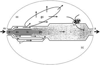
Movement of seawater into the non-muscular and blind-ending gastric caecum appears at first sight rather improbable. However, since the coelomic fluid surrounding the gastric caecum has a significantly higher osmolarity than that of the caecal fluid (Figure 3), an osmotic gradient exists across the relatively thin, almost transparent caecal wall. This leads us to hypothesize that osmotic pressure drives water molecules across the caecal wall into the coelomic cavity. In turn, seawater from the lumen of the anterior stomach flows into the gastric caecum in order to replace water lost to the coelomic cavity (Figure 5). In order to balance this influx into the coelomic cavity, a similar volume of fluid must return to the digestive tract lumen (Fechter, Reference Fechter1972). This process could take place via the rectum and the intestinal caecum. In these parts of the digestive tract, molecules from the coelomic cavity have been shown to move from the coelomic cavity towards the digestive tract lumen, before escaping through the anus (Warnau et al., Reference Warnau, Temara, Ameye and Jangoux1998).
Schematic drawing of the hypothetical trajectory of water in Echinocardium cordatum. The digestive tract is drawn uncoiled. Small arrows correspond to internal seawater pathways, large arrows to water flowing into and out of the animal. The ingested sediment is represented in grey: dark grey corresponds to the mucus-moulded sediment column and light grey to scattered sediment. a, anus; as, anterior stomach; cc, coelomic cavity; e, oesophagus; gc, gastric caecum; I, intestine; ic, intestinal caecum; is, intermediate stomach; m, mouth; ps, posterior stomach; r, rectum; s = siphon.

Detrital particles are abundant in coastal waters and are used as a source of nutrients by a well-represented feeding guild in the marine realm—the suspension feeders (see, for example, Margalef, Reference Margalef and Lauff1967; Turquier, Reference Turquier1989). In order to catch suspended particles, these animals have developed a variety of specialized mechanisms that involve two basic mechanical processes: use of water currents (whether generated or not by the organism itself) and particle interception (the organism places specialized organs or appendages across these water currents). Based on the observations presented here, we hypothesize that the gastric caecum in E. cordatum, and probably also the gastric caecum found in other irregular sea urchins, acts as an internal trap that intercepts suspended organic matter in seawater. Being trapped in the caecal lumen, these particles could then undergo digestion by bacterial activity (Thorsen, Reference Thorsen1998, Reference Thorsen1999) and/or the secretions of glandular enterocytes that are abundant in the gastric caecum (De Ridder & Jangoux, Reference De Ridder and Jangoux1993). The general morphology of the gastric caecum fits an absorptive function. The well-developed microvilli, the occurrence of primary and secondary lysosome-like vacuoles in the enterocytes, and the extensive network of haemal lacunae within the caecal wall in spatangoids (De Ridder & Jangoux, Reference De Ridder and Jangoux1993; Ziegler et al., Reference Ziegler, Mooi, Rolet and De Ridder2010) are indicative of nutrient exchange in this part of the digestive tract.
Many irregular sea urchins and almost all spatangoids possess a well-developed gastric caecum (Ziegler et al., Reference Ziegler, Mooi, Rolet and De Ridder2010). Since it is precisely these sea urchin taxa that have acquired a predominantly infaunal lifestyle, we hypothesize that the development of the gastric caecum could be an adaptation of the irregular sea urchin digestive tube to optimize detritus exploitation, presumably as a response to restricted access to food sources. Through the gastric caecum acting as an internal trap, partially as well as fully infaunal spatangoids could gain additional access to suspended particulate organic matter in seawater. Spatangoid species could consequently have adopted a generalized diet made of two complementary feeding strategies (i.e. deposit and suspension feeding) and would therefore be particularly well adapted to exploit a wide range of detritus. Suspended organic particles in the water column constitute a complementary food source in a clypeasteroid species (Dendraster excentricus) as demonstrated by Chia (Reference Chia1969) and Timko (Reference Timko1976), and in two spatangoid species as suggested by Hollertz (Reference Hollertz1999) for Brissopsis lyrifera and by Cramer (Reference Cramer1991) for E. cordatum.
However, this additional feeding mode and its importance in terms of food supply remain to be demonstrated. The evaluation of the quantitative importance of the uptake of suspended organic matter, observation of its extracellular and intracellular digestion within the caecum, investigation on the absorption of nutrients by enterocytes, and the identification of the bacterial community found within the caecal fluid as compared to the bacterial community found in the surrounding seawater and the sediment are aspects that remain to be studied in order to fully demonstrate that E. cordatum uses suspended particles as a complementary food source.
ACKNOWLEDGEMENTS
We are grateful to the Station Marine at Wimereux (Nord-Pas-de-Calais, France), the Biologische Anstalt Helgoland (Helgoland, Germany), and to André Péqueux (Université de Liège, Belgium) for providing facilities. We also thank Véronique Rousseau and Jean-Yves Parent (Université Libre de Bruxelles, Belgium) for help with the identification of diatoms. We are grateful to Susanne Mueller (Charité-Universitätsmedizin Berlin, Germany) for MRI support. Rich Mooi (California Academy of Sciences, San Francisco, USA), John Lawrence (University of South Florida, Tampa, USA), and Michel Jangoux (Université Libre de Bruxelles, Belgium) are acknowledged for their encouraging comments on previous drafts of this paper. This study was supported by an FRFC grant (No. 2.4594.07) to Chantal De Ridder, and constitutes a contribution of the Centre Inter-universitaire de Biologie Marine (CIBIM).