





Non-technical Summary
This contribution describes Keimeliopteridae, a new family of insects containing two new species, Keimelioptera solensis and Keimelioptera rara, from the exceptionally preserved Solite deposit of Virginia and North Carolina. These insects lived during the Late Triassic Period, about 220 million years ago. A phylogenetic analysis determined that Keimeliopteridae is an early relative of modern bark lice, true lice, thrips, and hemipterans on the basis of morphological characteristics, including primitive features of their wing venation. Keimeliopteridae is unique, and similar insect groups are unknown from elsewhere during the late Paleozoic and early Mesozoic eras, which may be expected given the general rarity of shared taxa between the Solite deposit and other Triassic insect deposits. This new family further demonstrates the significance of the Solite deposit as a window into the evolutionary history of insects following the End Permian Mass Extinction.
Introduction
Crown Paraneoptera Martynov, Reference Martynov1923 (= Acercaria Börner, Reference Börner1904) is a diverse group of insects comprising one extinct and three extant higher taxa: †Permopsocida Tillyard, 1926 (Tillyard, Reference Tillyard1926a) (= Tetrastigmoptera Kluge, Reference Kluge2019), Psocodea Hennig, Reference Hennig1966 (barklice, booklice, and parasitic lice), Thysanoptera Haliday, Reference Haliday1836 (thrips), and Hemiptera Linnaeus, Reference Linnaeus1758 (e.g., aphids, scales, cicadas, hoppers, true bugs). The evolutionary history of this clade is reflected in the morphology of their mouthparts, from generalized “picking” or chewing mouthparts in Psocodea to the distinctive piercing-sucking “beaks” of Hemiptera (Grimaldi and Engel, Reference Grimaldi and Engel2005). The clade Paraneoptera is also referred to as Acercaria, which translates to “without cerci” or “not cerci-bearing,” due to their loss of these paired abdominal appendages. A recent phylogenetic analysis of Paraneoptera (Prokop et al., Reference Prokop, Pecharová, Garrouste, Beattie, Chintauan-Marquier and Nel2017) grouped the extinct taxa Hypoperlidae Martynov, Reference Martynov1928 and Miomoptera Martynov, Reference Martynov1927 with Paraneoptera; however, because of some plesiomorphic traits including the retention of cerci, hypoperlids and miomopterans do not fit within crown Paraneoptera and, instead, fall as sister to the crown group. Paraneoptera therefore includes crown taxa Psocodea + (†Permopsocida + (Hemiptera + Thysanoptera)) plus the stem-group taxa Miomoptera and Hypoperlidae (Prokop et al., Reference Prokop, Pecharová, Garrouste, Beattie, Chintauan-Marquier and Nel2017), herein referred to as Paraneoptera sensu lato (s.l.).
Hypoperlidae were originally described by Martynov (Reference Martynov1928) on the basis of a series of unusual wings from the Permian of Russia. The type species was later revised by Huang et al. (Reference Huang, Bechly, Nel, Engel and Prokop2016), demonstrating the presence of paraneopteran forewing synapomorphies, including fused veins R + M + CuA, fused veins M + CuA separating distally from vein R, a cua-cup crossvein, and a two-branched CuA vein forming an areola postica (Nel et al., Reference Nel, Prokop, Nel, Grandcolas, Huang, Roques, Guilbert, Dostál and Szwedo2012a; Huang et al., Reference Huang, Bechly, Nel, Engel and Prokop2016, supplementary information).
Miomoptera shares the same paraneopteran forewing synapomorphies with Hypoperlidae, the main difference between the taxa being the structure of their cerci. In Hypoperlidae, the cerci are reduced and undivided, while they are long and likely dimerous in Miomoptera (Prokop et al., Reference Prokop, Pecharová, Garrouste, Beattie, Chintauan-Marquier and Nel2017). Since their initial descriptions, various fossils have been ascribed to these two taxa. Re-examination of these taxa, however, revealed that many differ from the typical paraneopteran venation and should be excluded from their respective clades; therefore, only the type families of each (Hypoperlidae and Palaeomanteidae) are recognized as belonging to Paraneoptera s.l. (Prokop et al., Reference Prokop, Pecharová, Garrouste, Beattie, Chintauan-Marquier and Nel2017).
Paraneoptera have a long fossil history, stretching back to the Carboniferous, with stem-group and basal taxa of †Permopsocida (Carbonopsocus Prokop et al., Reference Prokop, Rosová, Pecharová, Sroka, Leipner and Nel2024), Psocodea (Westphalopsocus Nel et al., Reference Nel, Roques, Nel, Prokin and Bourgoin2013), Thysanoptera (Westphalothripides Nel et al., Reference Nel, Azar, Prokop, Roques, Hodebert and Nel2012b), and Hemiptera (Protoprosbole Laurentiaux, Reference Laurentiaux1952; Aviorrhyncha Nel et al., Reference Nel, Roques, Nel, Prokin and Bourgoin2013; Carbonatura Boderau et al., Reference Boderau, Schubnel, Fu, Mottequin, Robin, Roques, Kundura, Garrouste and Nel2026; Carbopsyllidium Boderau et al., Reference Boderau, Schubnel, Fu, Mottequin, Robin, Roques, Kundura, Garrouste and Nel2026) known from the Pennsylvanian of Europe. The stem-group paraneopterans Mazonopsocus Prokop et al., Reference Prokop, Pecharová, Garrouste, Beattie, Chintauan-Marquier and Nel2017 (Miomoptera, Palaeomanteidae) and Bruayaphis Nel et al., 2012 (Nel et al., Reference Nel, Prokop, Nel, Grandcolas, Huang, Roques, Guilbert, Dostál and Szwedo2012a) are also known from the Moscovian, while Hypoperlidae appear slightly later during the middle Permian (Martynov, Reference Martynov1928; Prokop et al., Reference Prokop, Pecharová, Garrouste, Beattie, Chintauan-Marquier and Nel2017). Although one miomopteran was reported from the Triassic of Argentina (Miomina riojana Martins-Neto et al., Reference Martins-Neto, Brauckmann, Gallego and Carmona2006), the wing is incomplete; therefore, neither hypoperlids nor miomopterans are definitively known from the Mesozoic. Herein, we describe Keimeliopteridae n. fam., the first stem-group Paraneoptera from the Mesozoic, specifically from the Upper Triassic (Norian) Solite deposit of Virginia and North Carolina. Like Hypoperlidae and Miomoptera, these insects share many forewing venation characters with Paraneoptera sensu stricto (s.s.). However, a phylogenetic analysis indicates placement outside of the crown group and as sister to Paraneoptera s.s. due to a series of retained plesiomorphies, including pentamerous maxillary palpi, pentamerous tarsi, and probable cerci.
Materials and methods
Locality and materials
The insects described in this study were collected from the Upper Triassic Cow Branch Formation, located in the Dan River Basin of southern Virginia and northern North Carolina, United States. The fossiliferous portions of this formation comprise part of the Solite deposit, or Solite Konservat-Lagerstätte, outcropping in the former Solite Corporation Quarries in Cascade, Virginia. Most of the Solite insects come from a 34 mm thick layer referred to as the “insect layer” (cycle 2; Olsen, Reference Olsen1979), which is approximately 220 million years old (Norian; Olsen et al., Reference Olsen, Reid, Taylor, Whiteside and Kent2015; Kent et al., Reference Kent, Olsen and Muttoni2017). The Solite insects are preserved as two-dimensional, silvery, carbonaceous films in a matrix of fine-grained black mudstone. Their preservation is often exceptional, with many specimens nearly complete and fully articulated; microscopic features such as setae and microtrichia are common. In addition to abundant insects from at least nine orders, plants, clam shrimp (“conchostracans”), occasional fish, and the aquatic reptile Tanytrachelos ahynis Olsen, Reference Olsen1979 have also been recovered from the insect layer (Fraser et al., Reference Fraser, Grimaldi, Axsmith, Heckert, Liuktus-Pierce, Smith, Dooley, Fraser and Sues2017).
Methods
Specimens were wetted with 70% ethanol to increase contrast and viewed using a Nikon SMZ1500 stereomicroscope. A fiber-optic ring light provided the non-directional, diffuse light source required to illuminate the silvery, carbonaceous film in which the insects are preserved. Photographs were taken using a Nikon 16MP camera with Nikon Elements NIS software. Because of the minute size of these insects (1–2 mm body length), a Nikon petrographic microscope fitted with 4×, 10×, 20×, and 40× PlanApo lenses having epioptic, polarized illumination was used for higher-resolution study. This allowed for examination of the microscopic details of the wing venation, setae, and microtrichia. Specimens were illustrated using Adobe Illustrator, and measurements were taken using ImageJ software. Venation nomenclature follows that of Nel et al. (Reference Nel, Prokop, Nel, Grandcolas, Huang, Roques, Guilbert, Dostál and Szwedo2012a).
A phylogenetic analysis using morphological data was performed using Asado version 1.61 (formerly WinClada) (Nixon, Reference Nixon1999–2004). The taxon list (Appendix 1) and character list (Appendix 2) were modified from those used by Huang et al. (Reference Huang, Bechly, Nel, Engel and Prokop2016) and Prokop et al. (Reference Prokop, Pecharová, Garrouste, Beattie, Chintauan-Marquier and Nel2017) with character additions and minor coding corrections where necessary (Appendix 3). A total of 16 terminal taxa were included: three outgroups in Polyneoptera and Holometabola and 13 ingroup taxa. Due to the fragmentary preservation of many fossil taxa, two species of each genus were coded and combined as generic composites for most terminals. Character 36 (location where BSc and 2Ax separate in wing articulation), character 38 (sclerotization of forewing relative to hind wing), character 41 (location where vein M separates from vein R), character 43 (angle of radial stem at point of re-emergence of veins CuA and M), and character 46 (whether vein RP is forked or unforked) from the matrices of Huang et al. (Reference Huang, Bechly, Nel, Engel and Prokop2016) and Prokop et al. (Reference Prokop, Pecharová, Garrouste, Beattie, Chintauan-Marquier and Nel2017) were omitted for lack of clarity or for being uninformative within the present coded taxon set. In addition, 11 characters were added to those used in the matrices of Huang et al. (Reference Huang, Bechly, Nel, Engel and Prokop2016) and Prokop et al. (Reference Prokop, Pecharová, Garrouste, Beattie, Chintauan-Marquier and Nel2017). Characters were treated as non-additive (unordered). The phylogenetic tree was created using the program’s ratchet function with 200 iterations/rep and the remaining options set to their defaults.
Repositories and institutional abbreviations
The specimens described herein are housed at the Virginia Museum of Natural History (VMNH) in Martinsville, Virginia, USA; the American Museum of Natural History (AMNH) in New York, New York, USA; and the Yale Peabody Museum of Natural History in New Haven, Connecticut, USA.
Systematic paleontology
Clade Clareocercaria Prokop et al., Reference Prokop, Pecharová, Garrouste, Beattie, Chintauan-Marquier and Nel2017
Clade Paraneoptera Martynov, Reference Martynov1923 sensu lato
Order Incertae sedis
Family Keimeliopteridae new family
Type genus
Keimelioptera, n. gen. by present designation.
Diagnosis
Antennal flagellum with base swollen and distal half stylate; lacking bulbous clypeus and bulging eyes; palpi well developed, multi-articled; wings heteronomous, hind wing ~0.7× length of forewing, venation reduced; forewing with pigmented pterostigma between branches of RA, perpendicular crossvein below middle of pterostigma, RP three-branched; long M + CuA fusion, M three-branched; areola postica present (its peak fused with M for a short distance); setulae present along stem M, CuA, and CuP; M + CuA not emerging from R + M + CuA near wing base; cua-cup very short; margins of fore- and hind wings with fringe of setae; male gonostyli stout, well developed.
Etymology
From the Greek keimēlion (κειμήλιον), meaning “relic” or “heirloom,” and pteron (πτερόν), meaning “wing,” referring to the relict nature of the forewings.
Genus Keimelioptera new genus
Types species
Keimelioptera solensis n. gen. n. sp. by present designation.
Diagnosis
Body small (1.4–2.2 mm); antennal flagellum with base bulbous and distal half stylate; mouthparts hypognathous, palpi present; thorax compact, short and deep, segments integrated, mesothorax large; wings heteronomous, hind wing ~0.7× length of forewing, with reduced venation; forewing slender with three or four internal cells; Sc tightly adjoining R + M + CuA proximally, then diverging to anterior wing margin in proximal fifth of wing length; radial field with two closed cells and pigmented pterostigma between branches of RA, crossvein below middle of pterostigma perpendicular to RA2 and RP1+2, RP with three terminal branches, rp2-rp3 and rp3-m1 present; long M + CuA fusion, M + CuA diverging from R in proximal fifth of wing length; M diverging from M + CuA in proximal quarter of wing length, M with three pectinate branches; areola postica (ap) formed between two branches of CuA and cua1-cua2 crossvein, anterior corner of ap fused with stem M for a short distance; CuP reduced, reaching posterior wing margin in proximal ~third of wing; cua-cup crossvein and two anal veins present; setulae along CuA, CuP, and part of stem M; margins of fore- and hind wings with fringe of setae; legs relatively long and slender, especially tibiae; tarsi pentamerous; male terminalia large, gonocoxites stout, gonostyli well developed; female with short, setulose terminal appendages, probably cerci.
Occurrence
Former Solite Quarry, Eden, Rockingham County, North Carolina, USA (36°32’29.7"N, 79°40’12.8"W); Norian, Upper Triassic, Cow Branch Formation.
Etymology
From the Greek keimēlion (κειμήλιον), meaning “relic” or “heirloom,” and pteron (πτερόν), meaning “wing,” referring to the relict nature of the forewings.
Remarks
Keimelioptera n. gen. shares a variety of characters with various paraneopteran taxa, which has caused their identity to remain elusive. They were originally suggested as members of the extinct hemipteran family Archescytinidae Tillyard, Reference Tillyard1926b because of venation similarities (Fraser et al., Reference Fraser, Grimaldi and Olsen1996), but reexamination of the mouthparts revealed that the structures originally assumed to be parts of a rostrum were palpi, precluding their placement within Hemiptera. Furthermore, their plesiomorphic retention of pentamerous maxillary palpi, pentamerous tarsi, and probable cerci suggest that they belong in a stem-group paraneopteran clade, outside of Paraneoptera s.s.
Keimelioptera solensis new species
Keimelioptera solensis n. gen. n. sp., holotype, VMNH 49652: (1) part; (2) counterpart. Scale bar = 0.5 mm.

Figure 1. Long description
A two-panel grayscale photograph of a fossilized insect specimen against a dark, textured rock matrix.
Panel 1, located on the left, shows the part of the holotype V M N H 49652. The insect is oriented vertically with its head at the top. The head features two thin antennae extending upward. The thorax is broad and light-colored due to mineralization. Two pairs of wings are spread horizontally. The forewings are long and narrow with visible venation patterns, while the hindwings are partially obscured. The abdomen is segmented, tapering slightly toward the posterior end. Thin legs are visible extending from the thoracic region.
Panel 2, located on the right, shows the counterpart of the same specimen. It presents a mirror-image impression of the insect described in panel 1. The mineralization appears brighter and more concentrated on the head and thorax in this view. The wing venation is more distinct on the left side of this panel.
A vertical white scale bar representing 0.5 mm is located in the bottom-right corner of panel 2.
Keimelioptera solensis n. gen. n. sp., type series: (1) VMNH 50096; (2) VMNH 94561; (3) VMNH 50530; (4) VMNH 49655; (5) AMNH 04-38; (6) AMNH 04-100. Scale bars = 0.5 mm.

Figure 2. Long description
A multi-panel photo containing six individual panels labeled 1 through 6. Each panel features a white scale bar in the bottom-right corner representing 0.5 mm. The fossils appear as light-colored, detailed impressions against a dark, textured matrix.
* Panel 1: Top-left. A dorsal view of V M N H 50096 showing a symmetrical insect with wings spread wide, a segmented abdomen, and a distinct head and thorax.
* Panel 2: Top-right. V M N H 94561 is shown in a slightly oblique dorsal view with the abdomen curved to the left and wings partially overlapping.
* Panel 3: Middle-left. V M N H 50530 displays a ventral or dorsal view with a very clear, elongated segmented abdomen and thin, spindly legs extending from the thorax.
* Panel 4: Middle-right. V M N H 49655 shows a lateral view of the insect, highlighting the profile of the head and the hind wing stretched vertically above the insect’s body.
* Panel 5: Bottom-left. A M N H 04-38 is a lateral view showing a robust, segmented body and the venation pattern of a single prominent wing.
* Panel 6: Bottom-right. A M N H 04-100 provides a high-magnification close-up of the wing venation and a portion of the thorax, showing the intricate network of veins.
Reconstruction of Keimelioptera solensis n. gen. n. sp., male. (1) Full body; (2, 3) forewing and hind-wing venation. A1, A2 = anal veins (first, second); CuA1, CuA2 = cubitus anterior veins (first, second); cua-cup = cua-cup crossvein; CuP = cubitus posterior; M1, M2, M3 = media (first, second, third); RA, RA1, RA2 = radius anterior veins (first, second); RP, RP1, RP2, RP3 = radius posterior veins (first, second, third); Sc = subcostal vein. Scale bars = 0.25 mm.

Figure 3. Long description
Panel 1 shows a dorsal line drawing of a male insect with short antennae, a segmented abdomen, and four wings. The forewings feature a dark pterostigma on the leading edge. Scale bar equals 0.25 mm.
Panel 2 displays the black-and-white venation of the forewing and hind-wing. Starting from the top leading edge and moving clockwise, labels include S c, R A sub 1, R A sub 2, R P sub 1, R P sub 2, R P sub 3, M sub 1, M sub 2, M sub 3, C u A sub 1, and C u A sub 2. Along the trailing edge are C u P, A sub 1, and A sub 2. A crossvein labeled c u a dash c u p connects the cubitus veins near the base. The hind-wing below shows partial venation with labels preceded by question marks: ? R A, ? R P, ? M sub 1, and ? M sub 2.
Panel 3 replicates the venation from Panel 2 but uses color-coding to differentiate vein groups. The R A group is red, R P is orange, M is green, C u A is blue, and the anal veins are purple. The vein at the base of the wing is labeled R plus M plus C u A.
Head and mouthpart morphology of Keimelioptera solensis n. gen. n. sp. (1, 2) VMNH 49655; (3, 4) AMNH 04-38; (5) VMNH 94514; (6) VMNH 94563. Scale bars = 0.1 mm.

Figure 4. Long description
A multi-panel figure containing four photographs and two line drawings.
* Panel 1 is a high-contrast photograph of specimen V M N H 49655 showing a lateral view of the head and elongated mouthparts against a dark matrix.
* Panel 2 is a line drawing corresponding to panel 1. It illustrates the head capsule and a pair of elongated palpi. A vertical scale bar at the bottom right represents 0.1 mm.
* Panel 3 is a photograph of specimen A M N H 04-38 showing another lateral view of the head and mouthparts. .
* Panel 4 is a line drawing corresponding to panel 3. It labels the pedicel, flagellum, and palpi. The palpi are shown as segmented structures at the bottom left. A vertical scale bar at the bottom right represents 0.1 mm.
* Panel 5 is a photograph of specimen V M N H 94514 showing the head with long, thin and bent antennae extending to the left and right. A white scale bar is at the bottom right.
* Panel 6 is a photograph of specimen V M N H 94563 providing another lateral view of the head morphology. A white scale bar is at the bottom right.
Thorax and leg morphology of Keimelioptera solensis n. gen. n. sp. (1, 2) AMNH 04-38; (3, 4) AMNH 04-130; (5, 6) VMNH 50096; (7) bristles on thorax, VMNH 93570. cx1 = procoxa; epm2 = epimeron 2; eps2 = episternum 2; epm3 = epimeron 3; eps3 = episternum 3; mscm = mesoscutum; mstlm = mesoscutellum. Scale bars = 0.25 mm.

Figure 5. Long description
A multi-panel display containing four photographs and three line drawings of insect fossils.
* Panels 1 and 2: A photograph and corresponding line drawing of the lateral view of the thorax of specimen A M N H 04-38. The drawing identifies the m s c m at the top, followed by the m s t l m. Below these are the e p s 2 and e p m 2, with the e p s 3 and e p m 3 positioned further back. The c x 1 is located at the lower right of the thoracic block.
* Panels 3 and 4: A photograph and line drawing of the lateral view of the thorax of specimen A M N H 04-130. The m s t l m and m s c m are at the dorsal peak. The lateral sclerites are labeled from right to left as c x 1, e p s 2, e p m 2, e p s 3, and e p m 3.
* Panels 5 and 6: A line drawing and photograph of the thorax of specimen V M N H 50096. The drawing shows a dorsal view of the thorax with the m s t l m centered and small bristles scattered across the surface.
* Panel 7: A photograph of specimen V M N H 93570 focusing on the fine bristles on the thorax.
Vertical scale bars in the bottom right of panels 2, 4, 5, 6, and 7 represent 0.25 mm.
Forewing and hind-wing morphology of Keimelioptera solensis n. gen. n. sp. (1) VMNH 94561; (2) VMNH 50096; (3) VMNH 49744; (4) forewing microtrichia, VMNH 49744; (5) reduced hind-wing venation and marginal fringe, VMNH 49655; (6) bristles, or setulae, along CuA vein, VMNH 49636. (1–3, 5, 6) Scale bars = 0.25 mm; (4) scale bar = 25 μm.

Figure 6. Long description
A collection of six black and white micrographs of fossilized insect wings.
* Panel 1: Shows two overlapping wings of specimen V M N H 94561. The wings are elongated with a complex network of light-colored veins against a dark, textured rock matrix. A vertical scale bar is in the bottom right.
* Panel 2: Displays specimen V M N H 50096, showing a single forewing with clearly defined longitudinal and cross veins forming several closed cells. A vertical scale bar is in the bottom right.
* Panel 3: Features specimen V M N H 49744, a well-preserved forewing showing the branching pattern of the veins toward the outer margin. A vertical scale bar is in the bottom right.
* Panel 4: A high-magnification close-up of specimen V M N H 49744, revealing a dense covering of tiny hair-like structures called microtrichia on the wing surface. A vertical scale bar representing 25 micrometers is in the bottom right.
* Panel 5: Shows specimen V M N H 49655, a narrow hind-wing with reduced venation and a distinct fringe of fine hairs along the posterior margin. A vertical scale bar is in the bottom right.
* Panel 6: Displays specimen V M N H 49636, focusing on the C u A vein which is lined with distinct bristles or setulae. A vertical scale bar is in the bottom right.
Abdomen and terminalia morphology of Keimelioptera solensis n. gen. n. sp. (1–4, 10) Female terminalia, most likely cerci. (5–9) Male terminalia. (1) YPM IP 036453; (2) VMNH 93570; (3) VMNH 51743; (4) VMNH 50096; (5, 6) VMNH 50530, part and counterpart; (7) VMNH 94561, oblique view; (8) VMNH 49652; (9) male terminalia, lateral view, VMNH 51807; (10) female terminalia, lateral view, VMNH 94563. Scale bars = 0.1 mm.

Figure 7. Long description
A collection of 10 high-contrast micrographs of fossilized insect remains.
* Panels 1 through 4 and 10 show female terminalia. In panels 1 to 4, the abdomen tapers toward the bottom of the frame, ending in paired, elongated structures identified as cerci. Panel 10 provides a lateral view of the entire abdomen, showing segmented sclerites and the terminal cerci pointing to the left.
* Panels 5 through 9 show male terminalia. Panels 5 and 6 are part and counterpart of the same specimen, showing a broader, more robust terminal segment compared to the females. Panel 7 shows an oblique view of the terminalia. Panel 8 shows a dorsal or ventral view of the posterior abdomen. Panel 9 provides a lateral view of the male abdomen, showing the segmented structure and the specialized terminal apparatus.
Each panel includes a white vertical scale bar in the bottom right corner representing 0.1 mm. The fossils appear as light, textured mineralizations against a dark rock matrix.
Holotype
VMNH 49652, male, part and counterpart. Paratypes: VMNH 50096 (female), VMNH 94561 (male with excellent venation), VMNH 50530 (male with tarsal segments and terminalia), VMNH 49655 (lateral view with hind wing, mouthparts, and antennae), AMNH 04-38 (lateral view with mouthparts), AMNH 04-100 (lateral view with overlapping fore- and hind wings).
Materials
Approximately 240 specimens. Notable specimens include AMNH 04-101, 04-130; VMNH 49626, 49636, 49649, 49743, 49744, 51743, 51807, 93570, 94514, 94545, 94563; YPM IP 036453.
Diagnosis
Distinguished from the very similar Keimelioptera rara n. gen. n. sp. by the absence of a basal rp3-m crossvein and the sinuous CuA1 vein.
Occurrence
Former Solite Quarry, Eden, Rockingham County, North Carolina, USA (36°32’29.7"N, 79°40’12.8"W); Norian, Upper Triassic, Cow Branch Formation.
Description
Body small, mean length 1.8 mm (range 1.4–2.2 mm, N = 24; excluding antennae and terminalia), males slightly smaller (mean 1.6 mm, N = 10) than females (mean 1.8 mm, N = 9); forewings nearly the same length as body (mean BL/FWL 1.05, N = 13).
Head
Short, relatively deep, head capsule ovoid in lateral view, somewhat trapezoidal in dorsoventral view; mouthparts hypognathous, antennae held forward; eyes (e.g., size, shape) and presence/absence of ocelli not discernable. Frons and vertex with scattered, stiff setulae. Clypeal region and face not inflated or bulging, gena not divided.
Antennae
Stylate, positioned near mouthparts, bases nearly touching. Scape and pedicel seemingly short, ringlike; flagellum bulbous at base, tapered to narrow stylus. Flagellum with fine, scattered setulae, including at tip of stylus. Stylus portion of flagellum slightly greater than length of bulbous portion, stylus width ~0.25× bulbous portion; stylus possibly dimerous, sometimes each stylus with distinct elbow in basal third (no definitive subdivision observed; e.g., VMNH 49655, 94514).
Mouthparts
Hypognathous, suspended beneath head capsule; feeding appendages (e.g., mandibles) obscure (mouthparts not rostrate or siphonate), best seen in specimens preserved in lateral view (e.g., VMNH 49655, AMNH 04-38). Palpi present, with very fine setulae/microtrichia on some parts; articles very faint: in VMNH 49655, one palpus appears pentamerous, corresponding to a maxillary palpus, another palpus with three thicker palpomeres (labial palpus?). Labrum not observed.
Thorax
Short, very compact, with some sutures preserved between pro- and mesothoracic segments, although thoracic segments and sclerites highly integrated, no constrictions between segments apparent. In lateral view, thorax much deeper than long. Pro-, mesothoracic pleural suture apparent (e.g., VMNH 94614), prothorax short; mesoscutum occupies most of thoracic dorsum but differs in size (cf. AMNH 04-38, AMNH 04-130), with scattered, fine, stiff setulae; mesoscutellum small, slightly rectangular in dorsal view (width ~0.3× width of thorax). Episternum2 large, narrow, with definitive sulcus between it and epimeron2; episternum3 and epimeron3 well developed.
Legs
Relatively long and slender, without spines, large bristles, or apical/preapical spurs on any podomeres. Procoxa relatively large, long, laterally positioned, separated by gap from ipsilateral cx2; cx2 and cx3 small, entirely ventral, very close, touching mesally or nearly so. Metafemur (f) shorter than metatibia (tb), mean f/tb 0.6 (N = 4, range 0.5–0.7); tb much longer than metatarsomere1 (ta1), mean tb/ta1 2.3 (N = 4, range 1.9–2.5). Tarsi pentamerous, very long, tarsomeres decreasing in length from proximal to distal one; pulvilli present but small, empodium obscure, pretarsal claws small, no longer than pulvilli.
Wings
Forewing slender, length/greatest width 2.5–4.3 (mean 3.2, N = 10), apex rounded (not narrowed); hind wing length 0.7× that of forewing, venation very reduced. Membrane of fore- and hind wings entirely covered with dense, minute microtrichia. Margins of wings with fringe of fine setae; setae longest on anal lobe area, gradually decreasing to tip of wing, where very short. Forewing venation: vein C circumambient. Sc very short, tightly adjoining R + M + CuA proximally (not fused), then diverging to anterior wing margin in proximal fifth of wing length, no crossveins or branches, apex meeting C not far from wing base. RA apically forked, RA1 very short, RA2 much longer; heavily sclerotized pterostigma bounded between RA1, RA2, and C. RP branching from near middle of RA, RP with three terminal branches; short crossvein below middle of pterostigma, running perpendicular between RA2 and RP1+2; RP3 branching from RP1+2 at near right angle, just proximal to ra2-rp1+2 crossvein; rp2-rp3 crossvein forming closed cell rp. Long stem M + CuA not emerging from R + M + CuA near to wing base, M branching at virtually right angle from M + CuA in proximal fifth of wing length; M with three terminal branches, short crossvein connecting M1 and RP3. CuA with two terminal branches forming areola postica (ap), upper corner of ap fused with stem M for a short distance; narrow cell mcu formed by short distal crossvein between branches of CuA. CuA1 slightly sinuous, CuA2 straight, except at apex where it bends toward wing margin. CuP straight, with short, basal cua-cup crossvein. Two anal veins present, parallel to each other and to CuP. Setulae along CuA, CuP, and part of stem M. Hind-wing venation: very reduced, anterior margin of C flat (probably fits into and couples with posterior margin of forewing; no coupling structure observed); no pterostigma, vein R simple, except for short, crossvein-like vein (RA?) connecting R to C. R diverging basally from a second vein (M?) with two terminal branches. No other veins preserved.
Abdomen
Relatively slender, with seven well defined tergites, terminal tergite short; pleural membrane exposed. Male terminalia: large, as broad as terminal tergites, composed of pair of stout gonocoxites with well developed, recurved gonostyli. Female abdomen tapered to slender apex, ending in pair of short, setulose appendages located medially, probably cerci; these appendages with blunt, rounded tips, apparently undivided (unimerous), and not held tightly against each other (as in ovipositor valves).
Etymology
The specific epithet is named for the former Virginia Solite Corporation quarries, from which the specimens were recovered.
Remarks
Keimelioptera n. gen. shares similar traits with various crown paraneopteran orders. The forewing venation of Keimelioptera n. gen. (Figs. 3, 6) somewhat resembles that of †Permopsocida (e.g., Psocorrhyncha burmitica Huang et al., Reference Huang, Bechly, Nel, Engel and Prokop2016) in the pterostigma bounded by RA1 and RA2, the crossvein ra2-rp1 below the middle of the pterostigma, and the forked RP. However, Keimelioptera n. gen. differs significantly from this order in the structure of the antennae (long and filiform in †Permopsocida compared with short and stylate with a bulbous base in Keimelioptera n. gen.; Fig. 4), proximal constriction of the abdomen in †Permopsocida (compared with no constriction in Keimelioptera n. gen.), and both the venation (especially the short cua-cup crossvein in Keimelioptera n. gen. compared with a long one in †Permopsocida) and relative sizes of the fore- and hind wings (of similar size and venation in †Permopsocida compared with smaller hind wings with reduced venation and without pterostigma in Keimelioptera n. gen.). The wings of Keimelioptera n. gen. also possess a marginal fringe of setae like those on the wings of thysanopterans; although significantly shorter than those of thrips, these fringes are likely a convergence due to the minute size of these insects. Finally, the distinct bristles along the forewing cubital veins resemble those on the veins of the extinct hemipteran family Protopsyllidiidae Becker-Migdisova, Reference Becker-Migdisova1960 (e.g., Postopsyllidium emilyae Grimaldi, Reference Grimaldi2003).
Keimelioptera rara new species
Keimelioptera rara n. gen. n. sp., holotype, VMNH 49625. (1) Habitus; (2) forewing venation. CuA1, CuA2 = cubitus anterior veins (first, second); M1, M2, M3 = media (first, second, third); RA1, RA2 = radius anterior veins (first, second); RP1, RP2, RP3 = radius posterior veins (first, second, third); Sc = subcostal vein. Scale bars = 0.25 mm.

Figure 8. Long description
Panel 1 is a grayscale photograph of the holotype fossil. It shows a dorsal view of the insect body with a segmented abdomen and a partially preserved forewing extending to the left. A white vertical scale bar is in the bottom right.
Panel 2 is a technical line drawing of the forewing venation. Starting from the proximal wing base on the right, an arrow points to the junction of R plus M plus C u A. Moving along the anterior margin (top), the S c vein runs parallel to the edge for a short distance. A shaded pterostigma is located between R A sub 1 and R A sub 2. Continuing clockwise around the distal apex and posterior margin, the labels are R P sub 1, R P sub 2, R P sub 3, M sub 1, M sub 2, M sub 3, C u A sub 1, and C u A sub 2. Dotted lines indicate inferred or incomplete vein segments. A black vertical scale bar representing 0.25 mm is in the bottom right.
Holotype
VMNH 49625, part and counterpart.
Diagnosis
Distinguished from the very similar Keimelioptera solensis n. gen. n. sp. by the presence of a basal rp3-m crossvein and the linear CuA1 vein.
Occurrence
Former Solite Quarry, Eden, Rockingham County, North Carolina, USA (36°32’29.7"N, 79°40’12.8"W); Norian, Upper Triassic, Cow Branch Formation.
Description
Single specimen known from partial body with one forewing, preserved in oblique view. Body small, 2.2 mm long; forewing length (as preserved) ~0.8× body length. Head short, somewhat ovoid; eyes, mouthparts, and antennae not preserved. Thorax compact, sutures between segments not visible. Fragments of at least three legs present, details not discernable. Single forewing visible, length/greatest width 2.5, apex rounded (not narrowed); at least posterior margin with fringe of fine setae, setulae visible only along part of CuA1, microtrichia on wing surface not preserved; venation as in Keimelioptera solensis n. gen. n. sp. except for short crossvein between right angle of RP3 and stem M forming a fourth closed cell, and CuA1 linear; anal area not preserved. Only anterior margin of hind wing visible beneath overlapping areola postica of forewing. Abdomen slender, with at least six segments visible, terminalia not preserved, sex unknown.
Etymology
The specific epithet is derived from Latin rara, meaning “rare” or “uncommon,” in reference to the singleton status of this species.
Remarks
This species is known from a single specimen that is more poorly preserved than those of the type species. In Keimelioptera solensis n. gen. n. sp., the area directly below the near-right-angle bend in RP appears to be desclerotized and is missing (or there is a hole in the matrix) in every specimen of the type species. Desclerotization seems less pronounced in this species—the single specimen of Keimelioptera rara n. gen. n. sp. is the only one in which a crossvein is evident between RP3 and M in this location. This in addition to a distinctly linear CuA1 (compared with a sinuous CuA1) separate this species from Keimelioptera solensis n. gen. n. sp.
Results
Phylogenetic analysis
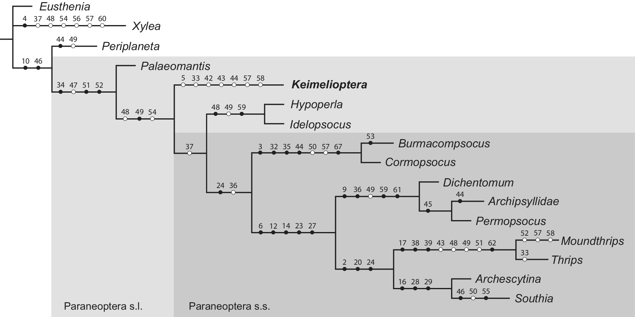
An equally weighted parsimony analysis using the ratchet function in Asado (formerly WinClada) produced a single most parsimonious tree with length = 114, consistency index (CI) = 71, and retention index (RI) = 77 (Fig. 9). Keimeliopteridae n. fam. was recovered outside of crown Paraneoptera, as sister to Hypoperlidae + Paraneoptera s.s.
Cladogram of the single most parsimonious tree showing paraneopteran relationships. Tree length = 114, consistency index (CI) = 71, and retention index (RI) = 77. Circles indicate coded characters. Black circles show non-homoplasious character states (synapomorphies or autapomorphies); white circles indicate homoplasious character states. Character numbers are noted above each circle.

Figure 9. Long description
A phylogenetic tree diagram with a root on the left branching into several lineages.
At the top, Eusthenia and Xylea form an outgroup, with Xylea showing character markers 4, 37, 48, 54, 56, 57, and 60.
Below this, a major clade labeled Paraneoptera s l (sensu lato) begins, marked by characters 10 and 46. This clade splits into Periplaneta (characters 44, 49) and a larger group.
The larger group contains Palaeomantis and a nested clade labeled Paraneoptera s s (sensu stricto). Within Paraneoptera s s, the first branch leads to Keimelioptera (characters 5, 33, 42, 43, 44, 57, 58) and a sister group containing Hypoperla and Idelopsocus.
A deeper branch within Paraneoptera s s leads to a group containing Burmacompsocus and Cormopsocus (characters 3, 32, 35, 44, 50, 57, 67).
The final major cluster branches into three main lineages:
1. A group containing Dichentomum, Archipsyllidae, and Permopsocus.
2. A group containing Moundthrips and Thrips, marked by characters 17, 38, 39, 43, 48, 49, 51, and 62.
3. A group containing Archescytina and Southia, marked by characters 16, 28, and 29.
Black circles represent non-homoplasious states, while white circles represent homoplasious states. Character numbers are positioned above each circle along the horizontal branches.
Discussion
Although Keimeliopteridae n. fam. cannot be assigned to a known order, they can be definitively placed near Paraneoptera because of the fusion of R + M + CuA into a common stem, the presence of an arched cua-cup crossvein, a simple CuP vein, a CuA vein with two branches forming an areola postica, a three-branched M vein, and a general lack of crossveins. A phylogenetic analysis of 13 paraneopteran taxa confirmed this family’s placement outside of crown Paraneoptera, as sister to Hypoperlidae + Paraneoptera s.s. This assignment is based on a series of plesiomorphic characters, namely, the pentamerous maxillary palpi, the pentamerous tarsi, the well developed male gonostyli, and the probable presence of cerci in the female. Due to the two-dimensional preservation of the Solite insects, fine details of these “cerci” are difficult to discern. However, these structures are setulose, have rounded tips, and appear not to be divided into multiple cercomeres. Moreover, they are not held tightly against each other as would be the case if they were ovipositor valves. Their structure is therefore consistent with that of cerci. This, in combination with the other retained plesiomorphies, supports Keimeliopteridae’s placement between Miomoptera and Hypoperlidae + Paraneoptera s.s.
Hypoperlidae, Miomoptera, and Keimeliopteridae n. fam. all share the basic paraneopteran forewing venation. However, certain aspects of their venation easily separate the Solite insects from these taxa, most notably in the branching of the RP vein and the structure of the pterostigma. In both hypoperlids and miomopterans, vein RP usually splits into a series of parallel branches, compared with the single fork of RP in the Solite insects. In both hypoperlids and miomopterans, the pterostigma is also more “primitive,” covering the zone surrounding the apices of veins ScP, RA, and the area between RA and RP. By contrast, Keimeliopteridae n. fam. has a distinct pterostigma that is bounded by the fork of vein RA, similar to that of the crown paraneopteran Archipsyllidae Handlirsch, Reference Handlirsch1906 (order †Permopsocida). Another difference between the Keimeliopteridae n. fam. and these taxa is the structure of the antennae. In both Hypoperlidae and Miomoptera, the antennae are long and filiform; Keimeliopteridae n. fam. has modified antennae that are much shorter, with a swollen base and a stylate distal half. Finally, these insects are also unique within Paraneoptera s.l. in the size and venation of the hind wings. Unlike the other stem-group paraneopteran taxa, Keimeliopteridae n. fam. possesses hind wings with reduced venation that are significantly smaller than the forewings. In both Hypoperlidae and Miomoptera, the hind and forewings share a similar size and venation, resembling those of the Psocodea. Each of these morphological differences supports the placement of Keimeliopteridae n. fam. in a distinct lineage of stem-group Paraneoptera.
Fossil record and paleobiogeography
Paleozoic and Mesozoic insect-bearing deposits are uncommon in North America, making the Solite fauna particularly valuable for understanding insect evolution in western Laurasia during this time. The recognition of the Solite insects as belonging to Paraneoptera s.l. is particularly significant because of the sparse Mesozoic record of this clade from the New World. Stem-group paraneopterans are quite rare in North America with only Palaeomanteidae Handlirsch, Reference Handlirsch1906 (Miomoptera; Carpenter, Reference Carpenter1933) known primarily from the Permian Wellington Formation of Kansas and Oklahoma. This deposit also preserves a few early crown Paraneoptera, including Archescytinidae (e.g., Permopsylla Carpenter, Reference Carpenter1933), †Permopsocida (e.g., Dichentomum, Permopsocus; Carpenter Reference Carpenter1933, Reference Carpenter1939), and early members of Thysanoptera (Lophioneuridae, Cyphoneura Carpenter, Reference Carpenter1932); however, these taxa are unknown in eastern North America, perhaps due to a lack of Permian deposits. The Norian Solite deposit contains some of the next oldest paraneopterans: in addition to the stem-group paraneopterans and thysanopterans (Triassothrips virginicus Grimaldi et al., Reference Grimaldi, Shmakov and Fraser2004), nepomorphan Heteroptera (Belostomatidae and ?Notonectidae; Criscione and Grimaldi, Reference Criscione and Grimaldi2017) and a few “homopteran” Hemiptera (Ipsviciidae, Hylicellidae; Criscione-Vastano and Grimaldi, Reference Criscione-Vastano and Grimaldi2024a) are also represented. South America has also produced a few Triassic insect deposits in and around Argentina (e.g., Carnian Los Rastros Formation, Potrerillos Formation), but paraneopterans other than Hemiptera (predominantly “Homoptera”) are unknown from these sites.
Keimeliopteridae n. fam. is known from about 240 specimens from the deposit’s Cow Branch Formation, comprising about 4% of identifiable insect specimens and making them a comparatively common member of the Solite fauna. Despite their abundance here (which may be partially due to the deposit’s bias toward small-bodied insects as well as its extremely high-fidelity preservation), similar taxa are unknown from elsewhere during the late Paleozoic and early Mesozoic. However, this might be expected given the general rarity of shared taxa between Solite and coeval deposits. For example, of 100 beetle morphotypes from the Solite deposit’s two insect-bearing formations, only a handful are similar to those found in other deposits, mostly from the Upper Triassic–Lower Jurassic Lias Group of the United Kingdom (Criscione-Vastano and Grimaldi, Reference Criscione-Vastano and Grimaldi2024b). This uniqueness of the Solite fauna may be at least partially due to the site’s paleolatitude and climate. During the Triassic, global monsoonal conditions dominated due to the configuration of the continents into the Pangaean supercontinent and the resulting differential heating of each hemisphere between seasons. Unlike most other Upper Triassic lagerstätten, the Solite deposit alone was located near the Equator in the Pangaean interior (~15°N paleolatitude; Scotese, Reference Scotese2014; Fig. 10). Climate reconstructions suggest that the Solite deposit in eastern North America and the Lias Group in the United Kingdom (~30°N) would have had tropical, humid climates because of their proximity to the Tethys Sea (Miller and Baranyi, Reference Miller, Baranyi, Alderton and Elias2019). By contrast, the Denmark Hill Insect Bed of Australia (~60°S) and the Madygen Formation of Kyrgyzstan (~45°N) would have had more temperate climates, perhaps explaining some of the entomofaunal differences between the Solite deposit and other Triassic lagerstätten. Regardless of the cause, the Solite deposit preserves a unique window into the evolutionary history of insects following the End Permian Mass Extinction and expands the paraneopteran fossil record of early Mesozoic North America.
Paleogeographic map showing approximate locations of the major Upper Triassic insect deposits. Modified from Carnian Map #45 of the PALEOMAP Project (Scotese, Reference Scotese2014) and Criscione-Vastano and Grimaldi (Reference Criscione-Vastano and Grimaldi2024b, fig. 1).

Figure 10. Long description
A global paleogeographic map with a dark blue ocean and a central green and brown landmass representing Pangea. Latitude lines are marked at 60, 30, 0, -30, and -60 degrees. Six white circular markers indicate Triassic insect deposits.
* In the Northern Hemisphere, the Madygen Formation is located in the Northeast at approximately 60 degrees North.
* Moving Southwest toward the center, the Lias Group is positioned near the Tethys Ocean coast at roughly 30 degrees North.
* Further West, the Solite Deposit is located on the Western edge of the landmass near the equator.
* In the Southern Hemisphere, the Los Rastros Formation is situated on the Southwest coast at approximately 30 degrees South.
* To the Southeast of Los Rastros, the Molteno Formation is located further inland.
* In the far Southeast, the Denmark Hill Insect Bed is positioned near the coast at approximately 60 degrees South.
Acknowledgments
We are sincerely grateful to L. Treado, A. Pritchard, and the staff of the Virginia Museum of Natural History for arranging specimen loans and hosting J.C.-V.’s visit to their collections. Thank you to C. Martin, S. Alpert, D. Ebel, and the staff of the AMNH Earth and Planetary Sciences Department for enabling access to the petrographic microscope for higher-resolution photomicrography of the Solite specimens and to P. Nascimbene for his assistance with trimming select specimens for study with the petrographic microscope. Thank you also to D. Shcherbakov for his thoughts on the identity of these insects and to T. Wappler and an anonymous reviewer for their helpful comments on this manuscript. This research was funded in part by the Paleontological Society’s Bearded Lady Project/Currano Scholarship, and this manuscript is based on a chapter of J.C.-V.’s doctoral dissertation completed at Rutgers University, 2025.
Author contribution
A.N.: validation, writing—review and editing. D.A.G.: conceptualization, supervision, writing—review and editing. J.C.-V.: investigation, visualization, writing–initial draft. M.S.E.: investigation, validation, writing–review and editing.
Competing interests
The authors declare no competing interests.
Taxa used in the phylogenetic analysis. Outgroups are highlighted in gray

Appendix 1. Long description
The table consists of three columns: Order, Family, and Species.
* Plecoptera: Family Eustheniidae includes Eusthenia costalis Banks, Reference Banks1913 and Eusthenia spectabilis Westwood, Reference Westwood1832.
* Blattodea: Family Blattidae includes Periplaneta americana Linnaeus, Reference Linnaeus1758.
* Hymenoptera: Family Xyelidae includes Xyela julii Brébisson, Reference Brébisson1818.
* Hypoperlida: Family Hypoperlidae includes Hypoperla elegans Martynov, Reference Martynov1928; Hypoperla vaulevi Novokshonov, Reference Novokshonov2001; Idelopsocus splendens Zalessky, Reference Zalessky1948; and Idelopsocus mutovinus Rasnitsyn and Aristov, Reference Rasnitsyn and Aristov2013.
* Miomoptera: Family Palaeomanteidae includes Palaeomantis aestiva Novokshonov, Reference Novokshonov2000 and Palaeomantis laeta Novokshonov and Zhuzhgova, Reference Novokshonov and Zhuzhgova2002.
* Permopsocida: Family Archipsyllidae includes Archipsylla sinica Huang et al., Reference Huang, Nel, Azar and Nel2008 and Psocorrhyncha burmitica Huang et al., Reference Huang, Bechly, Nel, Engel and Prokop2016. Family Psocidiidae includes Dichentomum tinctum Tillyard, 1926 and Dichentomum grande Carpenter Reference Carpenter1933. Family Permopsocidae includes Permopsocus latipennis Tillyard, 1926 and Permopsocus ovatus Carpenter Reference Carpenter1933.
* Psocodea: Family Compsocidae includes Burmacompsocus perreaui Nel and Waller, Reference Nel and Waller2007 and Burmacompsocus ojancano Álvarez-Parra et al., Reference Álvarez-Parra, Peñalver, Nel and Delclòs2023. Family Cormopsocidae includes Cormopsocus groehni Yoshizawa and Lienhard, Reference Yoshizawa and Lienhard2020 and Cormopsocus baleoi Hakim et al., Reference Hakim, Azar and Huang2021.
* Thysanoptera: Family Moundthripidae includes Moundthrips beatificus Nel et al., Reference Nel, Azar and Nel2007. Family Thripidae includes Thrips tabaci Lindeman, Reference Lindeman1889 (extant).
* Hemiptera: Family Archescytinidae includes Archescytina species (Huang et al., Reference Huang, Bechly, Nel, Engel and Prokop2016) and Archescytina gunderseni Becker-Migdisova, Reference Becker-Migdisova1960. Family Kinnaridae includes Southia opposita Fabricius, Reference Fabricius1803 (extant) and Southia iridescens Fennah, Reference Fennah1945 (extant).
Order insertae sedis: Family Keimelioteridae includes Keimeloptera solensis new genus new species
Characters used in the phylogenetic analysis. H numbers (e.g., H4) indicate that character’s number in the character lists of Huang et al. (Reference Huang, Bechly, Nel, Engel and Prokop2016) and Prokop et al. (Reference Prokop, Pecharová, Garrouste, Beattie, Chintauan-Marquier and Nel2017)

Appendix 2. Long description
The figure is divided into three panels labeled A, B, and C.
* Panel A is a line drawing of the forewing and hind wing venation. The forewing shows a distinct pterostigma labeled P t, limited by the costal margin and vein R A. The radial vein R A and R P fork in the proximal half of the wing. A common stem R plus M plus C u A is present. The M vein forks into three pectinate branches. An areola postica is present, labeled a p, which is longer than it is high. A neutral crossvein c u a minus c u p is visible between the concave C u P and convex C u A. The hind wing is significantly reduced in size and venation compared to the forewing.
* Panel B is a photograph of the fossil specimen preserved in amber. The insect is shown in a lateral view, revealing a compact thorax and a head with short, stylate antennae. Distinct bristles are visible on the head and thorax. The legs show five-segmented hind tarsomeres and paired tarsal plantulae.
* Panel C is a close-up photograph of the forewing from the fossil. It highlights the presence of setae along the wing veins and a prominent fringe of hairs along the posterior edge of the wing. The structural details of the veins correspond to the mapping in Panel A.
Coded matrix used in the phylogenetic analysis. Column numbers indicate the character numbers as listed in Appendix 2. A question mark (?) indicates that the character state is unknown in that taxon; a dash (-) indicates that the character does not apply to that taxon

Appendix 3. Long description
The matrix is divided into six tables representing character ranges 1-11, 12-22, 23-33, 34-44, 45-55, and 56-68.
* Taxa listed in the first column include Eusthenia, Periplaneta americana, Xyela julii, Hypoperla, Idelopsocus, Palaeomantis, Archipsyllidae, Dichentomum, Permopsocus, Burmacompsocus, Cormopsocus, Moundthrips beatificus, Thrips tabaci, Archescytina, Southia, and Keimeliopteridae.
* Character states are represented by integers 0, 1, 2, 3, or 4.
* Unknown states are marked with a question mark and non-applicable characters with a dash.
* Eusthenia and Periplaneta americana consist primarily of 0 states across most characters.
* Thrips tabaci and Southia show higher frequency of state 1 and 2 in later character columns (34-68).
* Hypoperla contains the highest density of unknown (?) values across all panels.
* Character 44 shows the highest diversity of states, including 0, 1, 2, 3, 4, and unknown.




 Open access

