Introduction
The mammalian testis consists of a complex multicellular system that is divided into two compartments: the seminiferous tubules and the interstitial tissue. Two existing types of cells in the testicular tissue, germ cells (undifferentiated and differentiated cells) and somatic cell (including Sertoli cells, Leydig cells and peritubular myoid cells) carry out the male reproduction tasks, that is transmission of genetic information to the subsequent generation (Azizi et al., Reference Azizi, Niazi Tabar and Skutella2021a). Spermatogonia cells [also known as spermatogonial stem cells (SSC)], which are localized along the basement membrane of the seminiferous tubules, initiate one of the most important biological process during the male lifetime, spermatogenesis, which finally results in sperm-cell production (Azizi et al., Reference Azizi, Niazi Tabar, Mohammadi and Skutella2021b). As the in vitro study of different model systems in reproduction biology may not be fully possible and convenient, organ culture allows modelling of testicular conditions in vitro, making it a powerful tool with which to study tissue-specific cell–cell interactions and may provide a platform with which to study biological process precisely, including spermatogenesis. All spermatogenesis stages are controlled by the stem cell factor (SCF) signalling pathway (Zheng et al., Reference Zheng, Feng, Zhang, Lei, Li and Zeng2020).
In mammals, the testis is composed of complex networks of tubes that are unique and responsible for the expression of male reproductive potential. Germ cells and somatic cells collaborate in the testis (Kanbar et al., Reference Kanbar, Vermeulen and Wyns2021). In addition, germ cells are responsible for the production of spermatids and then sperm during spermatogenesis (Rezaei Topraggaleh et al., Reference Rezaei Topraggaleh, Rezazadeh Valojerdi, Montazeri and Baharvand2019). First, spermatogenesis is initiated by the main germ cells, known as spermatogonia (Spg), which are located on the base membrane of seminiferous tubules. Spgs have two functions after the division, the first function is to renew the main germ cells to retain the pool of progenitor cells and the second function is to produce primary and secondary spermatocytes (Azizi et al., Reference Azizi, Niazi Tabar, Skutella and Govahi2020b; Niazi Tabar et al., Reference Niazi Tabar, Sojoudi, Henduei and Azizi2022b). During the final division in spermatogenesis, secondary spermatocytes become spermatids, which differentiate into sperm as male fertility cells. Normal spermatogenesis needs not only normal germ cells but also an appropriate environment in which to provide sufficient nutrition and other chemical factors (Azizi et al., Reference Azizi, Niazi Tabar and Mohammadi2020a; Niazi Tabar et al., Reference Niazi Tabar, Azizi, Hashemi Karoii and Skutella2022a).
The VASA gene was first found to be essential for the development of female germ stem cells (GSCs) in Drosophila (Khadivi et al., Reference Khadivi, Koruji, Akbari, Jabari, Talebi, Ashouri Movassagh, Panahi Boroujeni, Feizollahi, Nikmahzar, Pourahmadi and Abbasi2020). In mice with systematic genetic deletions of the VASA gene, males exhibit a reproductive deficiency with a loss of sperm production. The male GSCs die during the zygotene step in meiosis, whereas the ovarian function appears to be normal (Abofoul-Azab et al., Reference Abofoul-Azab, Lunenfeld, Levitas, Zeadna, Younis, Bar-Ami and Huleihel2019). It has been observed that VASA is localized in PGCs in mice from embryonic day 12.5 onwards, directly after entering the gonadal anlage (Kanatsu-Shinohara et al., Reference Kanatsu-Shinohara, Ogonuki, Matoba, Morimoto, Shiromoto, Ogura and Shinohara2022). Previous studies have demonstrated the essential role of PLZF as another marker in the direct repression of the transcription of Kit, a marker of spermatogonia differentiation (Moraveji et al., Reference Moraveji, Esfandiari, Sharbatoghli, Taleahmad, Nikeghbalian, Shahverdi and Baharvand2019; Zheng et al., Reference Zheng, Feng, Zhang, Lei, Li and Zeng2020). It has also been demonstrated that loss of the encoding the PLZF gene results in limited numbers of normal spermatozoa, which leads progressively to a lack of the respective germline after birth (Kanbar et al., Reference Kanbar, Vermeulen and Wyns2021). During embryogenesis, PLZF regulates the gene expression stage for limb and axial skeletal patterning. In the present study we analyzed the co-expression of PLZF and Oct4 in two types of cell populations present in seminiferous tubules (Rahmani et al., Reference Rahmani, Movahedin, Mazaheri and Soleimani2019).
Infertility in humans is often caused by defective spermatogenesis. For the development of human subfertility and infertility, understanding normal spermatogenesis is essential. Some RNA-binding proteins are needed for germ cell formation. VASA expression in germ cells has been seen in rhesus macaques, goats, cattle, pigs, and other animals. An RNA-binding protein and an ATP-dependent RNA helicase are both encoded by the VASA gene. VASA protein expression may be utilized to identify spermatogonia, spermatocytes, and spherical spermatids in human testicular tissues. Understanding the expression patterns of these proteins in diverse germ cells at different stages might aid in the comprehension of human spermatogenesis (Amirian et al., Reference Amirian, Azizi, Hashemi Karoii and Skutella2022).
Several studies have demonstrated that the Vasa protein, which functions as an RNA chaperone and is connected to the chromatoid body, is dispersed evenly throughout the cytoplasm of Drosophila cells. According to different research, when the genome is inactive, VASA acts as the CB and transcribes mRNA that is still present in spermatozoa. VASA is necessary for the differentiation of embryonic stem cells into primordial germ cells and spermatogonium stem cells, in addition to spermatogenesis (Shukalyuk et al., Reference Shukalyuk, Golovnina, Baiborodin, Gunbin, Blinov and Isaeva2007; Gustafson and Wessel, Reference Gustafson and Wessel2010; Amirian et al., Reference Amirian, Azizi, Hashemi Karoii and Skutella2022).
PLZF may play an important role in SSC development; however, it is uncertain if the PLZF intermediate filament is required during differentiation in vitro, and one study on stage association in the rat seminiferous epithelium has been undertaken (Onohara et al., Reference Onohara, Fujiwara, Yasukochi, Himeno and Yokota2010). Finally, there have been few investigations on the expression of PLZF in male germ cells. In this investigation, we looked at the expression of PLZF in seminiferous tubules and germ cells in vivo and in vitro.
Materials and methods
For the present study, animal experiments were approved by the Ethical Committee of Amol University of Special Modern Technologies (Ir.ausmt.rec.1400.05). C57BL/6 mice used in this study were purchased from the Institute for Anatomy and Cell Biology at the University of Heidelberg (Heidelberg, Germany).
Isolation of spermatogonial stem cells
Testes from 6-day-old mice were collected, decapsulated and digested in an enzyme digestion solution that contained DNase (0.5 mg/ml) (Sigma-Aldrich), dispase (0.5 mg/ml), and collagenase IV (0.5 mg/ml) (Sigma-Aldrich) in Hank’s balanced salt solution (HBSS) buffer (PAA, USA).
Characterization of testicular cells
After enzyme digestion, mice testicular cells were fixed using 4% paraformaldehyde, placed on slides and underwent Cytospin™ centrifugation. The slides were washed with phosphate-buffered saline (PBS), blocked with 1% bovine serum albumin (BSA)/PBS, and incubated overnight with anti-PLZF to label spermatogonia and anti-OCT4. The slides were then incubated overnight with fluorochrome species-specific secondary antibodies. The nuclei were stained with 0.2 µg/ml 4′,6-diamidino-2-phenylindole (DAPI), and the cells were analyzed using confocal laser scanning fluorescence microscopy.
Testicular culture on STO feeder layer
SSCs were grown on a feeder layer that was generated from a SIM mouse embryo and was thioguanine and ouabain resistant (STO). The culture medium consisted of 1% l-glutamine (PAA, USA), 1% N2-supplement (Invitrogen, USA), StemPro-34 medium, 5 µg/ml BSA (Sigma-Aldrich, USA), 6 mg/ml d+-glucose (Sigma-Aldrich, USA), 1% penicillin/streptomycin (PAA, USA), 30 ng/ml estradiol (Sigma-Aldrich, USA), 1% non-essential amino acids (PAA, USA), 0,1% β-mercaptoethanol (Invitrogen, USA), 10 ng/ml FGF (Sigma-Aldrich, USA), 60 ng/ml progesterone (Sigma-Aldrich, USA), 100 U/ml human LIF (Millipore), 8 ng/ml GDNF (Sigma-Aldrich, USA), 1% MEM vitamins (PAA, USA), 30 µg/ml pyruvic acid (Sigma-Aldrich, USA), 20 ng/ml epidermal growth factor (EGF) (Sigma-Aldrich, USA), 1% ES cell qualified FBS, 1 µl/ml dl-lactic acid (Sigma-Aldrich, USA) and 100 µg/ml ascorbic acid (Sigma-Aldrich, USA) at 37°C and 5% CO2 in air. This method followed a one-step enzymatic digestion protocol (Azizi et al., Reference Azizi, Hashemi Karoii and Skutella2022; Hashemi Karoii and Azizi, Reference Hashemi Karoii and Azizi2022; Hashemi Karoii et al., Reference Hashemi Karoii, Azizi and Skutella2022; Karoii et al., Reference Karoii, Azizi and Amirian2022).
Immunohistofluorescence staining
Testicular tissue were picked up after decapsulation of the tunica albuginea, washed with PBS, and fixed in 4% paraformaldehyde. Tissue was dehydrated during tissue processing and surrounded in Paraplast Plus. Then, the tissue was cut with a microtome (usually ∼8–10 µm thickness). Sections from testis tissue were mounted on Hydrophilic Plus slides and stored at room temperature until use. During the immunohistofluorescence staining process, slides were washed in xylene and water was slowly replaced through a series of increasing concentrations of ethanol. Before staining, antigen retrieval was carried out by heat-induced epitope retrieval (HIER) methods at 95°C for 20 min and the non-specific binding site in the tissue sections was blocked with 10% serum/0.3% Triton X-100 in PBS. The characterization of immunohistofluorescence and immunocytofluorescence staining for these sections was followed as described in a previous study (Azizi et al., Reference Azizi, Conrad, Hinz, Asgari, Nanus, Peterziel, Hajizadeh Moghaddam, Baharvand and Skutella2016).
Immunocytofluorescence staining
Cells isolated from the testis were fixed in 4% paraformaldehyde, permeabilized with 0.1% Triton X-100/PBS, blocked with 1% BSA/PBS, and incubated with primary antibodies to PLZF and VASA. The process was continued with an overnight incubation (usually ∼16 h) of fluorochrome species-specific secondary antibody at 4°C. The labelled cells were identified by simple nuclear counterstain with 0.2 µg/ml DAPI dye. Antibody-labelled positive cells were examined using a Zeiss LSM 700 confocal laser scanning microscope, and images of cells were obtained using a Zeiss LSM-TPMT camera.
Cell viability
The testicular cells were planted onto culture plates (100 mm), cultured for 4 days, and trypsinized for viability determination. Trypan blue solution was added on day 4, and cells incubated for 5 min at room temperature to test the proliferative effect of the growth factor. Finally, the ratio of viable/dead cells was counted and determined.
Fluidigm BioMark system
To determine the level of expression of the CD117 gene, SSCs and trophoblast stem cells (TSC) cells were examined using the Fluidigm BioMark system. SSCs and TSC cells were picked up using a micromanipulator, lysed in a solution of lysis buffer containing 9 μl RT-PreAmp Master Mix [5.0 μl Cells Direct 2× Reaction Mix (Invitrogen, USA), 0.2 μl RT/Taq Superscript III (Invitrogen, USA), 2.5 μl 0.2× assay pool and 1.3 μl Tris-EDTA (TE) buffer]. We then examined the amount of amplified product of RNA-targeted copies with TaqMan Fluidigm real-time PCR on the Fluidigm BioMark system. Samples were analyzed in two technical repeats. The Ct values were calculated using GenEx software and MS Excel.
Search strategy and data preparation for network analysis
Spermatogenesis-related datasets were explored from the gene database (https://www. ncbi.nlm.nih.gov/gene/). The search strategy was (spermatogenesis) AND “Mus musculus” [porgn: _txid10090]. Then, the gene expression profiles were collected in an Excel file. A P-value < 0.05 was considered for the selection of gene interactions and clusters.
Protein–protein interactions (PPI) network analysis
The Retrieval of Interacting Genes (STRING v.11) online tool was applied to predict protein–protein biological and functional interactions (https://stringdb.org/) (Szklarczyk et al., Reference Szklarczyk, Gable, Nastou, Lyon, Kirsch, Pyysalo, Doncheva, Legeay, Fang, Bork, Jensen and von Mering2021). The spermatogenesis genes with a significant role in vimentin were uploaded in the STRING tool. Predicted PPIs were highlighted to identify the master regulator of vimentin and the spermatogenesis-related signalling pathway. The highlighted genes were imported to Cytoscape (version 3.8.2) with the CentiScape plugin for further analysis and PPI network visualization. The Kyoto Encyclopedia of Genes and Genomes (KEGG) and the Reactome enrichment pathway were investigated using Enrichr, an online software tool for functional gene annotation (http://amp.pharm.mssm.edu/Enrichr/).
Statistical analysis
Expression of PLZF and VASA in two populations of differentiated and undifferentiated cells was analyzed using the independent sample t-test. Statistical analysis was performed using IBM SPSS Statistics for Windows, v.25.0 (IBM Corp., Armonk, NY, USA) (P < 0.05).
Results
PLZF expression in seminiferous tubules by immunohistochemistry
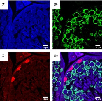
In the first step, we examined the expression of PLZF and VASA in adult testis through immunohistochemistry (Figure 1). Immunohistochemistry with confocal microscopy revealed that the PLZF protein was expressed in the spermatogonial cells that were localized on the basal membrane of seminiferous tubules (Figure 1A,B). In the adult testis sections, VASA-positive cells were distributed throughout the spermatogonia, spermatocytes, and spermatids with the exclusion of SSCs located in the cell layer directly connected to the base membrane of the seminiferous tubule and were also abundant in sperm (Figure 1C).
Immunohistofluorescence analysis in testis section. DAPI (A), immunohistofluorescence analysis for expression of PLZF in the testis (B) and VASA (C). Merged image with blue DAPI (D); (scale bars: 10 μm).

Testicular cells isolation and vimentin’s expression by immunocytochemical analysis
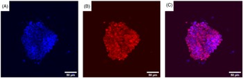
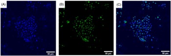
As the next step, by immunocytochemistry we evaluated the expression level of PLZF in adult SSCs after isolation and culture on a feeder layer. ICC images revealed that the generated SSCs were positive for PLZF, similar to findings under in vivo conditions (Figure 2), whereas the expression level of this marker was extremely low in differentiating germ cells (Figure 3).
Immunocytofluorescence analysis of PLZF in spermatogonial stem cells. DAPI (A), immunocytofluorescence analysis showing final colonies from the expansion of our extracted spermatogonial stem cells, and expressing PLZF (B). Merged with blue DAPI (C); (scale bars: 50 μm).

Immunocytofluorescence analysis of PLZF in differentiating spermatogonia. DAPI (A), immunocytofluorescence analysis showing low expression of PLZF in differentiating germ cells (B). Merged with blue DAPI (C); (scale bars: 50 μm).

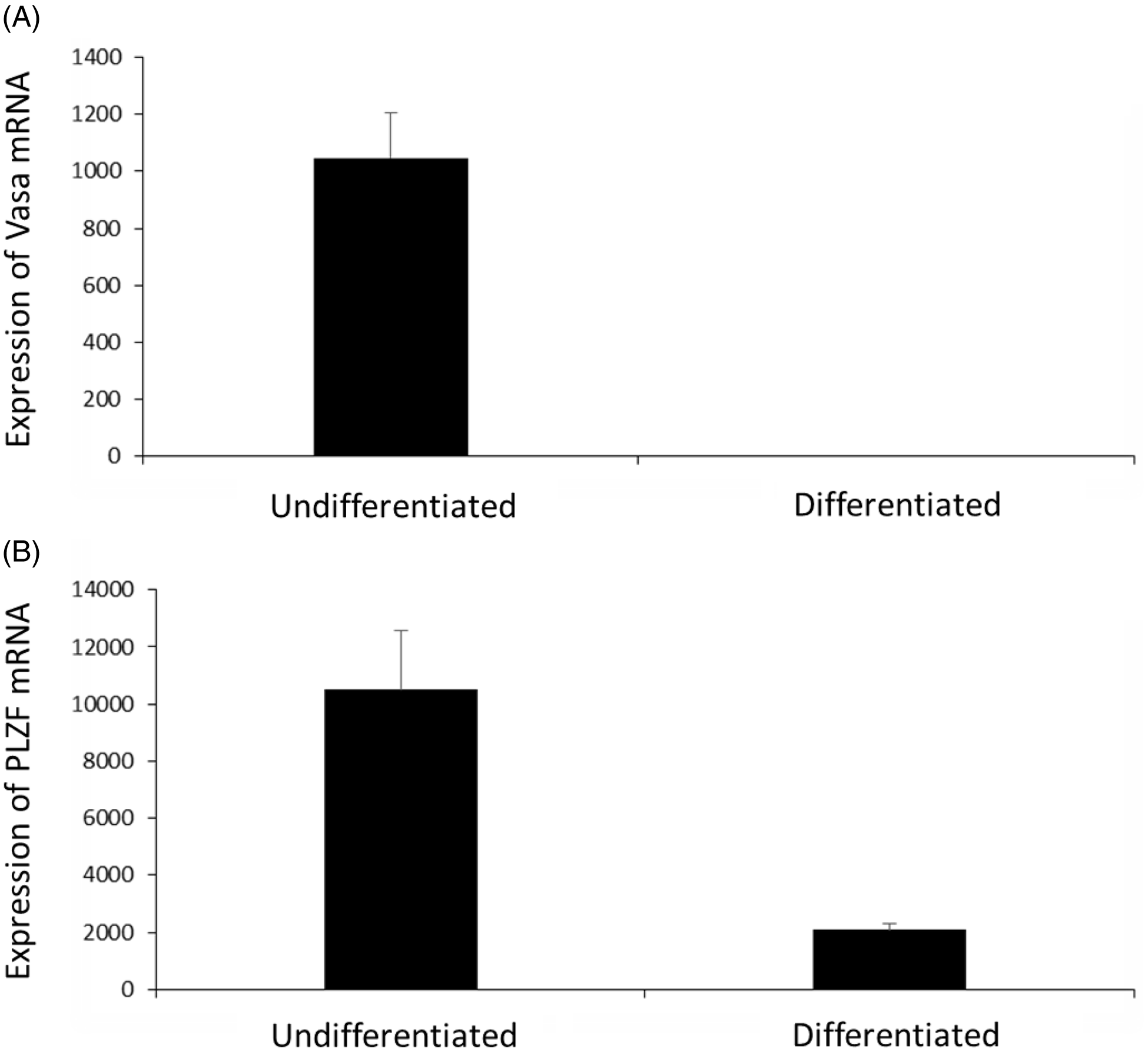
Our real-time PCR analysis as a support for our immunostaining revealed that the expression of PLZF mRNA in SSCs was significantly higher than in other germ cells (P < 0.05) in contrast with VASA mRNA that showed no significant difference in undifferentiated and differentiated spermatogonia (Figure 4).
mRNA expression of Vasa and PLZF. Fluidigm real-time PCR analysis of Vasa expression in two populations of differentiated and undifferentiated cells (A). Analysis of PLZF expression in two populations of differentiated and undifferentiated cells (B).

Protein–protein interaction visualization of PLZF in spermatogenesis
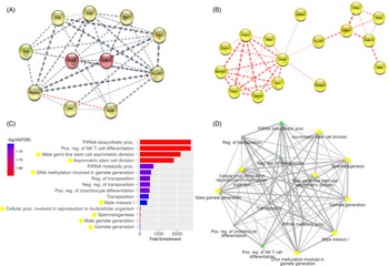
The protein–protein interaction network was visualized with 650 genes using the STRING (v.11) database. It demonstrated that there was a close relationship between interaction and regulated PLZF in the spermatogenesis process. We observed a high level of interaction between Tert, Nanog, vimentin, Sox2, Sox9, Gfra1, Zbtb16, POU5f1, Klf4, DAZL, DDX4 and PLZF. In addition, there was a clear association among Tert, Nanog, and vimentin. Reactome (https://reactome.org/) and KEGG (https://www.genome.jp/kegg/) selected any spermatogenesis-related signalling pathway to highlight the master regulator of the spermatogenesis pathways. There was a strong correlation between the highlighted genes, as shown in Figure 5.
PPI visualization of PLZF and VASA functions in spermatogenesis. The PPI network was visualized using 945 genes from the STRING database. (A) Protein–protein interaction PLZF with spermatogenesis genes. (B) Protein–protein interaction VASA with spermatogenesis genes. (C) Gene ontology of DAZL and VASA involved in spermatogenesis. (D) Gene ontology of DAZL and VASA networks.

Functional enrichments in the PPI network
To identify the enriched biological processes and molecular activities connected to VASA (DDX4), enrichment analysis was conducted (Figure 5C). Control of the reproductive process, piRNA binding, fertilization, male meiosis I, cell cycle, RNA binding, and other tasks exhibiting hub genes, was among the biological processes we chose depending on the goals of our investigation (Figure 5D).
Discussion
Spermatogonia cells (also known as spermatogonial stem cells, SSC), which are localized along the basement membrane of seminiferous tubules, initiate one of the most important biological process during male lifetimes, spermatogenesis, which finally results in sperm-cell production (Wei et al., Reference Wei, Yang, Du, Yu, Zhang, Tang, Ma, Li, Bai, Li and Hua2021). Immunohistochemistry analysis indicated the presence of PLZF-positive cells on the basal membrane of seminiferous tubules. It seems that the PLZF germ cell marker was expressed specifically in spermatogonial cells in the testis. Confocal microscopy characterization for the testis section demonstrated the localization of VASA-positive cells near the basal compartment.
The interaction and regulation of PLZF in the spermatogenesis process were shown to be closely related. Tert, Nanog, vimentin, Sox2, Sox9, Gfra1, Zbtb16, POU5f1, Klf4, DAZL, and DDX4 are all important regulators of chromosomal segregation in meiosis. Vimentin is the key regulator of meiotic spermatogenesis pathways. Therefore, PLFZ coregulation may involve the meiotic phase.
In the spermatogenesis process, there was a tight association between interaction and regulated PLFF. Tert, Nanog, vimentin, and PLZF interacted extensively. To highlight the master regulator of the spermatogenesis pathways, Reactome and KEGG chose any spermatogenesis-related signalling pathway. There was a strong link between the highlighted genes.
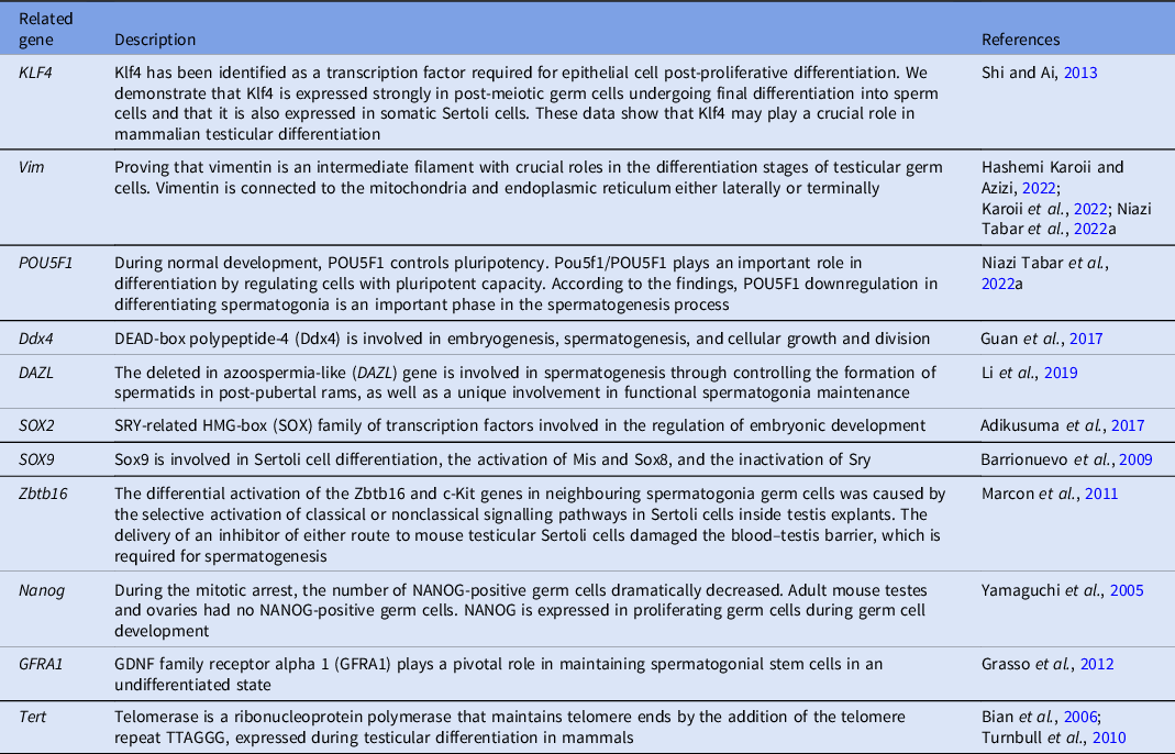
We used datasets relating to protein–protein interaction members in this investigation because of a paucity of information about PLZF expression at various spermatogenic stages. The gene expression ontology study revealed that multiple biological and functional pathways were involved in PLZF expression at different stages of spermatogenesis. This protein’s increasing expression, however, is linked to cell differentiation. Recently, PLZF expression has been linked to SSC differentiation, localization, and signalling pathways through cell surface receptors. Biological and functional investigation revealed that the Stat3, Mmp2, Trp53, Casp7, AURKB, Pik3r1, Ctnnb1, Lgals3, Cdkn1a, Snai1, and Pou5f1 genes increased PLZF expression (Table 1). The table describes genetic and biological roles in germ cell differentiation. There was a close relationship between interaction and regulated vimentin in the spermatogenesis process. There was high interaction among Stat3, Mmp2, Trp53, Casp7, AURKB, Pik3r1, Ctnnb1, Lgals3, Cdkn1a, Snai1, and Pou5f1. In addition, there was a clear association between Trp53, Mmp2, Casp7, Stat3, and Pik3r1. Reactome and KEGG selected any spermatogenesis-related signalling pathway to highlight the master regulator of the spermatogenesis pathways. There was a powerful correlation among the highlighted genes.
Close relationship protein–protein interaction between differentiation and regulation in spermatogenesis

In the present experiment after SSCs generation under stimulation with growth factors FGF, EGF and GDNF, our immunocytochemistry staining demonstrated the sharp expression of PLZF and VASA in SSCs in contrast with in vivo conditions in which VASA had a low expression in these cells. Data obtained from IHC analysis indicated that VASA was expressed in the centre of the testicular cords. We observed the expression of the VASA protein in spermatocytes located above the spermatogonial cell layer in the seminiferous tubule of the adult mouse testis, and a decrease in VASA protein expression during spermiogenesis. This might be due to histological changes in this compartment, including separation from Sertoli cells and the feeder cells. In fact, germ cell fate requires key gene regulation, hormones regulators and other chemical and physical support that remains poorly understood for 2D and 3D culture medium conditions (Kang et al., Reference Kang, Niu, Wu, Hua and Wu2020; Zheng et al., Reference Zheng, Feng, Zhang, Lei, Li and Zeng2020). Two-dimensional culture has played an important role for reproductive biology studies (Richer et al., Reference Richer, Baert and Goossens2020). As in other 2D cultural systems, co-culture systems of germ cells and somatic cells allowed insights into how these cells and extracellular matrix proteins in testis remain together in close contact (Sakib et al., Reference Sakib, Goldsmith, Voigt and Dobrinski2020). Recently, studies used three-dimensional culture or organoid culture that made their results more translatable to the situation in vivo (Alves-Lopes and Stukenborg, Reference Alves-Lopes and Stukenborg2018; Sakib et al., Reference Sakib, Voigt, Goldsmith and Dobrinski2019).
Our results agreed with those of previous studies, suggesting that PLZF is a specific marker for SSCs both in vivo and in vitro and VASA is a germline marker during spermatogenesis and also in proliferating spermatogonia that are expressed more specifically in SSCs under in vitro conditions (Strange et al., Reference Strange, Zarandi, Trivedi, Atala, Bishop, Sadri-Ardekani and Verma2018).
These findings not only conclusively demonstrated the importance of PLZF as a particular marker for SSCs, but they also suggested that further research into the in vitro differentiation of SSCs into functional sperm may benefit from these findings.
Acknowledgements
The present article was extracted from a PhD dissertation from the Animal Biology (Cellular Developmental) at the Department of Biology, Qaemshahr Branch, Islamic Azad University, Qaemshahr, Iran in 2022. I first should express my gratitude to my supervisor Farkhondeh Nemati. This research was supported by the Faculty of Biotechnology of Amol University of Special Modern Technologies.
Author contributions
Mohammad Babatabar Darzi: designed and carried out experiments, and assembled and analyzed data. Hossein Azizi: designed and carried out experiments, and edited the manuscript. Farkhondeh Nemati: provided critical feedback, analyzed data, and edited the manuscript. All authors read and approved the final manuscript.
Funding
This research received no external funding.