Introduction
The water buffalo (Bubalus bubalis) is important to the economy of several countries in Asia and South America, and there are isolated herds in Europe. In India, buffalo is the main dairy animal. Coccidiosis due to Eimeria is an important cause of diarrhoea in livestock, including buffalo. Buffalo and cattle (Bos taurus, Bos indicus) are considered to have more than 10 common species of Eimeria (Levine and Ivens, Reference Levine and Ivens1970); of these Eimeria bovis, Eimeria zuernii, Eimeria auburnensis and Eimeria alabamensis are considered the most pathogenic for cattle (Levine, Reference Levine1973) but I am not aware of any report of histologically confirmed clinical coccidiosis in buffalo associated with these five Eimeria species. Instead, Eimeria bareillyi has been documented to be the main pathogenic species in buffaloes; it is not transmissible to cattle (Sanyal et al. Reference Sanyal, Ruprah and Chhabra1985).
Eimeria bareillyi was first described from buffalo in India (Gill et al. Reference Gill, Chhabra and Lall1963). Since then its oocysts have also been reported in buffalo feces from Brazil, Italy and Turkey (Sayin, Reference Sayin1973; Barbosa et al. Reference Barbosa1992; Cringoli et al. Reference Cringoli1998). Eimeria bareillyi is pathogenic in experimentally infected buffaloes (Shastri et al. Reference Shastri, Krishnamurthi and Ghafoor1973; Shastri and Ghafoor, Reference Shastri and Ghafoor1982; Sanyal et al. Reference Sanyal, Ruprah and Chhabra1985). Clinical coccidiosis in buffalo has been reported from India (Shastri et al. Reference Shastri, Krishnamurthi and Ghafoor1974, Reference Shastri, Ghafoor and Krishnamurthy1976; Shastri and Krishnamurthi, Reference Shastri and Krishnamurthi1975), Brazil (Bastianetto et al. Reference Bastianetto2008; de Meireles et al. Reference de Meireles2012) and the Netherlands (Dubey et al. Reference Dubey, Wouda and Muskens2008). Although endogenous development of E. bareillyi has been reported previously (Pande et al. Reference Pande, Bhatia and Chauhan1971; Shastri et al. Reference Shastri, Krishnamurthi and Ghafoor1974, Reference Shastri, Ghafoor and Krishnamurthy1976; Shastri and Krishnamurthi, Reference Shastri and Krishnamurthi1975; Shastri and Ghafoor, Reference Shastri and Ghafoor1982; Dubey et al. Reference Dubey, Wouda and Muskens2008) many details are missing.
The purpose of the present communication is to describe in detail asexual and sexual development of E. bareillyi in a naturally infected buffalo.
Materials and methods
Materials and methods were same as described previously (Dubey et al. Reference Dubey, Wouda and Muskens2008). An outbreak of coccidiosis occurred in a well-managed buffalo farm in Limburg, the Netherlands. Nine of 22 calves born in November–December 2007 died after clinical illness with diarrhoea. The calves were born on slatted floor and then separated from dams. For one month, calves were housed individually in an igloo and then housed with other calves. Each calf received colostrum from its dam for the first 2 days and then were fed milk replacer. Diarrhoea was noticed in 3–6 weeks old calves and the calves died after 1 or 2 days after the farmer noticed diarrhoea. The present study is from a 22-day-old calf that was euthanized. A complete necropsy was performed and the cause of diarrhoea investigated. Other than Eimeria, bacterial, viral and other parasitic agents were not detected. A section of the jejunum and ileum was used for the present study. Numerous 3–4 µm sections stained with haematoxylin and eosin (HE), periodic acid Schiff (PAS) reaction with haematoxylin as counter stain, were examined for parasitic stages. Pieces of the jejunum from the paraffin block were deparaffinized and processed for transmission electron microscopy (TEM) examination (Trupkiewicz et al. Reference Trupkiewicz2016). For this, numerous 1 µm sections were stained with Toluidine blue and examined by light microscope. For light microscopy, all sections of intestine were examined at 1000× magnification, photographed and endogenous stages were measured using a digital camera (DP 73) fitted on an Olympus AX 70 microscope.
Results
The preservation was sub-optimal but still allowed certain aspects to be examined but not the ultrastructural details. Profuse asexual multiplication occurred both in the jejunum and ileum, but more in the jejunum than the ileum (Figs 1 and 2). Organisms were seen in enterocytes throughout the villus, from the tip to the tunica muscularis mucosae. Crypts were heavily infected, and the lumens were packed with schizonts and merozoites. Even in 1 µm sections, there was overcrowding making it difficult to discern individual parasite outlines.
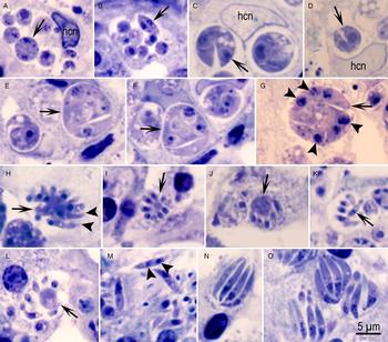
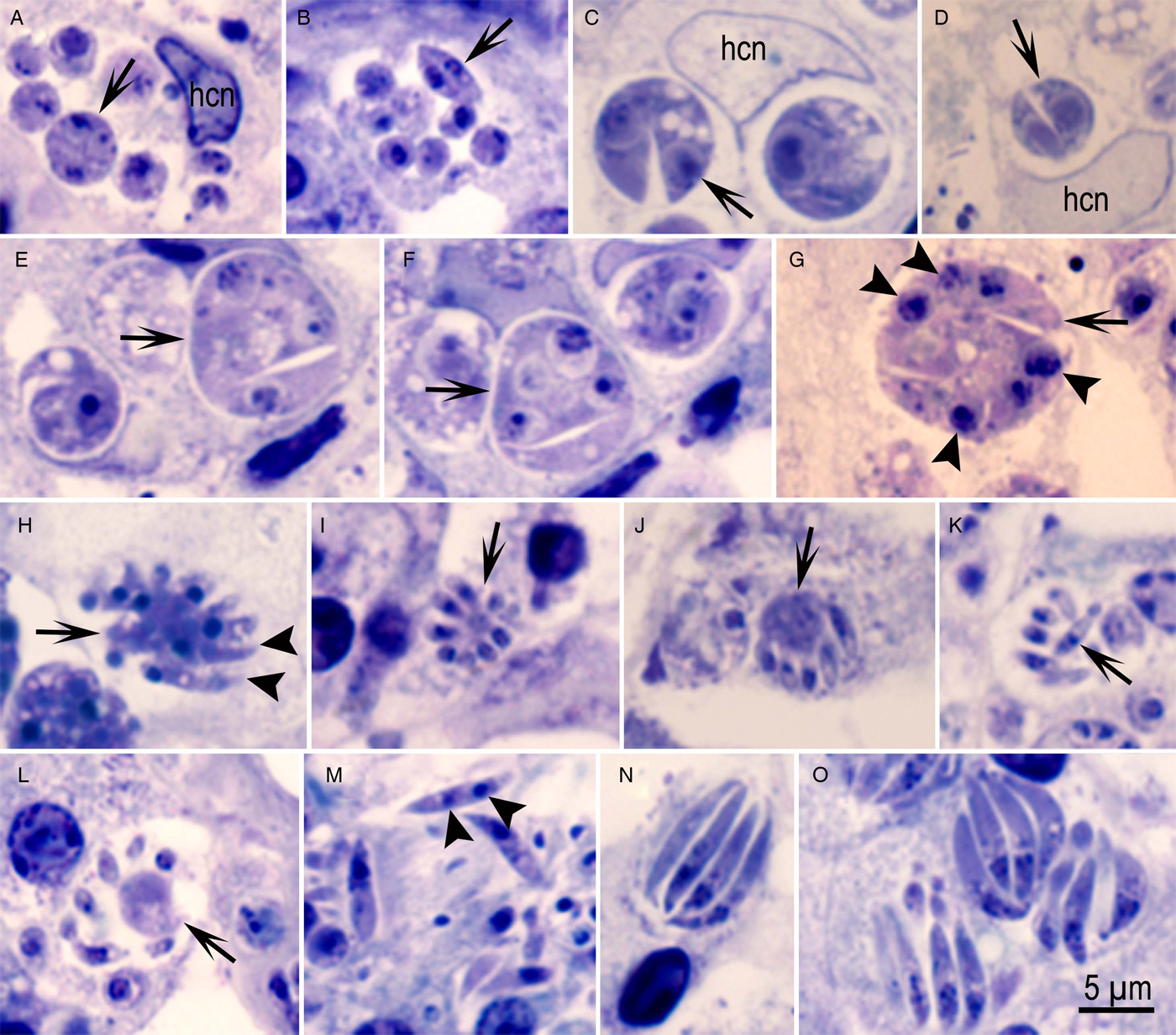
Asexual stages of Eimeria bareillyi in the jejunum of a naturally infected 22-day-old buffalo. One micrometre sections stained with Toluidine blue. Bar applies to all parts. hcn = host cell nucleus. (A) A group of six different sized globular zoites in a parasitophorous vacuole of an enterocyte. Arrow points to a zoite with two or three nuclei. (B) A group of zoites with one or two nuclei. Arrow points to an organism divided into two. (C, D) Organisms dividing into two (arrows). (E, F) Groups with a merozoite separating (arrows) from the multinucleated mass. (G) A schizont with at least six nuclei (arrowheads) and merozoite (arrow). (H) Schizont (arrow) with merozoites (arrowheads) forming/separating at the periphery. (I) Schizont with small-sized merozoites attached to a central residual body (arrow). (J) Schizont with large residual body (arrow). (K) Small-sized merozoites with a central nucleus (arrow). (L) Schizont with a large residual body (arrow) and loosely arranged merozoites. (M) Elongated merozoites. Note two nuclei-like structures (arrowheads) in a merozoite. (N) Schizont with four longitudinally cut merozoites. (O) A group of schizonts with relatively large merozoites.
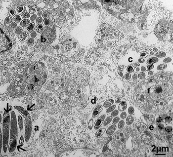
Transmission electron microscopic observations on asexual and sexual stages of Eimeria bareillyi in the jejunum of a naturally infected 22-day-old buffalo. Note four schizonts (a–d), a microgamont (e) and a macrogamont (f). Also, note arranged head to tail arrangement of merozoites (arrows) in schizont (a). Merozoites in schizonts (b–d) are cut in cross-section or obliquely.

Numerous individual uninucleated structures were seen, but it was not possible to discern if they were merozoites or gamonts. By light microscopy, three types of merozoites/division were seen. Figure 1A and B show groups of organisms with different sized globular parasites in individual parasitophorous vacuoles (Fig. 1A and B). Figure 1B shows an organism dividing into two, whereas others in the same vacuole are uninucleated or binucleated; the two merozoites in division are 5.0 × 1.0 µm and have a central nucleus. Figure 1C and D shows globular organisms dividing into two zoites that are much larger (7 × 4 µm) than the merozoites in Fig. 1B. Other globular organisms contained up to eight nuclei and some merozoites appeared to split off the main mass (Fig. 1E–G). Figure 1H–L shows merozoites arising from different-sized residual bodies; the merozoites are small, 3–4 µm. Figure 1M shows a 7 µm long merozoite apparently with two nuclei. The merozoite formation in small schizonts appeared to be asynchronous; some merozoites were protruding at the periphery of a multinucleated mass/residual body while others were not (Fig. 1H–L). Small-sized merozoites were 3–5 µm long and had a central nucleus (Fig. 1K).
Most schizonts contained elongated sickle-shaped merozoites (Fig 1N and O); these schizonts were often 3–5 µm wide and contained 8–11 µm long merozoites (Fig. 1N and O). In these sickle-shaped merozoites, the nucleus was located at the broader end.
In the present buffalo, there were more than two generations of schizonts. The number and sequence of schizont generation was not known in this naturally infected calf.
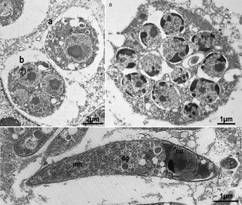
By TEM, numerous individual and groups of merozoites were identified (Fig. 2). The maximum dimension of schizonts was 12 µm in any direction and a maximum of 16 merozoites were identified in any schizont. The merozoites were sometimes arranged with their apical ends in opposite direction (Fig. 2). Early schizonts were difficult to distinguish from early microgamonts (Fig. 3A). Although merozoites were not well preserved, numerous micronemes, few dense granules and a nucleus located at the narrow (non-conoidal) end can be seen in Fig. 3B. The stages in asexual division were not observed that would have helped answer question on the mechanism of division.
Transmission electron microscopic observations on asexual stages of Eimeria bareillyi in the jejunum of a naturally infected 22-day-old buffalo. (a) One-nucleated with unidentified stage (a), and a four-nucleated organism (b), probably an early schizont. (B) A schizont-like structure with 12 nuclei. (C) Higher magnification of a merozoite showing the posteriorly located nucleus (nu), few dense granules (dg) and numerous micronemes (mn) towards the conoidal end.

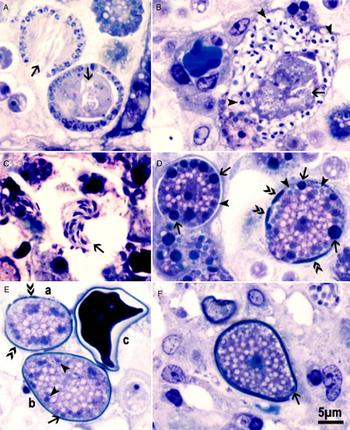
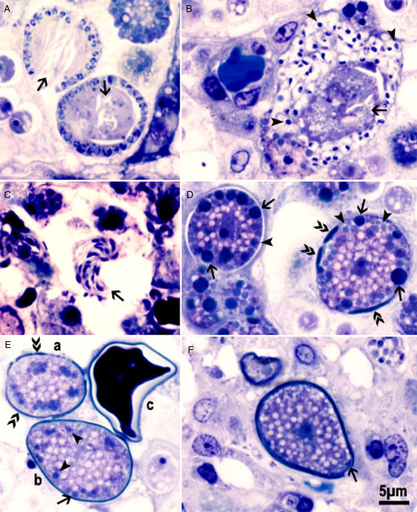
Gamonts were seen both in the jejunum and ileum, but more so in the ileum (Figs 4–7). By light microscopy, the earliest recognizable microgamont identified had eight peripherally located nuclei and had a residual body. Large microgamonts with peripherally located nuclei were up to 12–40 × 8–29 µm (average 26.4 × 20, n = 12) and contained up to 30 nuclei and one or two residual bodies in one plane of focus (Fig. 4A and B); however, some nuclei were located towards the centre. Mature microgametes were up to 2.5 µm long and had two flagella (Fig. 4C).
Gametogony of Eimeria bareillyi in the jejunum of a naturally infected 22-day-old buffalo. One micrometre sections stained with Toluidine blue. Bar applies to all parts. (A) Two developing microgamonts with peripherally located nuclei and residual bodies of different density (arrows). (B) A nearly mature microgamont with numerous microgametes (arrowheads) and a large residual body (arrow). (C) Free microgametes with two flagella (arrow). (D) Two macrogamonts. The macrogamont on the left has both types of wall-forming bodies (WFB1, WFB2). Type WFB1 bodies (arrows) stain more intensely than the type 2 (arrowheads). The empty white areas are probably polysaccharide granules. The macrogamont on the right shows wall formation in patches (double arrowheads). WFB1 are fewer than WFB2. (E) Three oocysts in different stages of maturation. (a) Wall formation in patches (double arrowheads). (b) One oocyst wall (arrow) has formed and only WFB2 are visible (arrowheads). (c) Cross-section of a mature oocyst. (F) Longitudinal section of a fully formed oocyst with central nucleus, numerous amylopectin granules (empty spaces) and the micropyle (arrow).
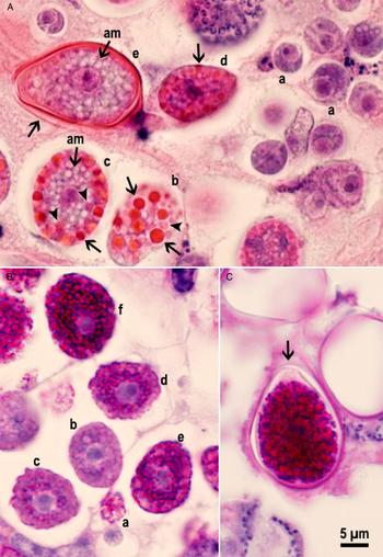
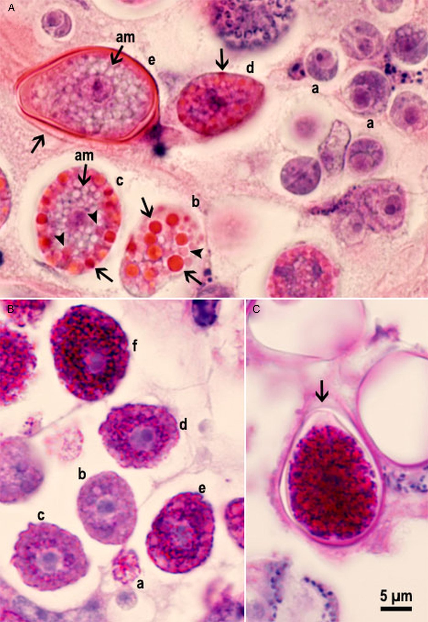
Macrogamonts and oocysts of Eimeria bareillyi in the jejunum of a naturally infected 22-day-old buffalo. The 3–5 µm sections stained with haematoxylin and eosin (A) and (B, C) periodic acid Schiff (PAS) reaction counter stained with haematoxylin. Bar applies to all parts. (A) Macrogamonts in presumed order of development (a–e). Earliest macrogamont (a) with a large nucleus and without wall-forming bodies (WFB) and amylopectin granules. (b, c) Macrogamonts and developing oocysts with WFB1 (arrows) and WFB2 (arrowheads). An immature oocyst (d) with part of the wall formed (arrow). (e) Longitudinally cut oocyst with darkly stained oocyst wall and an outer covering (arrow). Also, note the centrally located nucleus with a nucleolus, and numerous polysaccharide granules (empty spaces) and no WFB. (B) Macrogamonts with varying intensity (a–f) of PAS positivity. The earliest macrogamont (a) is PAS-negative, (b, c, d, e) have PAS-positive granules around the nucleus and (f) entire gamont is PAS-positive. (C) Longitudinally cut pear-shaped oocyst with a truncated anterior end (arrow). The sporont occupies the entire width of the oocyst and is intensely PAS-positive.
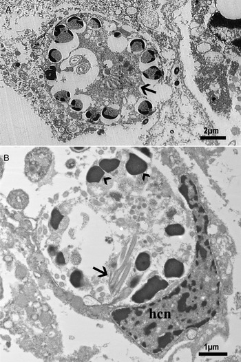
Transmission electron microscopic observations on microgamonts of Eimeria bareillyi in the jejunum of a naturally infected 22-day-old buffalo. (A) Nearly mature microgamont with peripherally located nuclei and a residual body (arrow). (B) An intracellular degenerating microgamont. Note flagella (arrow), peripheral nuclei (arrowheads). Also, note the host cell nucleus (hcn).

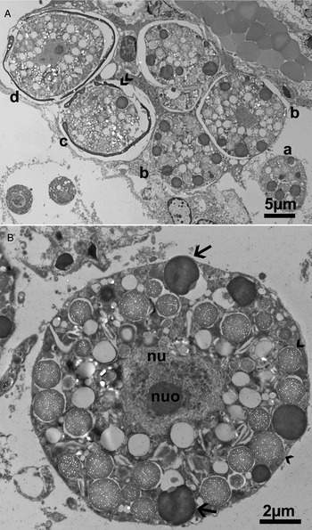
Transmission electron microscopic observations on gamonts and oocysts of Eimeria bareillyi in the jejunum of a naturally infected 22-day-old buffalo. Note, nucleus (nu), nucleolus (nuo), wall-forming bodies (WFB)1 (arrows) and 2 (arrowheads). (A) Gametogony and oocyst wall formation in presumed order of development (a–d). (a,b) developing macrogamonts with numerous WFB1, 2. (c) Oocyst wall formation in patches (double arrowheads) with WFB1 and several WFB2. (d) Almost completed oocyst wall. (B) Higher magnification of a macrogamont. Note a centrally located nu and nuo, electron-dense WFB1 (arrows), electron-lucent WFB2 (arrowheads).

By light microscopy, the earliest macrogamonts could be recognized by the presence of PAS-positive granules (amylopectin granules) and the two types of wall-forming bodies (WFB, also called plastic bodies) (Fig 4A and D). The PAS-positive granules were initially located centrally around the nucleus. Subsequently, more PAS-positive granules appeared as the gamonts became larger (Fig. 5B). The WFB appeared after the appearance of PAS-positive granules, and WFB2 appeared sooner than WFB1. The WFB1 were stained darker with HE and Toluidine blue than WFB2 (Fig. 5A). The oocyst wall laid down in patches, and the WFB were not seen in fully formed oocysts (Figs 4 and 5).
TEM confirmed the light microscopic observations on microgamonts (Fig. 6). Even by TEM, it was difficult to distinguish young microgamonts from schizonts. The peripheral location of nuclei and the presence of flagella-like structures helped in definitive identification (Fig. 4A and C).
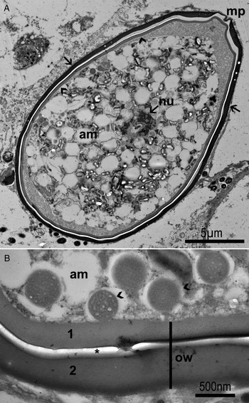
The light microscopic findings on macrogamonts were confirmed by TEM (Fig. 7). The WFB1 were electron dense, whereas WFB2 were spongy, both were 1–2 µm in diameter (Fig. 7). Fully formed oocysts had two oocyst layers with the total thickness of 1 µm (Fig. 8). The inner oocyst layer was homogenous, and approximately 375 nm thick. The outer oocyst layer was denser and approximately 500 nm thick. The two oocyst layers were separated by an electron lucent space, probably an artefact. Both oocyst layers were truncated at the anterior end, with no interruption of the inner oocyst layer. The outer oocyst layer was thinned out at the conical (micropylar end) (Fig. 8A). Remnants of WFB were still recognizable in fully formed oocysts (Fig. 8B). Longitudinally cut oocysts in sections were 22–29 × 14–18 µm (average 25.8 × 18.7, n = 10).
Transmission electron microscopic observations on an Eimeria bareillyi oocyst in the jejunum of a naturally infected 22-day-old buffalo. Note outer oocyst layer (arrows), inner oocyst layer (arrowheads), wall-forming bodies (1, 2) and empty spaces (amylopectin/lipid bodies). (A) The oocyst is 27 × 15 µm. Note the sporont occupying almost of the oocyst interior. The two oocyst layers differ in density, the outer layer (arrow) is denser than the inner layer (arrowhead). The oocyst is truncated at the micropylar end (mp) with thinning of the outer layer. (B) Higher magnification of oocyst layers. The two oocyst walls (1,2) are separated by an empty space (*), probably fixation artefact. Also, note WFB1, 2 (arrowheads).

Discussion
By light microscopy, the results described here are broadly similar to those reported previously (Pande et al. Reference Pande, Bhatia and Chauhan1971; Shastri and Ghafoor, Reference Shastri and Ghafoor1982; Sanyal et al. Reference Sanyal, Ruprah and Chhabra1985; Dubey et al. Reference Dubey, Wouda and Muskens2008). Shastri and Ghafoor (Reference Shastri and Ghafoor1982) orally inoculated 2-day-old buffaloes with one million E. bareillyi oocysts and necropsied them 5, 10 or 14 days post-inoculation (p.i.). Endogenous development occurred in the jejunum and ileum. On day 5 p.i., only uninucleated, 3.2–5.7 µm in diameter organisms (trophozoites) were seen in enterocytes in crypts of Lieberkühn in the mid-jejunum. On day 10 p.i., immature and mature schizonts were seen in lower 2/3 of the small intestine. Mature schizonts were 11.5–20.7 µm and contained 24–36 slender 10–15 µm long merozoites. On day 14 p.i., only gamonts and oocysts were found. Macrogamonts were 12.5–23.3 × 11.1–16.7 µm, and microgamonts were 15–30 × 10–23 µm. Whether the measurements of schizonts and gamonts were from smears or histological sections was not stated. Unfortunately, the photographs of schizonts and gamonts are very poor, and therefore, it is impossible to judge the accuracy of description. The line drawings appear to be schematic. In the present study, the schizonts were small and contained no more than 16 merozoites. Additionally, the size of merozoites varied, indicating several generations.
Pande et al. (Reference Pande, Bhatia and Chauhan1971) first reported E. bareillyi gamonts and oocysts in specimens of small intestine obtained from an abattoir; nothing was known of clinical status or age of the buffalo; this animal also had concurrent infection with other Eimeria. They observed characteristic E. bareillyi oocysts in scrapings made from polyp-like raised areas. Only gamonts and oocysts were seen. The oocysts were 23.3–27 × 13.3–17 µm, and the mature microgamonts were 20–37 × 18–25 µm in histological sections; these measurements are similar to those in the present study.
In the present study, both asexual and sexual stages were seen concurrently. This is probably related to trickle infection. Only E. bareillyi oocysts were seen in feces. Therefore, I assumed that endogenous stages observed belonged to E. bareillyi. Experimental E. bareillyi infections of newborn buffalo calves reared in coccidia-free conditions will be needed to confirm endogenous stages observed here. The present study has shown that newborn calves are highly susceptible to E. bareillyi infection.
The present study provided for the first time details of asexual and sexual multiplication of E. bareillyi. In most Eimeria species, the asexual development is considered by a divisional process of schizogony where sporozoites round up after invasion of host cells leading to a uninucleated organism called the trophozoite (Levine, Reference Levine1973). Subsequent development occurs by schizogony wherein the parasite nucleus divides in to four or more nuclei before merozoite formation occurs (Levine, Reference Levine1973). Some non-eimerid coccidia (Toxoplasma, Neospora) in the intermediate host divide by a process called endodyogeny where the mother cell divides into two daughter organisms, consuming the mother cell. To accommodate this divisional process, Levine (Reference Levine1973) used the term meront instead of the conventional term schizont. Here, I have used the term schizont loosely, although some organisms appeared to divide into two. To my knowledge, endodyogeny has not been demonstrated previously in any species of Eimeria. In the present study with E. bareillyi, some organisms divided in to two but the observations could not be confirmed by TEM. Confirmation of endodyogeny in Eimeria will require well-preserved specimens; only paraffin-embedded tissue was available for the present study.
The development of E. bareillyi oocyst wall is unknown. In Eimeria species, the oocyst wall is initiated by the secretions of WFB. Most Eimeria species have two types of WFB, type 1 and type 2. The WFB1 are electron dense and larger in size than WFB2, and their characteristics are species-specific (Ferguson et al. Reference Ferguson2003). The WFB arise during the maturation of the macrogamont and form the oocyst wall. In some Eimeria species, there are additional veil-forming bodies (VFB); they form a veil-like outer covering on the oocyst wall that disappears when the oocysts are excreted in feces (Ferguson et al. Reference Ferguson2003); VFB were not identified in the present study of E. bareillyi.
The prepatent period of E. bareillyi in experimentally infected buffaloes is 12–15 days, probably dependent on the age of buffaloes and the dose. The shortest prepatent period of 12 days was observed in 2-day-old buffaloes fed 115 000 oocysts (Shastri et al. Reference Shastri, Krishnamurthi and Ghafoor1973) and the 15th day prepatent period was observed in a 1-month-old calf fed 100 000 oocysts. In the present study, the calf was excreting numerous E. bareillyi oocysts in feces and had severe enteritis (Dubey et al. Reference Dubey, Wouda and Muskens2008). The buffalo calf was housed individually, and was fed milk replacer. It is unlikely that the calf ingested E. bareillyi oocysts as a single bolus of infection, but more likely infected over several days. The calf was only 22 days old, and considering the short prepatent period, E. bareillyi probably became infected soon after birth. The affected farm was well managed, the calf was not malnourished and no concurrent infectious agent was found. Nothing is known of the pathogenicity of different strains of E. bareillyi.
This is the first full description of asexual and sexual development of any Eimeria species in buffalo and the results should be helpful in differential diagnosis of Eimeria species in the intestines of buffaloes.
Acknowledgements
I would like to thank Camila Cezar, Fernando Antunes, Shiv Kumar Verma and Oliver Kwok for their help with this manuscript and Joseph Mowery for electron micrographs. I am grateful to Professor David Ferguson for his help with interpretations of electron micrographs.
Financial support
This research received no specific grant from any funding agency, commercial or not-for-profit sectors.
Conflict of interest
None.
Ethical standards
Not applicable