Introduction
The development of an embryo inside the chicken egg is a fascinating process of the transformation of chemical components into a living organism in 21 days. The increase in survival rate and viability of the hatchling are both very important as they relate to bird health, welfare and positive economic returns for producers. Several factors, including egg nutrient content, incubation temperature, relative humidity, pre-hatch storage and conditions can affect chick embryo development and these factors are well documented. Dramatic changes occur in lipid metabolism during the last trimester of embryonic development inducing oxidative stress. From this perspective, the pro-oxidant and antioxidant balance need to be protected during the incubation period. The purpose of this review is to provide an insight into the antioxidant defence systems that are involved in the growth and development of the chicken embryo into a healthy hatchling.
Requirements of embryo during the developmental process
The chick embryo develops within a closed system: the egg. Before laying, hens deposit the nutrients that are needed for the sustaining growth and development of the embryo in the egg. During early development, glucose and amino acids provide more than 90% of the total energy requirements of the embryo. In the latter half of development the embryo utilises fatty acids derived from yolk lipids for energy (Speake et al., Reference SPEAKE, SURAI and NOBLE1998b). During early incubation, the yolk sac membrane collects the O2 that diffuses through the shell and albumen and has a role in embryonic gas exchange (Mortola, Reference MORTOLA2009). Nevertheless, the main function of the yolk sac membrane (YSM) is for transferring lipids into the embryo's circulation. The uptake of yolk lipids is followed by their oxidation for energy and for the synthesis of structural membranes, steroid hormones and signal transduction (Noble and Cocchi, Reference NOBLE and COCCHI1990; Speake et al., Reference SPEAKE, MURRAY and NOBLE1998a). Transfer of yolk lipids is slow during the first half of incubation, and is accelerated during the second half because of increased energy requirement. (Yadgary et al., Reference YADGARY, YAIR and UNI2011). As the chick embryo grows, it needs more oxygen to satisfy increased energy metabolism. There are three principle reasons for increasing energy requirements during embryonic development:
1. Tissue maintenance, including cell repair, neurone excitability, excretory processes, cardiac and respiratory functions.
2. Energy loss from cell multiplication, tissue formation and organogenesis.
3. Increased body movements and thermogenesis towards the end of incubation (Vleck et al., Reference VLECK, VLECK and HOYT1980).
Gas transfer between the environment and the blood continues during the development of the embryo. It especially increases during the period of rapid growth (d10-d18), because of the growth of the chorioallantoic membrane and its increased vascularity and proximity to the inner membrane of the shell. Blood itself contributes to the rise in gas transfer because of the increase in blood volume and O2 carrying capacity (Mortola, Reference MORTOLA2009). Oxygen consumption is stable during the last days of embryogenesis (Vleck et al., Reference VLECK, VLECK and HOYT1980).
Reactive oxygen species (ROS) are the products of cellular metabolism. Generation of ROS is an inevitable outcome from the maintenance of physiological functions and cellular redox reactions (Misra and Fridovich, Reference MISRA and FRIDOVICH1972). The rate of ROS generation depends on the concentration of oxygen in the tissues and metabolic rate (Turrens, Reference TURRENS2003). Oxidative stress is defined as a disturbance in the balance between oxidants and antioxidants. High levels of ROS cause protein and lipid oxidation and DNA damage. External noxious sources such as ionising radiation, toxins, drugs, chemicals, and environmental pollutants can all cause oxidative stress. Some immune cells are specialised to produce ROS continually or in the form of an oxidative burst to destroy pathogens. In other words, metabolism produces free radicals even under healthy conditions, but they might have diversified useful effects (Siess, Reference SIESS1991). The levels of free radicals and other ‘reactive species’ in living organisms are controlled by a complex of antioxidant defences. While embryos are developing and consuming more oxygen, increased energy metabolism can make them susceptible to lipid peroxidation (LPO) caused by ROS. Reactive oxygen species play an important role in embryonic development because ROS production is essential for mediating apoptosis and cell elimination (Salas-Vidal et al., Reference SALAS-VIDAL, LOMELI, CASTRO-OBREGON, CUERVO, ESCALANTE-ALCALDE and COVARRUBİAS1998). Oxidative stress and redox reactions related to signalling are vital to normal developmental processes such as progression to the blastocyst stage, neuronal differentiation, and digit formation, and increased oxidative stress is associated with embryotoxicity (Phyllis, Reference PHYLLIS2007).
Yolk lipids are sources of energy and essential fatty acids. Egg yolk fatty acids are the major modifiable factor known to influence the lipid and fatty acid composition of the developing chick (Cherian and Sim, Reference CHERIAN and SIM1991; Cherian et al., Reference CHERIAN, GOPALAKRISHNAN, AKIBA and SIM1997). Essential fatty acids are the precursors of polyunsaturated fatty acids (Cherian and Sim, Reference CHERIAN and SIM2001, Cherian, Reference CHERIAN2011). Most lipids in chick embryo tissues are unsaturated (Noble and Cocchi, Reference NOBLE and COCCHI1990). Polyunsaturated fatty acids (PUFA) are vital to embryonic development and are obtained from the hen's diet and are accumulated in the egg yolk (Noble and Cocchi, Reference NOBLE and COCCHI1990). Polyunsaturated fatty acids are highly vulnerable to free radical-induced peroxidative damage. The intense accretion of PUFA in embryonic tissues during late incubation makes them highly susceptible to lipid peroxidation and free radicals (Porter et al., Reference PORTER, CALDWELL and MILLS1995) and require the protective effect of the natural antioxidants such as vitamin E (Gaal et al., Reference GAAL, MEZES, NOBLE, DIXON and SPEAKE1995; Surai et al., Reference SURAI, NOBLE and SPEAKE1996), vitamin C (Surai et al., Reference SURAI, NOBLE and SPEAKE1996), vitamin A (Surai et al., Reference SURAI, IONOV, KUKLENKO, KOSTJUK, MACPHERSON, SPEAKE, NOBLE and SPARKS1998) and antioxidant enzymes SOD (Surai et al., Reference SURAI, NOBLE and SPEAKE1996); CAT and GSH-PX (Wilson et al., Reference WILSON, LUI and DEL MAESTRO1992). Some of these protective antioxidants are produced by the organism itself (e.g. cholesterol, uric acid and glutathione; Surai, Reference SURAI2002).
Large amounts of antioxidants are transferred from the yolk to the embryo during the last few days of embryonic development. Most of these antioxidants are stored in the embryonic liver just before hatching, from where they are readily mobilised into the circulation during the neonatal period, thus preventing lipid peroxidation by scavenging singlet oxygen or by quenching free radicals (Surai and Speake, Reference SURAI and SPEAKE1998). Studies in poultry have shown that egg antioxidants positively influence egg hatchability, the rate of body mass and osteometric growth, immunity, behaviour and viability at least in the early post-hatch period, which is a critical time for chick survival (Lin et al., Reference LIN, CHANG and HSU2004).
Natural antioxidants interact with each other to build the antioxidant systems of the cell which protects the cell from the detrimental effect of free radicals and the toxic effect of LPO products. Maternal diet composition is a major determinant of the antioxidant system that develops in the chick during embryogenesis (Gaal et al., Reference GAAL, MEZES, NOBLE, DIXON and SPEAKE1995; Surai, Reference SURAI1999a) and in post-natal development (Surai, Reference SURAI1999a). Antioxidant protection via maternal or egg nutrient supply is potentially crucial for early survival, potentially affecting the somatic and neural development of the offspring as well as for other major functions, including immunity (Gaal et al., Reference GAAL, MEZES, NOBLE, DIXON and SPEAKE1995; Surai, Reference SURAI1999a). Therefore, strategies that will enhance antioxidant capability during pre- and early post-hatch period will ultimately lead to improvement in bird health, welfare and increase in economic returns to the poultry industry.
There are three levels of antioxidant defence in the living cells. The first is responsible for the prevention of radical formation, including SOD, GSH-PX, CAT, glutathione and thioredoxin systems and metal-binding proteins like transferrin, lactoferrin, myoglobin, ceruloplasmin and albumin. The second is the prevention and restriction of chain formation and propagation and is mediated through vitamins A, E, C, carotenoids, ubiquinols, glutathione (GSH), and uric acid. The final level of defence is excision and repair of damaged parts of molecules which is mediated through lipases, peptidases, proteases, transferases and DNA repair enzymes (Surai, Reference SURAI2002).
Avian embryo and some of the natural antioxidants
carotenoids
Carotenoids are lipid-soluble secondary plant compounds, and since they are not produced by animals, they must be supplied in feed (Dua et al., Reference DUA, DAY, HILL and GROGAN1967). Most carotenoids are converted into vitamin A in the intestinal mucosa, while the rest of them escape conversion and enter the circulation to be deposited in yolk and skin (Williams et al., Reference WILLIAMS, BOILEAU and ERDMAN1998). During embryonic development, the lipids and associated components are transferred from the yolk to the surrounding YSM by phagocytosis. Within the YSM, lipids are assembled with vitamin E, carotenoids, and apoproteins to form lipoproteins, which are released into circulation (Noble and Cocchi, Reference NOBLE and COCCHI1990). Delipidation by the action of lipase in the capillaries of the embryo's adipose tissue and muscle follows this stage (Speake et al., Reference SPEAKE, NOBLE and MCCARTNEY1993).
Since the lymphatic system is not developed in birds, carotenoids are delivered directly to the liver and other tissues by large proteins (portomicrons) that the lipid-rich lipoproteins release from the intestinal cells into the portal vein. Portomicrons partly undergo some transformation in the liver, but, most likely, they are largely released into the circulation unchanged or with minor changes (Traber et al., Reference TRABER, LANE, LAGMAY and KAYDEN1992). Portomicron remnants are cleared from circulation by the liver and in this way carotenoids are delivered to this organ. Thus, high levels of vitamin E and carotenoids accumulate in the liver, particularly during the final few days of development, whereas lower amounts of these components are distributed to the other tissues (Surai et al., Reference SURAI, NOBLE and SPEAKE1996).
Previous studies have shown that carotenoids are useful for promoting cell differentiation, regulating cell proliferation (Rock et al., Reference ROCK, KUSHUSKI, GALVEZ and ETHIER1995), and for enhancing immune functions (Rice-Evans et al., Reference RICE-EVANS, SAMSON, BRAMLEY and HOLLOWAY1997). Carotenoids show activity as scavengers of singlet oxygen and peroxyl radicals (Terao et al., Reference TERAO, BOEY, OJIMA, NAGAO, SUZUKI and TAKAMA1992), sulphur radicals (Chopra et al., Reference CHOPRA, WILSIN and THURNHAM1993), thiyl, sulphonyl/NO2 radicals (Everett et al., Reference EVERETT, DENNIS, PATEL, MADDIX, KUNDU and WILSON1996) and provide protection of lipids against superoxide/hydroxide radicals (Chopra et al., Reference CHOPRA, WILSIN and THURNHAM1993). Thus, it prevents lipid peroxidation as quenched singlet oxygen and can interrupt the propagation stage of lipid peroxidation (Rice-Evans et al., Reference RICE-EVANS, SAMSON, BRAMLEY and HOLLOWAY1997).
vitamin A
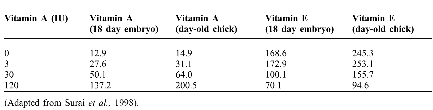
Since poultry are incapable of vitamin A synthesis, all retinoids must be obtained from the diet as preformed vitamin A (retinyl esters, retinol and a very small amount of retinoic acid) either from animal products or as provitamin carotenoids from fruits and vegetables (Goodman, Reference GOODMAN1984). The requirements of vitamin A in chicks are met by liver storage. The liver secretes retinol into the circulation and which is delivered from the circulatory system to the yolk by serum retinol binding proteins and preferentially transported from the yolk to the embryo before day 14 of incubation, the period of intensive transfer of lipid (Noble and Cocchi, Reference NOBLE and COCCHI1990). Vitamin A supplementation in laying hen diets is an important factor in development and antioxidant regulation during pre- and early post-hatch periods (Surai et al., Reference SURAI, IONOV, KUKLENKO, KOSTJUK, MACPHERSON, SPEAKE, NOBLE and SPARKS1998). Depending on its concentration and experimental conditions, vitamin A can serve as a pro-oxidant (Tesoriere et al., Reference TESORIERE, CIACCIO, BONGIORNO and FRIESLEBEN1994). However, increased quantities of fat soluble vitamin into the diet of the laying hen's causes their concentrations to increase in the liver (Table 1) and egg yolk. Conversely, an excess of vitamin A in the maternal diet can cause the opposite effect when the concentration of other natural antioxidants, such as vitamin E and carotenoids and ascorbic acid are decreased (Surai et al., Reference SURAI, IONOV, KUKLENKO, KOSTJUK, MACPHERSON, SPEAKE, NOBLE and SPARKS1998). Similarly, the intubation of large amounts of vitamin A to one-day-old chicks was associated with the inhibition of vitamin E and carotenoid utilisation from the liver during chick postnatal development (Surai et al., Reference SURAI, KUKLENKO, IONOV, NOBLE and SPARKS2000).
Hen diet and vitamin A and E (µg/g) in the liver of the embryo and hatched chick.

(Adapted from Surai et al., Reference SURAI, IONOV, KUKLENKO, KOSTJUK, MACPHERSON, SPEAKE, NOBLE and SPARKS1998).
vitamin E
Vitamin E is a major lipid soluble antioxidant which breaks the chain reaction of LPO, and is transported from the yolk to the embryonic tissues during development (Gaal et al., Reference GAAL, MEZES, NOBLE, DIXON and SPEAKE1995; Surai et al., Reference SURAI, NOBLE and SPEAKE1996). Alpha tocopherol is the most active form of vitamin E (80%) in the embryonic tissues (Surai et al., Reference SURAI, NOBLE and SPEAKE1996; Surai, Reference SURAI2002); other isomers are present in egg yolk and embryonic tissues, but their concentrations as high as α-tocopherol. Surai et al. (Reference SURAI, SPARKS and NOBLE1999b) reported that although high levels of β+γ-tocopherols may appear in the diet, α-tocopherol is predominantly accumulated in the egg yolk, and the efficiency of transfer of α-tocotrienol from the feed to the egg yolk is twice that for other tocotrienols.
Vitamin E transfers from the feed into the egg yolk and into the tissues of the embryo. In the avian embryo, yolk-derived lipid droplets containing α-tocopherol is assembled into triacylglycerol-rich lipoproteins by the endodermal cells of the YSM (Noble and Cocchi, Reference NOBLE and COCCHI1990). The release of these lipoproteins into the embryonic circulation is followed by the hydrolysis of the triacylglycerol components by lipoprotein lipase (Surai et al., Reference SURAI, NOBLE and SPEAKE1996) and the subsequent uptake of the resultant lipoprotein remnants by the liver (Shand et al., Reference SHAND, WEST, NOBLE and SPEAKE1994) between days 18 and 21 of development (Surai et al., Reference SURAI, NOBLE and SPEAKE1996). Vitamin E accumulation in the embryonic liver reaches its maximum at hatching (Surai et al., Reference SURAI, IONOV, KUKLENKO, KOSTJUK, MACPHERSON, SPEAKE, NOBLE and SPARKS1998). During the first two weeks post-hatch, the concentration of vitamin E in the liver decreases by 10 times in chickens (Surai, Reference SURAI2000). It is suggested that this is an adaptive mechanism in birds to deal with overproduction of free radicals at the time of hatching (Surai, Reference SURAI2002). The chick brain has low levels of vitamin E because of the absence of lipoprotein lipase in this tissue (Speake et al., Reference SPEAKE, NOBLE and MCCARTNEY1993; Gaal et al., Reference GAAL, MEZES, NOBLE, DIXON and SPEAKE1995).
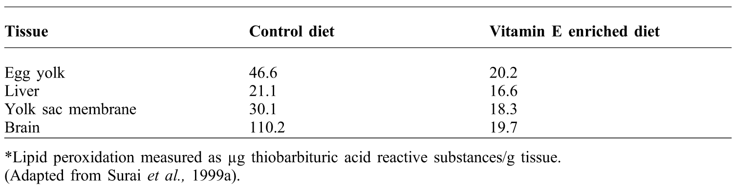
Increased vitamin E supplementation in the maternal diet can substantially increase vitamin E concentration in the developing tissues of the chick and significantly decrease their susceptibility to lipid peroxidation (Surai et al., Reference SURAI, NOBLE and SPEAKE1999a; Yih-Fwu et al., Reference YIH-FWU, HSIU-LING, YI-CHUN and SUE-JOAN2005) (Table 2). Increased vitamin E concentration in the yolk is associated with its accumulation in the liver of the newly-hatched chick (Surai, Reference SURAI1999a; Surai et al., Reference SURAI, KOSTJUK, SPEAKE, NOBLE and SPARKS1997).
Effect of dietary supplementation of the parent hen with vitamin E on the lipid peroxidation in day old chick tissues*.
*Lipid peroxidation measured as µg thiobarbituric acid reactive substances/g tissue.
(Adapted from Surai et al., Reference SURAI, NOBLE and SPEAKE1999a).
ascorbic acid
Vitamin C or L-ascorbic acid is the most important antioxidant and free radical scavenger in extracellular fluids. Its monovalent anion is called ascorbate. The best-known function of ascorbate is its ability to act as an electron donor. It is highly effective against peroxyl- and hydroxyl-radicals, superoxide, singlet oxygen, and peroxynitrites (Sies et al., Reference SIES, STAHL and SUNDQUIST1992). Although ascorbate reactions occur in the aqueous phase, this can prevent oxidation of lipid soluble vitamin E (α-tocopherol), which in turn prevents peroxidation of cell membrane (Niki, Reference NIKI1991). Oxidized ascorbate, both semi-dehydroascorbate and dehydroascorbate, can be reduced and recycled by GSH and other intracellular thiols (Meister, Reference MEISTER1994), and in some cells by a GSH-dependent dehydroascorbate reductase (Fornai et al., Reference FORNAI, SAVIOZZI, PIAGGI, GESI, CORSINI, MALVALDI and CASINI1999). Ascorbate acts in concert with other low-molecular weight substances such as GSH, vitamin E, SOD and GSH-PX (Meister, Reference MEISTER1994). The suppressive effect of corticoids in chickens is alleviated with vitamin C supplementation (Pardue et al., Reference PARDUE, THAXTON and BRAKE1985).
Ascorbic acid is not present in the yolk at the early stage of embryonic development. In contrast to vitamin E and carotenoids, vitamin C is synthesised in the yolk sac membrane and reaches the maximal level at day eight, and is subsequently transported to various tissues, particularly to the brain (Surai et al., Reference SURAI, NOBLE and SPEAKE1996). It is shown that, except for ascorbic acid, levels of α tocopherol, carotenoids (Surai et al., Reference SURAI, NOBLE and SPEAKE1996), selenium (Se) (Gaal et al., Reference GAAL, MEZES, NOBLE, DIXON and SPEAKE1995), GSH-PX and CAT (Gaal et al., Reference GAAL, MEZES, NOBLE, DIXON and SPEAKE1995) are very low in the brain, in comparison with the liver and other major embryonic tissues. Ascorbic acid may play a key role in the antioxidant defence system of the avian brain. Its concentration is markedly increased between days eight and 13 and is maintained at a high level throughout development. The liver contains much lower levels of ascorbic acid than the brain. It is possible that ascorbic acid in the brain may function in the regeneration of vitamin E by promoting the recycling of the tocopheroxyl radical back to an active antioxidant. The brain is far more susceptible than the other tissues of the chick to in vitro lipid peroxidation (Surai et al., Reference SURAI, NOBLE and SPEAKE1996).
antioxidant enzymes
The first line of defence against ROS consists of three antioxidant enzymes: SOD, GSH-PX and CAT. Superoxide dismutase enzymes have two forms, one contains copper and zinc (CuZn SOD) and appears to be predominantly cytoplasmic. The copper is involved in catalysis while the zinc provides the stability of the enzyme. The second contains manganese (MnSOD), and it is originated from mitochondria. Both of them require their particular metal ions for activity (Ose and Fridovich, Reference OSE and FRIDOVICH1976).
Glutathione peroxidase is primarily located in the cytosol. Glutathione is considered to be one of the most important water soluble antioxidants in the cell. Surai (Reference SURAI2000) reported that 200 mg/kg vitamin E dose in the maternal diet increased the concentration of glutathione in the liver of newly-hatched chicks and its elevated concentration could be considered an indication of increased antioxidative protection of tissues. Similar results were obtained with rats fed a high dose of vitamin E (Lii et al., Reference LII, KO, CHIANG, SUNG and CHEN1998). GSH-PX contains Se as a constituent of the amino acid selenocysteine, located at the active site of the enzyme, where it plays a crucial role in a redox reaction (Burk and Hill, Reference BURK and HILL1993).
Catalase is an intracellular antioxidant enzyme that is mainly located in cellular peroxisomes and catalyses the reaction of hydrogen peroxide to water and molecular oxygen. It indirectly detoxifies superoxide radicals, which are turned into hydrogen peroxide by SOD. The enzyme is very effective in oxidative stress and protects cells from hydrogen peroxide produced within the cell (Wassmann, Reference WASSMANN2004).
When each embryonic stage was examined, it has been reported that SOD activity increases in the liver between days 10 and 11 of development, then decreases significantly up to day 15 and remains with the same activity during the rest of the developmental period (Surai, Reference SURAI1999b). Similarly, Wilson et al. (Reference WILSON, LUI and DEL MAESTRO1992) showed that there was no difference in SOD specific activity in the embryonic liver between days 12 and 18. In the liver, GSH-PX probably plays a primary role in antioxidant defence while CAT might be less important. CAT reaches the maximal activity at day 10 of embryonic development and on the first day of chicks post hatch. After hatching liver CAT activity is still high, e.g. in the kidney, where it is much higher than in lung, brain and heart tissues (Surai, Reference SURAI1999b).
In addition to ascorbic acid (Surai et al., Reference SURAI, NOBLE and SPEAKE1996), SOD is an important protector against LPO in the embryonic brain, particularly during the last days of incubation, while GSH-PX and CAT activities are low during the period. This timing is associated with neuronal and glial proliferation and differentiation (Wilson et al., Reference WILSON, LUI and DEL MAESTRO1992). It has been suggested that effective recycling of vitamin E by ascorbic acid is one of the main protective mechanisms in the brain in vivo, but is insufficient in providing protection to the brain under increased stress conditions during in vitro incubation, SOD may be another important protective element in the embyonic brain. (Surai et al., Reference SURAI, NOBLE and SPEAKE1996). The embryonic lung has been characterised by high SOD activity, which increases at day 15 and thereafter decreases (Surai, Reference SURAI1999b). In the heart, GSH-PX activity is half of that in seen the liver or lungs, but activity slightly increases before hatching. Heart CAT activity is low and does not change during embryo development (Surai, Reference SURAI1999a).
In the YSM, all the antioxidant enzymes reach their maximal activity on day 15 of incubation. Thereafter, levels of GSH-PX decrease gradually until the end of the incubation period (Surai, Reference SURAI1999b). Probably, this decrease depends on the increasing levels of vitamin E, carotenoids (Surai et al., Reference SURAI, NOBLE and SPEAKE1996) and vitamin A (Gaal et al., Reference GAAL, MEZES, NOBLE, DIXON and SPEAKE1995) and therefore decreasing LPO susceptibility during the same interval. (Surai et al., Reference SURAI, NOBLE and SPEAKE1996).
Antioxidant enzymes, including GSH-PX, CAT and SOD, are synthesised and regulated endogenously but require dietary supplies of the appropriate nutrients. The components of this defence system work in a cooperative manner; thus, changes in the status of one component may affect that of the others (Yih-Fwu et al., Reference YIH-FWU, HSIU-LING, YI-CHUN and SUE-JOAN2005).
trace elements in antioxidant protection
Due to the diversity of proteins and enzymes containing zinc (Zn), copper (Cu), manganese (Mn), and selenium (Se), these trace minerals play critical roles in embryo development (Wilson, Reference WILSON1997). They are important for apolipoprotein, the major structural protein of lipoprotein, and participate in lipid metabolism. Cu, Zn, Mn, and Se are essential for catalytic activity and proper spatial conformation of antioxidant enzymes such as SOD and GSH-PX and hence play important roles in antioxidant capacity (Sun et al., Reference SUN, GUO, MA, YUAN, AN and LI2012). In general, trace minerals function either as catalytic or structural cofactors in metal-containing enzymes and proteins that are contained in the cells of the embryo and its extraembryonic membranes (Richards and Steele, Reference RICHARDS and STEELE1987).
Owing to the role of plasma protein synthesis, the yolk sac has the ability to regulate the export of trace minerals to the embryo during development. It regulates the metalloproteins in inter-organ transport, cellular uptake, and intracellular storage of trace minerals. Thus, embryonic trace mineral homeostasis is established through the coordinated actions of the yolk sac, which mobilises and exports trace minerals derived from egg stores; the vitelline circulation, which transports them to the embryo; and the liver, which accumulates trace minerals and distributes them to the rest of the tissues of the embryo via embryonic circulation (Richards, Reference RICHARDS1997). While the largest portion of most trace minerals is deposited in the yolk, much smaller quantities are deposited in the albumen portion of the egg (Romanoff, Reference ROMANOFF1967).
Trace elements may interact at the levels of absorption, distribution and retention. Deficiency or supplementation of one of them may alter their balance and induce an antagonistic or synergistic effect. The alteration of a trace-element balance may affect the antioxidant defence system since several trace elements, like Se, Cu, iron (Fe) and Zn, are integral parts of various antioxidant enzymes. There is a positive correlation between Se and Cu and Zn (Pappas et al., Reference PAPPAS, ZOIDIS, GEORGIOU, DEMIRIS, SURAI and FEGEROS2011).
Selenium is a trace element that plays a key role in the antioxidant defence as one selenium atom is required at the active site of all selenoproteins in the form of selenocysteine. These selenoproteins include GSH-PX and thioredoxin reductase, which have important antioxidant and detoxification functions (Burk and Hill, Reference BURK and HILL1993). Depending on geographic location, many agricultural soils produce crops with limited Se content. As a result, most poultry feeds contain supplemental Se. In general inorganic sources of Se (such as Na2SeO3) are used in feeds, but organic forms of Se (such as high-Se yeast) can also be used (Kelly and Power, Reference KELLY and POWER1995). However embryonic and extra-embryonic Se concentrations were found higher in eggs from hens fed organic Se than eggs from hens fed inorganic selenium (Paton et al., Reference PATON, CANTOR, PESCATORE, FORD and SMITH2002). Inclusion of organic selenium in breeder diets significantly increased vitamin E concentration in egg yolk (Surai et al., Reference SURAI, KUKLENKO, IONOV, NOBLE and SPARKS2000).
Selenium is deposited in the egg and distributed among the developing tissues during embryogenesis (Gaal et al., Reference GAAL, MEZES, NOBLE, DIXON and SPEAKE1995; Surai, Reference SURAI2000; Paton et al., Reference PATON, CANTOR, PESCATORE, FORD and SMITH2002). The largest increase in embryonic Se concentration is observed during days 10 to 15 of incubation (Paton et al., Reference PATON, CANTOR, PESCATORE, FORD and SMITH2002). Supplementary Se in the diet of the hen increases its concentration in the egg (Surai, Reference SURAI2000; Paton et al., Reference PATON, CANTOR, PESCATORE, FORD and SMITH2002; Pappas et al., Reference PAPPAS, KARADAS, SURAI and SPEAKE2005) and tissues of the chick at hatch (Surai, Reference SURAI2000; Pappas et al., Reference PAPPAS, KARADAS, SURAI and SPEAKE2005) and elevate the expression of GSH-PX while reducing the generation of lipid peroxides in the liver of the day-old chick. Selenium can have an effect on aspects of vitamin E metabolism and transport to target tissues (Surai, Reference SURAI2000). Feeding a 200 mg/kg diet of vitamin E to hens led to increases the concentration of glutathione in the liver of newly-hatched chicks (Surai, Reference SURAI2000). Elevated concentrations of GSH-PX can be considered an indication of increased antioxidant protection of the tissues. There is tissue-specificity in selenium transfer from egg to the embryo. For example, in contrast to the liver, there was only a trend for selenium accumulation in the brain of chickens hatched from eggs enriched in selenium (Meydani et al., Reference MEYDANI, MACAULEY and BLUMBERG1988).
Cu and Zn join together in cellular defence against oxidants with Se to form a triad of trace elements that are involved in cytosolic antioxidant defence: hydroperoxides, including H2O2, are reduced to the respective alcohols or water in a reaction that is catalysed by the selenoenzyme GSH-PX with glutathione (GSH) as the electron donor (Klotz et al., Reference KLOTZ, KRÖNCKE, BUCHCZYK and SIES2003). Similarly to Cu and Zn, Fe's ability to accept or donate electrons makes it central to the catalytic activity of many redox-active enzymes (Pappas et al., Reference PAPPAS, ZOIDIS, GEORGIOU, DEMIRIS, SURAI and FEGEROS2011). One of the Cu-containing enzymes, ceruloplasmin, binds up to 95% of circulating Cu, regulates Fe availability, takes part in oxidation-reduction reactions and may regulate immune function (Healy and Tipton, Reference HEALY and TIPTON2007). Manganese is a component of metalloenzymes that take part in reduction reactions with copper. These metalloenzymes are involved in multiple processes including respiration, carbohydrate and lipid metabolism, antioxidant activities (as a cofactor of MnSOD), and collagen formation (Andrieu, Reference ANDRIEU2008).
Conclusions
Avian embryo development relies on natural antioxidants accumulated in the egg yolk to protect tissues during hatching against lipid peroxidation. Antioxidant protection at hatching is considered to be an important determinant of chick viability during the first few days of life. Dietary supplementation of certain antioxidants, such as vitamin E, C, A, carotenoids or trace elements which are essential in antioxidant enzyme formation (such as Se, Cu, Zn, Mn) significantly enhance the activity of the antioxidant systems of the embryo, decreasing the susceptibility of the tissue against ROS. It can be concluded that regulation of antioxidant activity in chicken embryos depends on the parent hen diet. Ensuring suitable dietary levels can offer protection to the chick against lipid peroxidation during the embryonic development and post-hatch period.
Acknowledgments
The authors wish to acknowledge support from The Turkish Council of Higher Education, for supporting A.A. Yigit and Indian Council of Agriculture Research, Hyderabad for supporting A.K. Panda.