




No CrossRef data available.
Article contents
Determinants for resistance and susceptibility to microfilaraemia in Litomosoides sigmodontis filariasis
Published online by Cambridge University Press: 12 July 2001
Abstract
Filarial infections of humans are chronic diseases. Despite an ongoing immune response, adult filariae continuously produce their offspring, the microfilariae (Mf), which are able to persist in sufficient numbers to ensure transmission. In this study, host- and parasite-derived factors, which contribute to persistence of Mf, were investigated using the filariasis model of Litomosoides sigmodontis in mice. Different strains of mice were found to differ widely in their capability to eliminate circulating Mf. Studies of congenic mouse strains showed that early and rapid clearance of Mf was mediated by activation pathways relevant to innate immunity, whereas late or delayed clearance of Mf was pre-determined by MHC-related factors. Genetic knock-out of genes for the MHC class-II molecules totally abrogated resistance. Most interestingly, the presence of only 1 adult female, but not male worms, renders all mice susceptible, irrespective of the genetic background, enabling Mf to circulate for extended periods of time. Such prolonged microfilaraemia was also observed in L. sigmodontis-infected animals challenged with heterologous Mf of Acanthocheilonema viteae. The use of cytokine gene knock-out mice showed that persistence of L. sigmodontis Mf was facilitated by IL-10, but not by IL-4 or IFN-γ. In conclusion, irrespective of a resistant or susceptible host genetic background, survival of Mf of L. sigmodontis in mice is decisively regulated by the presence of adult female L. sigmodontis which will skew and exploit immune responses to facilitate the survival and persistence of their offspring in the infected host.
Keywords
Information
- Type
- Research Article
- Information
- Copyright
- © 2001 Cambridge University Press
INTRODUCTION
Filarial parasites of man generally persist in their host for many years, and have developed strategies which will allow them to survive and propagate in a hostile environment. Microfilariae (Mf) are very important in the epidemiology and transmission of the parasite as well as for the host–parasite interaction, including pathogenesis. Provision of an adequate milieu for long-term persistence and the regulation of the numbers of microfilariae in the individual host play key roles in the population dynamics and control of the parasite. For investigation of such parasite–host relationship experimental filariasis models are required which permit qualitative and quantitative analysis of immune responses during distinct states of infection. Such detailed and longitudinal investigations have become possible with the establishment of Litomosoides sigmodontis in BALB/c mice (Petit et al. 1992); a filariasis mouse model with complete parasite development and where patent infections are generated in an immune competent host. In several aspects the L. sigmodontis–mouse model resembles human filariasis. L. sigmodontis belongs to the family of Onchocercidae, as human pathogenic filariae, and shares features including patterns of larval migration, genomic structure and extensive immunological cross-reactivity in common with Brugia spp., Wuchereria bancrofti, Loa loa and Onchocerca volvulus (Xie, Bain & Williams, 1994; Bain et al. 1994). In Mf-positive BALB/c mice Th2 type cytokine responses predominate together with an impaired cellular proliferative response to filarial antigens (Maréchal et al. 1997; Le Goff et al. 2000). In mice, depletion of CD4+-T cells results in higher L. sigmodontis worm burdens and increase and prolong microfilaraemia (Al-Qaoud et al. 1997), and IL-5 appeared to be involved in encapsulation and killing of adult worms mediated by neutrophil granulocytes (Al-Qaoud et al. 2000). An acquired immunity can be generated by inoculation of irradiated infective 3rd-stage larvae (L3) resulting in reduction of worm numbers after challenge infection by 50–80% (Le Goff et al. 1997), mediated by IL-5 and eosinophils in conjunction with parasite-specific antibodies. For analysis of stage- and site-specific immune responses, intravenous injection of Mf or implantation of adult worms into the peritoneal cavity of mice were found to be a suitable approach to generate a transient microfilaraemia (Pfaff et al. 2000a). Selection of different inbred, congenic and transgenic mouse strains with distinct parasitological features offers the opportunity to investigate, in more detail, immunological and genetic determinants for susceptibility or resistance, as well as factors controlling subclinical infections (Hoffmann et al. 2000). Data presented in this study show that irrespective of a resistant or susceptible host genetic background, survival of Mf of L. sigmodontis in mice is decisively regulated by the presence of adult female worms. The presence of a single adult female for a few hours in the ‘resistant' recipient host sufficed to allow injected Mf to circulate for extended periods of time. Furthermore, host IL-10 production seems to be important but not solely responsible for prolonged microfilaraemia. These results support the idea that immune activation pathways relevant to innate immunity, and cytokines acting in concert, are strongly involved in regulating the course and duration of microfilaraemia in L. sigmodontis filariasis.
MATERIALS AND METHODS
Mice and parasites
Mice were purchased from Harlan Winkelmann GmbH (Borchen, Germany), i.e. BALB/bOlaHsd (BALB/b), BALB/cOlaHsd (BALB/c) B10.D2/nOlaHsd (B10.D2), CBA/JHsd (CBA/J), C3H/HeNHsd (C3H/He), C3H/HeJOlaHsd-Lpsd, (C3H/HeJ-Lpsd), C57BL/6JOlaHSD (C57BL/6), C57BL/10ScSnOlaHsd (C57BL/10), DBA/1OlaHsd (DBA/1), DBA/2OlaHsd (DBA/2), and 129SvHsd (129Sv), SJL/JHanHsd (SJL/J), and subsequently kept at our own facilities in microisolator cages.
MHC-class IIAβ(−/−) knockout mice on C57BL/6 background (MHC II ko) were kindly provided by Dr B. Kyewski (Deutsches Krebsforschungszentrum, Heidelberg, Germany), DBA/1-Ifngtm1Ts (IFN-γ ko) mice were from Dr K. Klingel (University of Tübingen, Germany) and IL-4 and IL-10 knock-out mice both on C57BL/6 background (IL-4 ko and IL-10 ko) (Kopf et al. 1993) were kindly given by Dr H. Mossmann (Max–Planck–Institute for Immunobiology, Freiburg, Germany). Four to eight-week-old male mice were used in all experiments. Adult worms of L. sigmodontis were isolated from the pleural and peritoneal cavities of previously infected jirds (Meriones unguiculatus) or cotton rats (Sigmodon hispidus) under sterile conditions. For the isolation of Mf of L. sigmodontis and Acanthocheilonema viteae, blood of infected hosts was collected and the Mf purified using a Percoll® density gradient (Chandrashekar et al. 1984). L. sigmodontis antigen was prepared by homogenization and sonication of adult worms or isolated microfilariae in phosphate-buffered saline (Sigma, Deisenhofen, Germany). After centrifugation, supernatants were sterilized by filtration. The protein concentration was determined using the bicinchoninic acid (BCA) protein assay (Pierce, Rockford, IL, USA) (Keller & Neville, 1986).
Infection protocols
Mice were anaesthesized with a mixture of 15 mg/kg body weight (bw) of xylasine (Rompun®, Bayer, Leverkusen, Germany) and 100 mg/kg bw of ketamine (Ketanest®, Parke-Davis, Berlin, Germany). Mice were shaved and the skin disinfected with 80% ethanol at the site of injection. Adult worms were surgically implanted into the peritoneal cavity. In sham operations, short pieces of sterile surgical sutures were implanted. The techniques for isolation, injection and counting of L. sigmodontis Mf was as previously described (Pfaff et al. 2000a). Briefly, Percoll®-isolated Mf were injected into the vena jugularis of anaesthesized mice at a standard dose of 100000 in 70 μl of RPMI 1640 medium (Gibco, Eggenstein, Germany). Blood samples from the retro-orbital vein plexus of mice were taken at intervals as indicated. Mf were counted in 30 μl blood samples. To confirm a Mf-negative blood sample an additional 100 μl of blood was collected and examined.
Proliferation and antibody assays
At necropsy, the peritoneal cavity of mice was washed out with 3 ml of ice-cold RPMI. Cells were then washed and the number of viable cells was determined by Trypan blue exclusion (Seromed, Berlin, Germany). Then 100 μl of a 1×10⁶ cells/ml suspension were added to the wells of flat-bottom 96-well plates (Costar) and kept at 37 °C for 2 h, after which non-adherent cells were removed by washing with RPMI 1640. Spleens were removed aseptically, and a single cell suspension was prepared by repeated passage through a metal gauze into RPMI 1640. Spleen cells were washed and re-suspended in RPMI 1640 (supplemented with 10% foetal calf serum (Seromed, Berlin, Germany), antibiotic–antimycotic solution (100 U/ml penicillin, 100 μg/ml streptomycin, 2.5 μg/ml amphotericin, Gibco) and 20 mM glutamine (Flow Laboratories, Irvine, Scotland, UK)), to give a final concentration of 2×10⁶ cells/ml. Each assay was done in triplicate cultures of 100 μl cell suspension per well. For stimulation, cells were incubated with 4 μg/ml Concanavalin A (ConA, Seromed). Proliferation was assayed by adding [3H]thymidine (Amersham, Braunschweig, Germany) at 1 μCi/ well over the final 18 h of a 48 h incubation, after which samples were transferred to a filter mat (Wallac, Turku, Finland) and processed for liquid scintillation counting. Parasite-specific serum antibody levels were determined as previously described (Maréchal et al. 1997).
Statistical analysis
Comparison of groups was done with the non-parametrical Mann–Whitney U-test. P values [les ]0.05 were considered significant.
RESULTS
Microfilaraemia in different inbred mouse strains after intravenous injection of Mf of L. sigmodontis
The duration of microfilaraemia following intravenous injection of 100000 Mf of L. sigmodontis into previously unexposed (naive) recipient mice was investigated (Fig. 1A). In CBA/J (MHC haplotype H-2k), DBA/1 (H-2q), DBA/2 (H-2d), C3H/He (H-2k), and SJL (H-2s) Mf were eliminated in less than 3 days, intermediate Mf clearance was found in 129Sv (H-2b) and C57BL/6 (H-2b), whereas Mf persisted in BALB/c (H-2d) mice for more than 30 days. To investigate the potentially confounding effects of Mf-associated presence of bacterial endotoxin, e.g. LPS as found in symbiotic Wolbachia, L. sigmodontis Mf were injected into C3H/HeJ-Lpsd mice, these being non-responders for LPS, and also into C3H/He control mice. In both strains an equally rapid Mf elimination was observed with microfilaraemia being terminated after 2 days.

Fig. 1. Microfilaraemia in different inbred mouse strains following (A) intravenous injections of 100000 microfilariae or (B) intraperitoneal implantation of 5 fecund adult female worms of Litomosoides sigmodontis. Data points in (A) represent arithmetic means+S.D. of 5–6 mice per group. Lines in (B) represent microfilaraemia of individual mice.
Microfilaraemia in different inbred mouse strains after implantation of adult L. sigmodontis
Five fecund adult female L. sigmodontis were transplanted into the peritoneum of naive mice of different strains which previously have been shown to be resistant to prolonged circulation of injected Mf, i.e. DBA/1, C3H/He and C57BL/6. All mice developed microfilaraemia, which persisted for several months (Fig. 1B). The shortest duration of patent infections was seen in C57BL/6 mice with 2.5 months, while in some DBA/1 and C3H/He mice, Mf of L. sigmodontis circulated for more than 7 months.
Microfilaraemia in congenic strains and MHC II gene knock-out mice
To investigate the influence of MHC-related and non-related genetic host factors, Mf were injected intravenously into naive mice, and the development and persistence of microfilaraemia in MHC-congenic strains compared (Fig. 2 A). In B10.D2 mice, exhibiting background genes of C57BL/10, but which share the MHC haplotype H-2d with the susceptible BALB/c strain, injected Mf were eliminated as quickly from the peripheral circulation as in C57BL/10 mice. In BALB/b mice, which share the MHC haplotype H-2b with the resistant C57BL/10 strain injected Mf initially persisted but levels and duration of microfilaraemia were already significantly reduced by 10 days post-inoculation, as compared to the congenic BALB/c strain. In addition, in BALB/b, an earlier IgG response, mainly IgG2a and IgG3, was induced 2 weeks post-injection of Mf (data not shown).

Fig. 2. Microfilaraemia in (A) MHC-congenic mouse strains that differ either in their genetic background (BALB and C57BL) or their MHC-haplotype (H-2b and H-2d) and (B) MHC II knock-out mice on C57BL/6 genetic background. All mice received an intravenous (iv) injection of 100000 Mf of Litomosoides sigmodontis. Data points represent arithmetic means+S.D. of 5–6 mice per group. *P<0.05 (BALB/c versus BALB/b).
When 100000 Mf were injected intravenously into naive C57BL/6 MHC II gene knock-out (ko) mice, microfilaraemia persisted for several months (Fig. 2B). Interestingly, infection of C57BL/6 MHC II-ko mice with L. sigmodontis L3 resulted in maturation of the parasite and patent infection (data not shown).
Transplantation of immature adult L. sigmodontis in resistant mice and subsequent injection of Mf
Mf-resistant C57BL/6, DBA/1 and C3H/He mice were transplanted with immature adult L. sigmodontis (27–28 days post-infection). One week later, Mf of L. sigmodontis were injected. In all strains of recipient mice, injected Mf circulated at high levels for several weeks, showing that the presence of adult female worms will abolish resistance against Mf challenge (Fig. 3). The circulating Mf did not originate from the implanted L. sigmodontis because females will not start Mf release before 50 days post-infection (Hoffmann et al. 2000). Interestingly, when resistant DBA/1 mice were transplanted with adult male L. sigmodontis, subsequently injected Mf were eliminated even faster than in the absence of adult worms. In contrast, transplantation of adult male worms into BALB/c mice had no significant effect on microfilaraemia (data not shown).

Fig. 3. Microfilaraemia in different inbred mouse strains after intraperitoneal transplantation of 5 immature adult female Litomosoides sigmodontis followed by an intravenous injection of 100000 microfilariae of L. sigmodontis. Control animals received a sham surgery instead of transplantation of adult worms. Data points represent arithmetic means+S.D. of 5–6 animals per group.
Effect of transplanted worm load and duration of presence on subsequent microfilaraemia
One, 2, or 4 immature adult female L. sigmodontis were transplanted into resistant DBA/1 mice, and 4 days later mice were intravenously challenged with L. sigmodontis Mf (Fig. 4A). Irrespective of the number of adult L. sigmodontis transplanted, the magnitude of microfilaraemia was similar in all animals with 1 adult female being sufficient to allow for Mf persistence. To evaluate the minimum time required for the observed modulation of resistance against Mf, and also how long this effect lasts, recipient DBA/1 mice were transplanted with 5 adult L. sigmodontis and the subsequent intravenous challenge was conducted at various time-points post-transplantation. As shown in Fig. 4B, 6 hours were sufficient to allow for Mf persistence in otherwise resistant mice. The same retardation of Mf clearance was observed 2 months after transplantation.

Fig. 4. Microfilaraemia in DBA/1 mice after intraperitoneal transplantation of immature adult female Litomosoides sigmodontis followed by an intravenous injection of 100000 microfilariae of L. sigmodontis. Effect of (A) adult worm load, and (B) duration of adult worm presence (5 worms) on subsequent microfilaraemia. Data points represent arithmetic means+S.D. of 2–3 animals per group.
L. sigmodontis adult worm transplantation and microfilaraemia in IL-4, IL-10 and IFN-γ cytokine gene knock-out mice
Resistant C57BL/6 mice with a Th2-type cytokine gene knock-out mutation (IL-4 ko or IL-10 ko) were transplanted with 5 immature adult female L. sigmodontis and 1 week later challenged with 100000 Mf of L. sigmodontis. In IL-4 ko mice, intravenously injected Mf remained at the same high levels as in L. sigmodontis transplanted C57BL/6 wild-type animals (Fig. 5A). Similarly, sham-treated IL-4 ko and wild type controls cleared injected Mf within a few days. In contrast, in IL-10 ko mice transplanted with adult L. sigmodontis and then challenged with Mf, levels of microfilaraemia were significantly decreased compared to wild-type mice as soon as 4 days p.i. (Fig. 5B). However, L. sigmodontis transplanted and Mf-challenged IL-10 ko mice did not approach the dynamics of microfilaraemia clearance of only Mf-injected animals. For Th1-type cytokines, in transplanted and Mf challenged IFN-γ ko mice, dynamics of Mf clearance, remained similar to that of DBA/1 wild-type animals (Fig. 5C). Challenge of sham-treated wild-type and IFN-γ ko mice with L. sigmodontis Mf resulted in a similarly rapid clearance of Mf from the peripheral blood in both groups.

Fig. 5. Microfilaraemia in (A) interleukin 4 knock-out- (IL-4 ko), (B) IL-10 ko- and (C) interferon γ (IFNg) ko-mice after intraperitoneal transplantation of 5 immature adult female Litomosoides sigmodontis followed by an intravenous injection of 100000 microfilariae of L. sigmodontis 1 week later. Controls received a sham-surgery instead of transplantation of adult worms (sham-OP). Wild-type mice (wt) with the same genetic background were treated similarly. IL-4 ko and IL-10 ko-mice were on C57BL/6 genetic background, whereas IFNg ko-mice had a DBA/1 genetic background. Data points represent arithmetic means+S.D. of 5–7 mice per group. * P<0.05 (IL-10 wt versus IL-10 ko).
Heterologous Mf challenge
In order to investigate whether the presence of adult females of L. sigmodontis will affect the persistence of other filariae, resistant DBA/1 mice were transplanted with adult L. sigmodontis and subsequently challenged with 100000 Mf of Acanthocheilonema viteae (Fig. 6). As observed with an homologous L. sigmodontis Mf challenge, A. viteae microfilaraemia in transplanted DBA/1 mice persisted at significantly higher levels than in those animals without transplanted adult females of L. sigmodontis. The clearance of A. viteae Mf, injected into sham-treated DBA/1 mice, was noticeably slower than clearance of L. sigmodontis Mf.

Fig. 6. Microfilaraemia in DBA/1 mice after intraperitoneal transplantation of 5 immature adult female Litomosoides sigmodontis followed by an intravenous injection of 100000 microfilariae of Acanthocheilonema viteae 1 week later. Data points represent arithmetic means+S.D. of 5–6 animals per group. *P<0.05.
Cellular responses with L. sigmodontis transplantation and Mf challenge
Spleen cell responses to ConA were significantly increased in Mf challenged DBA/1 mice transplanted with adult L. sigmodontis when compared to sham-treated animals (Fig. 7). Reactivity of spleen cells from transplanted and Mf challenged DBA/1 mice was strongly abrogated when splenocytes were co-cultured with the adherent fraction of autologous peritoneal lavage exudate cells (PEC). Also, in IL-10 knockout and IL-10 wild-type mice, transplanted with L. sigmodontis and challenged with Mf, spleen cell reactivity to ConA was completely suppressed in the presence of autologous adherent PEC.

Fig. 7. Cellular responsiveness of immunocompetent DBA/1, interleukin 4-knock-out (IL-4 ko), IL-10 ko and their respective wild-type (wt) mice after intraperitoneal transplantation of 5 immature adult females of Litomosoides sigmodontis followed by an intravenous injection of 100000 microfilariae of L. sigmodontis 1 week later. Cytokine-gene knock-out and wild-type mice were all on C57BL/6 genetic background. Controls received a sham-surgery instead of transplantation of adult worms (sham-treated). Splenocytes of infected mice as well as peritoneal exudate cells (PEC) were recovered 18–28 days post-Mf injection and stimulated in vitro with Concanavalin A (4 μg/ml) for 48 h. Cellular proliferation was quantified by [3H]thymidine incorporation for the last 18 h of culture. Values are indicated as mean counts per minute (cpm) of triplicate cultures minus cpm from non-stimulated control cultures (net counts: delta cpm). Bars and vertical lines represent arithmetic means+S.E.M. of 5–6 animals per group. *P<0.05 (spleen cells versus spleen cells plus PEC). #P<0.05 (mice with implanted female worms versus sham-treated controls).
DISCUSSION
In experimental filariasis models, the determinants which contribute to resistance or reduction of developing L3 and adult filarial worms, in particular the involvement of Th1- and Th2-type cytokines, lymphocyte subpopulations as well as the genetic background of the host, have been investigated (reviewed by Lawrence, 1996 and Hoerauf & Fleischer, 1997). In various vaccination trials, the numbers of adult worms after challenge infections could be significantly reduced (Le Goff et al. 1997; Geiger et al. 1997). It has to be stressed, however, that even a few of the surviving filariae sufficed to produce patent infection with a persisting microfilaraemia for extended periods of time. The release and persistence of Mf is essential for transmission of all filarial parasites, and it is the main cause of pathogenesis in onchocerciasis. This study, therefore, addressed the determinants which regulate levels and duration of microfilaraemia in the L. sigmodontis filariasis.
Previously, Petit et al. (1992) and Maréchal et al. (1996) have shown that mouse strains differ widely in their responsiveness to injected L. sigmodontis L3. Several strains, e.g. C65BL/6 and DBA/1, were found to be strictly non-permissive for maturation of L3, while others, e.g. CBA and C3H/HeN mice, were found to be ‘semi-resistant' hosts allowing for maturation of L. sigmodontis, however, without development of microfilaraemia. Only in strains of the BALB background was full parasite development observed. In this study, intravenous injection of Mf generated transient or prolonged microfilaraemia with characteristic dynamic features which could be related to the genetic background and MHC haplotype of the recipient host. The observed wide range of responsiveness of different mouse strains to Mf clearance appeared to be regulated by immune mechanisms rather than due to a short life-span of Mf or non-specific physiological conditions of the murine hosts. This was supported by our observation that in MHC knock-out mice, which do not express MHC II class molecules and lack almost all CD4+-T cells, injected Mf of L. sigmodontis survived for more than 140 days. The dynamics of Mf clearance segregated mouse strains into susceptible, semi-resistant and refractory. In those mice with a rapid elimination of circulating Mf, clearance of Mf appeared to be entirely regulated by the genetic background and was independent of the MHC haplotype, e.g. B10.D2 (H-2d) versus BALB/c (H-2d). In mice with a susceptible genetic background, the MHC haplotype influences Mf elimination at later time points post-inoculation, e.g. BALB/b (H-2b) versus BALB/c (H-2d). This suggests that early and rapid clearance of Mf was mediated by activation pathways relevant to innate immunity, whereas late or delayed clearance of Mf was pre-determined by MHC-related factors. Also, as recently shown by us, nitric oxide seems not to be an important factor in innate immunity to Mf of L. sigmodontis (Pfaff et al. 2000b). A comparative approach with different inbred mouse strains was used to investigate immune responsiveness following injection of Mf of Brugia malayi (Fanning & Kazura, 1983) and implantation of adult A. viteae (Haque et al. 1980) where also different dynamics of Mf clearance were observed. Together, these observations are supportive of the idea that early and rapid Mf clearance is related to T cell-independent mechanisms. Previously, it has been shown that a switch in T-helper cell responses does not necessarily affect Mf clearance (Pearlman et al. 1995). T cell-independent immune mechanisms were suggested to be effective during early infection with L3 of L. sigmodontis (Al-Qaoud, Fleischer & Hoerauf, 1998) and Brugia species (Paciorkowski et al. 2000), with B1 lymphocytes being responsible for partial, but not sterile clearance of incoming L3, and the surviving worm burden sufficed to generate microfilaraemia. The latter study showed T cells to be necessary for long-term control of filarial infection, which is in line with our results and earlier studies (Vincent, Sodeman & Winters, 1980). Furthermore, Mf may elicit inflammatory immune responses induced by endotoxins (LPS) of the symbiotic Wolbachia bacteria in Mf of L. sigmodontis (Taylor & Hoerauf, 1999; Taylor, Cross & Bilo, 2000). However, our observation that an LPS non-responder mouse strain eliminated Mf as quickly as the corresponding control strain, does not support a decisive role of this pathway in Mf clearance.
Our approach to investigate the effects of implantation of immature adult female worms of L. sigmodontis on subsequently injected microfilariae provided several new aspects of the filaria–host interaction. Firstly, resistance to microfilaraemia was completely abrogated when adult female L. sigmodontis were present in the peritoneum. Such breakdown of resistance, mediated by adult female, but not adult male filariae, was observed in all mouse strains tested so far. To allow for patent infection, even in an otherwise resistant host, the parasite load requires only 1 adult female, and Mf clearance was also retarded in animals challenged with heterologous Mf of A. viteae. Consequently, the molecules involved in such modulatory pathways have to be effective at very low concentrations, and potentially being effective across filarial species barriers. Secondly, the time required to switch a ‘Mf-resistant' into a ‘Mf-permissive' host was only a few hours, suggesting that T cells, antigen presentation, and pathways of adaptive immunity are initially not required for the switch from resistance to susceptibility. Even though the molecules and mechanisms involved remain yet unknown, likely candidates for such activity are non-proteins, potentially phosphocholine-containing molecules (Harnett & Harnett, 1993) and repetitive carbohydrates previously shown to modulate immune responses independently of T-cells (Velupillai et al. 1997). Suppression of homologous immunity, comparable to our results and also mediated by adult worms was observed in mice infected with the gastro-intestinal nematode Nematospiroides dubius (Behnke, Hannah & Pritchard, 1983).
During patent L. sigmodontis infection, a Th2-type cytokine profile predominates associated with profound suppression of cellular proliferation (Schönfeld & Zahner, 2000). However, our observation that in L. sigmodontis-transplanted IL-4 ko mice injected Mf circulated at high levels similar to wild-type animals, suggested that IL-4 is not directly involved in suppression of Mf clearance. In contrast, in our L. sigmodontis-transplanted IL-10 ko animals, microfilaraemia decreased significantly faster than in the wild-type control, suggesting that host production of IL-10 is required for Mf persistence, and that this IL-10 production is elicited by the transplanted adult females. Despite the accelerated clearance of Mf in IL-10 ko animals, IL-10 gene knock-out alone did not suffice to restore dynamics of Mf clearance of wild-type mice, suggesting that additional components, or a complex concerted action of modulatory cytokines, are required for Mf persistence. The abrogation of spleen cell responses in the presence of autologous adherent PEC in female-implanted mice has also been observed in B. malayi-infected mice (Allen, Lawrence & Maizels, 1996). Suppression was induced by secretion products of adult filariae (Allen & MacDonald, 1998) and, interestingly, IL-4 was reported to be indispensable for the generation of suppressor cell but not the sole component mediating suppression of T-cells (MacDonald et al. 1998). However, in BALB/c mice infected with B. pahangi, IL-10 and antigen-presenting cells actively suppressed Th1 cells (Osborne & Devaney 1999). Recently, Doetze et al. (2000) reported that the antigen-specific cellular hyporesponsiveness in onchocerciasis patients was mediated by Th3-type cytokines IL-10 and transforming growth factor-beta.
Earlier, suppression of human lymphocyte proliferative responses was observed in vitro in the presence of secretory–excretory products of female but not male worms of O. volvulus (Elkhalifa et al. 1991).
In summary, adult female L. sigmodontis have evolved with powerful strategies to facilitate their offspring, i.e. microfilariae, survival and persistence in the infected host, and by doing such they ultimately assure transmission of their progeny. Most impressively, irrespective of the genetic background of the host, the presence of a single adult female L. sigmodontis for a few hours was enough to make a resistant host susceptible for microfilaraemia. Not only that such ‘host conditioning' may be effective across filarial species barriers, this powerful strategy of filarial parasites should be considered when selecting for and developing potential anti-filarial vaccines, and also when conducting vaccination trials.
This work was supported by the Fortüne-Programme from the University of Tübingen (Grant no. F.1490176), the Commission of the European Community Grant no. ICA4-CT-1999-10002, the Edna McConnell Clark Foundation (Grant no. 97138) and the Deutsche Forschungsgemeinschaft (DFG-So367/1-2).
References
ALLEN, J. E., LAWRENCE, R. A. & MAIZELS, R. M. (1996). APC for mice harbouring the filarial nematode, Brugia malayi, prevent cellular proliferation but not cytokine production. International Immunology 8, 143–151.CrossRefGoogle Scholar
ALLEN, J. E. & MACDONALD, A. S. (1998). Profound suppression of cellular proliferation mediated by the secretions of nematodes. Parasite Immunology 20, 241–247.CrossRefGoogle Scholar
AL-QAOUD, K. M., FLEISCHER, B. & HOERAUF, A. (1998). The Xid defect imparts susceptibility to experimental murine filariosis-association with a lack of antibody and IL-10 production by B cells in response to phosphorylcholine. International Immunology 10, 17–25.CrossRefGoogle Scholar
AL-QAOUD, K. M., PEARLMAN, E., HARTUNG, T., KLUKOWSKI, J., FLEISCHER, B. & HOERAUF, A. (2000). A new mechanism for IL-5-dependent helminth control: neutrophil accumulation and neutrophil-mediated worm encapsulation in murine filariasis are abolished in the absence of IL-5. International Immunology 12, 899–908.CrossRefGoogle Scholar
AL-QAOUD, K. M., TAUBERT, A., ZAHNER, H., FLEISCHER, B. & HOERAUF, A. (1997). Infection of BALB/c mice with the filarial nematode Litomosoides sigmodontis: role of CD4+ T cells in controlling larval development. Infection and Immunity 65, 2457–2461.Google Scholar
BAIN, O., WANJI, S., VUONG, P. N., MARECHAL, P., LE GOFF, L. & PETIT, G. (1994). Larval biology of six filariae of the sub-family Onchocercinae in a vertebrate host. Parasite 1, 241–254.CrossRefGoogle Scholar
BEHNKE, J. M., HANNAH, J. & PRITCHARD, D. I. (1983). Nematospiroides dubius in the mouse: evidence that adult worms depress the expression of homologous immunity. Parasite Immunology 5, 397–408.CrossRefGoogle Scholar
CHANDRASHEKAR, R., RAO, U. R., RAJASEKARIAH, G. R. & SUBRAHMANYAM, D. (1984). Isolation of microfilariae from blood on iso-osmotic Percoll® gradients. Indian Journal for Medical Research 79, 497–501.Google Scholar
DOETZE, A., SATOGUINA, J., BURCHARD, G., RAU, T., LOLIGER, C., FLEISCHER, B. & HOERAUF, A. (2000). Antigen-specific cellular hyporesponsiveness in a chronic human helminth infection is mediated by T(h)3/T(r) 1-type cytokines IL-10 and transforming growth factor-beta but not by a T(h)1 to T(h)2 shift. International Immunology 12, 623–630.CrossRefGoogle Scholar
ELKHALIFA, M. Y., GHALIB, H. W., DAFA'ALLA, T. & WILLIAMS, J. F. (1991). Suppression of human lymphocyte responses to specific and non-specific stimuli in human onchocerciasis. Clinical and Experimental Immunology 86, 433–439.CrossRefGoogle Scholar
FANNING, M. M. & KAZURA, J. W. (1983). Genetic association of murine susceptibility to Brugia malayi microfilaraemia. Parasite Immunology 5, 305–316.CrossRefGoogle Scholar
GEIGER, S. M., HOFFMANN, W., RAPP, J., SCHULZ-KEY, H. & EISENBEISS, W. F. (1997). Filariidae: cross-protection in filarial infections. Experimental Parasitology 83, 352–356.Google Scholar
HAQUE, A., WORMS, M. J., OGILVIE, B. M. & CAPRON, A. (1980). Dipetalonema viteae: Microfilariae production in various mouse strains and in nude mice. Experimental Parasitology 49, 398–404.CrossRefGoogle Scholar
HARNETT, W. & HARNETT, M. M. (1993). Inhibition of murine B cell proliferation and down-regulation of protein kinase C levels by a phosphorylcholine-containing filarial excretory-secretory product. Journal of Immunology 151, 4829–4837.Google Scholar
HOERAUF, A. & FLEISCHER, B. (1997). Immune responses to filarial infection in laboratory mice. Medical and Microbiological Review 185, 207–215.Google Scholar
HOFFMANN, W. H., PETIT, G., SCHULZ-KEY, H., TAYLOR, D. W., BAIN, O. & LE GOFF, L. (2000). Litomosoides sigmodontis in mice: reappraisal of an old model for filarial research. Parasitology Today 16, 387–389.CrossRefGoogle Scholar
KELLER, R. P. & NEVILLE, M. C. (1986). Determination of total protein in human milk: comparison of methods. Clinical Chemistry 32, 120–123.Google Scholar
KOPF, M., LE GROS, G., BACHMANNN, M., LAMERS, M. C. & BLUETHMAN, H. (1993). Disruption of the murine IL-4 gene blocks Th2 cytokine responses. Nature, London 362, 245–248.CrossRefGoogle Scholar
LAWRENCE, R. A. (1996). Lymphatic filariasis: what mice can tell us. Parasitology Today 12, 267–271.CrossRefGoogle Scholar
LE GOFF, L., MARECHAL, P., PETIT, G., TAYLOR, D. W., HOFFMANN, W. & BAIN, O. (1997). Early reduction of the challenge recovery rate following immunization with irradiated infective larvae in a filaria mouse system. Tropical Medicine and International Health 2, 1170–1174.CrossRefGoogle Scholar
LE GOFF, L., MARTIN, C., OSWALD, I. P., VUONG, P. N., PETIT, G., UNGEHEUER, M. N. & BAIN, O. (2000). Parasitology and immunology of mice vaccinated with irradiated Litomosoides sigmodontis larvae. Parasitology 120, 271–280.CrossRefGoogle Scholar
MACDONALD, A. S., MAIZELS, R. M., LAWRENCE, R. A., DRANSFIELD, I. & ALLEN, J. E. (1998). Requirement for in vivo production of IL-4, but not IL-10, in the production of proliferative suppression by filarial parasites. Journal of Immunology 147, 144–151.Google Scholar
MARÉCHAL, P., LE GOFF, L., HOFFMANN, W., RAPP, J., OSWALD, I. P., OMBROUK, C., TAYLOR, D. W., BAIN, O. & PETIT, G. (1997). Immune response to the filaria Litomosoides sigmodontis in susceptible and resistant mice. Parasite Immunology 19, 273–279.CrossRefGoogle Scholar
MARÉCHAL, P., LE GOFF, L., PETIT, G., DIAGNE, M., TAYLOR, D. W. & BAIN, O. (1996). The fate of the filaria Litomosoides sigmodontis in susceptible and naturally resistant mice. Parasite 3, 25–31.CrossRefGoogle Scholar
OSBORNE, J. & DEVANEY, E. (1999). Interleukin-10 and antigen-presenting cells actively suppress Th1 cells in BALB/c mice infected with the filarial parasite Brugia pahangi. Infection and Immunity 67, 1599–1605.Google Scholar
PACIORKOWSKI, N., PORTE, P., SHULTZ, L. D. & RAJAN, T. V. (2000). B1 B lymphocytes play a critical role in host protection against lymphatic filarial parasites. Journal of Experimental Medicine 191, 731–735.CrossRefGoogle Scholar
PEARLMAN, E., HEINZEL, F. P., HAZLETT, F. E. JR & KAZURA, J. W. (1995). IL-12 modulation of T helper responses to the filarial helminth, Brugia malayi. Journal of Immunology 154, 4658–4664.Google Scholar
PETIT, G., DIAGNE, M., MARECHAL, P., OWEN, D., TAYLOR, D. W. & BAIN, O. (1992). Maturation of the filaria Litomosoides sigmodontis in BALB/c mice; comparative susceptibility of nine other inbred strains. Annales de Parasitologie Humaine et Comparée 67, 144–150.CrossRefGoogle Scholar
PFAFF, A. W., SCHULZ-KEY, H., SOBOSLAY, P. T., GEIGER, S. M. & HOFFMANN, W. H. (2000a). Litomosoides sigmodontis: Dynamics of the survival of microfilariae in resistant and susceptible strains of mice. Experimental Parasitology 96, 67–74.Google Scholar
PFAFF, A. W., SCHULZ-KEY, H., SOBOSLAY, P. T., GEIGER, S. M. & HOFFMANN, W. H. (2000b). The role of nitric oxide in the innate resistance to microfilariae of Litomosoides sigmodontis in mice. Parasite Immunology 22, 397–407.Google Scholar
SCHÖNFELD, K. & ZAHNER, H. (2000). Immunomodulatory effects in Litomosoides sigmodontis-infected Mastomys coucha. Parasitology Research 86, 101–108.CrossRefGoogle Scholar
TAYLOR, M. J., CROSS, H. F. & BILO, K. (2000). Inflammatory responses induced by the filarial nematode Brugia malayi are mediated by lipopolysaccharide-like activity from endosymbiotic Wolbachia bacteria. Journal of Experimental Medicine 191, 1429–1435.CrossRefGoogle Scholar
TAYLOR, M. J. & HOERAUF, A. (1999). Wolbachia bacteria of filarial nematodes. Parasitology Today 15, 437–442.CrossRefGoogle Scholar
VELUPILLAI, P., SECOR, W. E., HOERAUF, A. M. & HARN, D. A. (1997). B-1 cell (CD5+B220+) outgrowth in murine schistosomiasis is genetically restricted and is largely due to activation by polylactosamine sugars. Journal of Immunology 158, 338–344.Google Scholar
VINCENT, A. L., SODEMAN, W. A. & WINTERS, A. (1980). Development of Brugia pahangi in normal and nude mice. Journal of Parasitology 66, 448.CrossRefGoogle Scholar
XIE, H., BAIN, O. & WILLIAMS, S. A. (1994). Molecular phylogenetic studies on filarial parasites based on 5S ribosomal spacer sequences. Parasite 1, 141–151.CrossRefGoogle Scholar
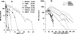
Fig. 1. Microfilaraemia in different inbred mouse strains following (A) intravenous injections of 100000 microfilariae or (B) intraperitoneal implantation of 5 fecund adult female worms of Litomosoides sigmodontis. Data points in (A) represent arithmetic means+S.D. of 5–6 mice per group. Lines in (B) represent microfilaraemia of individual mice.
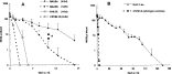
Fig. 2. Microfilaraemia in (A) MHC-congenic mouse strains that differ either in their genetic background (BALB and C57BL) or their MHC-haplotype (H-2b and H-2d) and (B) MHC II knock-out mice on C57BL/6 genetic background. All mice received an intravenous (iv) injection of 100000 Mf of Litomosoides sigmodontis. Data points represent arithmetic means+S.D. of 5–6 mice per group. *P<0.05 (BALB/c versus BALB/b).

Fig. 3. Microfilaraemia in different inbred mouse strains after intraperitoneal transplantation of 5 immature adult female Litomosoides sigmodontis followed by an intravenous injection of 100000 microfilariae of L. sigmodontis. Control animals received a sham surgery instead of transplantation of adult worms. Data points represent arithmetic means+S.D. of 5–6 animals per group.
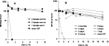
Fig. 4. Microfilaraemia in DBA/1 mice after intraperitoneal transplantation of immature adult female Litomosoides sigmodontis followed by an intravenous injection of 100000 microfilariae of L. sigmodontis. Effect of (A) adult worm load, and (B) duration of adult worm presence (5 worms) on subsequent microfilaraemia. Data points represent arithmetic means+S.D. of 2–3 animals per group.
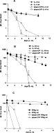
Fig. 5. Microfilaraemia in (A) interleukin 4 knock-out- (IL-4 ko), (B) IL-10 ko- and (C) interferon γ (IFNg) ko-mice after intraperitoneal transplantation of 5 immature adult female Litomosoides sigmodontis followed by an intravenous injection of 100000 microfilariae of L. sigmodontis 1 week later. Controls received a sham-surgery instead of transplantation of adult worms (sham-OP). Wild-type mice (wt) with the same genetic background were treated similarly. IL-4 ko and IL-10 ko-mice were on C57BL/6 genetic background, whereas IFNg ko-mice had a DBA/1 genetic background. Data points represent arithmetic means+S.D. of 5–7 mice per group. * P<0.05 (IL-10 wt versus IL-10 ko).

Fig. 6. Microfilaraemia in DBA/1 mice after intraperitoneal transplantation of 5 immature adult female Litomosoides sigmodontis followed by an intravenous injection of 100000 microfilariae of Acanthocheilonema viteae 1 week later. Data points represent arithmetic means+S.D. of 5–6 animals per group. *P<0.05.
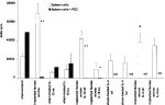
Fig. 7. Cellular responsiveness of immunocompetent DBA/1, interleukin 4-knock-out (IL-4 ko), IL-10 ko and their respective wild-type (wt) mice after intraperitoneal transplantation of 5 immature adult females of Litomosoides sigmodontis followed by an intravenous injection of 100000 microfilariae of L. sigmodontis 1 week later. Cytokine-gene knock-out and wild-type mice were all on C57BL/6 genetic background. Controls received a sham-surgery instead of transplantation of adult worms (sham-treated). Splenocytes of infected mice as well as peritoneal exudate cells (PEC) were recovered 18–28 days post-Mf injection and stimulated in vitro with Concanavalin A (4 μg/ml) for 48 h. Cellular proliferation was quantified by [3H]thymidine incorporation for the last 18 h of culture. Values are indicated as mean counts per minute (cpm) of triplicate cultures minus cpm from non-stimulated control cultures (net counts: delta cpm). Bars and vertical lines represent arithmetic means+S.E.M. of 5–6 animals per group. *P<0.05 (spleen cells versus spleen cells plus PEC). #P<0.05 (mice with implanted female worms versus sham-treated controls).
 You have
Access
- 41
- Cited by