Diagnosis of myocarditis in the paediatric population is particularly challenging as clinical presentation may overlap with different conditions such as early onset of genetic or metabolic cardiomypathies.Reference Lipshultz, Law and Asante-Korang1,Reference Foerster, Canter and Cinar2
The pathophysiologic relationship between cardiomyopathies and myocardial inflammatory process has been reinforced by finding viral genome in tissue samples of patients with supposed idiopathic cardiomyopathy.Reference Bouin, Gretteau and Wehbe3
The role of systematic and prospective electrocardiogram appraisal in patients with suspected myocarditis during admission and follow-up has been underevaluated so far. ST-T changes and conduction defect are reported in this clinical setting.Reference Buttà, Zappia, Laterra and Roberto4 Most importantly, cardiac magnetic resonance and biopsy cannot be routinely done in small critical babies; therefore, in this particular group of patients, additional insights from the electrocardiogram might be particularly warranted.Reference Lv, Han and Wang5
We have previously described the presence of QRS fragmentation correlating with evidence of late gadolinium enhancement in a series of adult patients with acute myocarditis.Reference Ferrero, Piazza, Grosu, Brambilla, Sironi and Senni6 QRS fragmentation, defined of multiples R spikes, has been widely reported as a marker of myocardial fibrosis in different heart disease and has a recognised prognostic value.Reference Redfors, Kosmidou and Crowley7-Reference Das, Maskoun and Shen9
This study aims to confirm this finding in a single-centre cohort of children who were eventually diagnosed with acute myocarditis.
Materials and methods
Patients younger than 18 years, discharged with the diagnosis of acute myocarditis between 2003 and 2019 were reviewed. Patients with valid and complete clinical records and high-quality electrocardiogram tracing were included. Diagnosis was based on clinical presentation, increase of myocardial necrosis markers, presence of oedema/late gadolinium enhancement on the cardiac magnetic resonance, and endomyocardial biopsy, in selected cases. All patients had undergone microbiologic workup at admission, guided by the presence of organ-specific symptoms, which included blood cultures, serology, and polymerase chain reaction assay for viral genome on nasopharyngeal aspirate. Standard 12-lead electrocardiogram was recorded with filter range 0.16–100 Hz, paper speed 25 mm/s and 50 mm/s and 10 mm/mV, taking particular care to optimise contact between the electrodes and the skin. QRS fragmentation was defined as the presence of an additional R wave (R′) or notching in the nadir of the S wave or the presence of >1 R′ in at least two contiguous leads. All electrocardiograms were reviewed independently and blindly by the authors. All patients had undergone follow-up, including clinical assessment, echocardiography, standard and 24 hours electrocardiogram recording. Not sustained arrhythmias were defined as any ventricular tachycardia consisting of at least four beats. Distribution of late gadolinium enhancement was categorised into three patterns according to the myocardial segment involved: antero-septal, infero-lateral, and spotted.
Statistical methods
Continuous variables were reported as median ± sd or median and interquartile ranges and compared by Student t-test or Wilcoxon rank sum test as appropriate. Normality of continuous variables was assessed by visually inspecting the distribution histograms. Categorical variables were presented as counts and percentages and compared by chi-square or Fisher exact test.
A p-value of 0.05 was assumed as cut-off for statistical significance. Analysis was performed with STATA 11.0 Stata Corp (College Station, TX).
Results
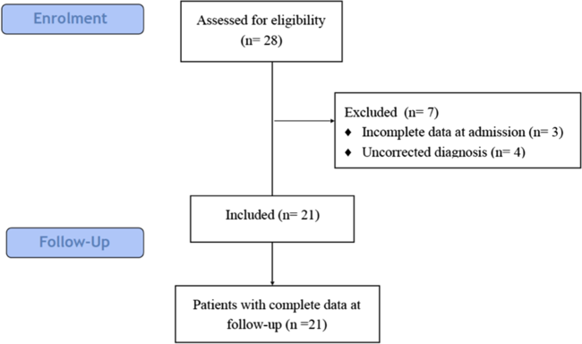
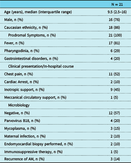
After excluding patients with incorrect diagnosis or incomplete data, 21 patients, 16 (76%) males, were eventually included in the analysis (Fig 1). Median age was 9.5 (2.5–16) years. All patients had prodromic symptoms: 17 (81%) had fever, associated with pharyngodynia in 6 (29%) and gastrointestinal syndromes in 4 (20%). Ten patients (50%) presented with chest pain, two (10%) had cardiac arrest secondary to pulseless fast ventricular tachycardia, nine (45%) needed inotropic support, and one (5%) underwent mechanical circulatory support.
Diagram showing the selection of patients included in the analysis.

In 12 patients, microbiologic workup was negative, in 4 (20%) parvovirus and in 3 (15%) mycoplasma were isolated. In two infants, maternal streptococcal infection transmission was documented (Table 1).
Demographic and clinical general characteristics.

AM=acute myocarditis.
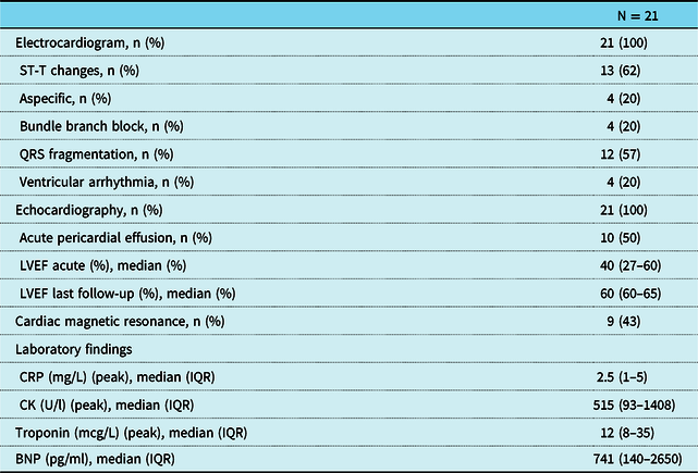
At admission, electrocardiogram was abnormal in 17 (81%) displaying transient ST-T changes in 13 (62%) and bundle branch block in 4 (19%). QRS fragmentation was seen in 12 (57%) patients, while 4 (19%) presented with not sustained ventricular arrhythmias.
Pericardial effusion was present in 10 (48%) and median ejection fraction at admission was 40% (27–60).
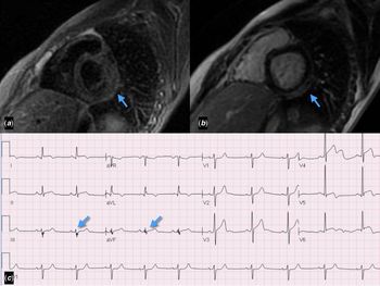
Cardiac magnetic resonance was performed in nine (43%) patients after a median time of 10.5 (range from 3 to 26) days after admission, displaying late gadolinium enhancement distributed in the anterior-sepal region in three patients, inferior-lateral in four and spotted in two. In six out of nine patients with late gadolinium enhancement, QRS fragmentation was also observed with a rough topographic correspondance (Fig 2). One patient underwent endomyocardial biopsy, after 10 days from the onset of symptoms, which disclosed the presence of inflammatory infiltrates, although not fulfilling Dallas criteria. Decision to perform the biopsy was motivated by persistent left ventricular dysfunction and haemodynamic impairment despite conventional therapy.
(a) CMR short-axis image showing oedema involving the inferior–posterior region of the left ventricle. (b) CMR short-axis image showing late gadolinium enhancement involving the inferior–posterior region of the left ventricle. (c) Electrocardiogram tracing showing QRS fragmentation in DIII and aVf. CMR, cardiac magnetic resonance.

All patients but one had C-reactive protein (CRP) elevation, and median peak was of 2.5 mg/dl (1–5) (<1 mg/dl). Median peak troponin was 12 (8–35) mcg/L, (<0.08) and median highest brain natriuretic peptide 741 (140–2650) ng/L, (<100) (Table 2). Patients with QRS fragmentation showed a trend towards a higher peak troponin: 26 (8–44) versus 8 (3–12) mcg/L p = 0.06.
Instrumental and laboratory data.

IQR=interquartile range; LVEF=left ventricle ejection fraction; BNP=brain natriuretic peptide.
All patients were treated with conventional heart failure medications, including beta blockers, angiotensin-converting-enzyme inhibitors (ACEi), Angiotensin II Receptor Blockers (ARBs), and mineralocorticoid receptor antagonists. One patient, with persistent left ventricular (LV) dysfunction, underwent a trial of immunosuppressive therapy with high-dose steroids and immunoglobulins, based on the results of the biopsy.
All patients were alive at a median follow-up of 600 (190–2343) days with a median ejection fraction of 60% (60–65). Three (14%) of patients had incomplete ventricular function recovery.
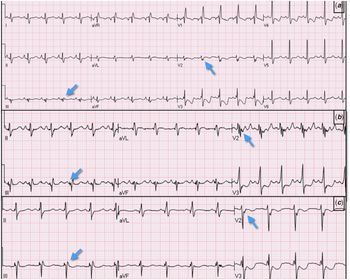
QRS fragmentation persisted in 7 patients out of the 12 in which was initially observed (Fig 3).
(a) ECG recorded at day 1. (b) ECG recorded at day 7 showing more evident signs of QRS fragmentation. (c) ECG recorded at day 30 showing persistence of subtle QRS fragmentation. ECG, electrocardiogram.

Discussion
Myocarditis in the paediatric population is associated with significant mortality, ranging between 7 and 17% and a need for mechanical circulatory support and/or heart transplant which can achieve 30%.Reference Abe, Tsuda, Miyazaki, Ishibashi-Ueda and Yamada10 A major challenge is clinical diagnosis which might require more than one clinical examinations.Reference Durani, Egan, Baffa, Selbst and Nager11 In this study, we investigated the presence of QRS fragmentation in children with suspected acute myocarditis. In agreement with previous series, infart-like pattern characterised by chest pain and ST-T wave change was the most common clinical presentation in this range of age.Reference Abe, Tsuda, Miyazaki, Ishibashi-Ueda and Yamada10,Reference Martinez-Villar, Gran and Sabaté-Rotés12
QRS fragmentation was acutely present in 57% and persisted in 33% of patients confirming previous preliminary observation in adult population.Reference Ferrero, Piazza, Grosu, Brambilla, Sironi and Senni6
QRS fragmentation is a simple electrocardiogram marker already described in other heart diseases and is deemed to be the expression of local slowing and anisotropic electrical conduction.Reference Haukilahti, Eranti, Kenttä and Huikuri13
In patients with myocarditis, similarly to other heart disease, in which QRS fragmentation has been well described, this sign might be the epiphenomenon of local slowed and anisotropic electrical conduction. This hypothesis is supported by the association between QRS fragmentation and late gadolinium enhancement on the cardiac magnetic resonance. QRS fragmentation has been recognised as an independent prognostic factor in ischaemic and not ischaemic heart disease.Reference Pietrasik and Zaręba14 This finding can be explained interpreting QRS fragmentation as a marker of more extensive myocardial damage and potential arrhythmic substrate. Arrhythmic burden has been recognised as an independent prognostic factor in both adults and children even though an electrocardiographic marker of risk on the standard electrocardiogram has not been described so far.
As for adult patients, QRS fragmentation was not observed in all patients with evidence of late gadolinium enhancement; furthermore, maximum QRS fragmentation evidence was observed after few days from admission.
To explain the not constant association between QRS fragmentation and late gadolinium enhancement, we hypothesised a threshold of substrate above which electrocardiogram changes are detectable; on the other hand, persistence of oedema, typical of the early stages of the disease, might mask QRS fragmentation. The association of QRS fragmentation with a more extensive myocardial damage is also suggested by a trend, towards a higher troponin peak in patients in the group of patients showing this particular sign. Because of the small sample, this finding was not statistically significant; therefore, it needs further confirmations.
Of interest in 3 patients out of 12, QRS fragmentation disappeared suggesting a role of this sign as a dynamic marker of tissue remodelling and pathologic evolution.Reference Małek, Kamińska and Barczuk-Falęcka15
Differently from other published series, we did not report mortality in our cohort, reflecting the retrospective nature of our study that includes patients discharged alive from hospital, while the percentage of incomplete recovery is in line with the literature.Reference Foerster, Canter and Cinar2,Reference English, Janosky, Ettedgui and Webber16,Reference Klugman, Berger, Sable, He, Khandelwal and Slonim17
This finding, if confirmed on larger multi centre samples, might turn out as an additional diagnostic and prognostic tools, particularly as far as the arrhythmic risk is concerned.Reference Miyake, Teele and Chen18,Reference Anderson, Silver, Richmond and Liberman19
Limitations
This study has several limitations. Firstly, it is based on a single-centre database with a limited number of observations; in particular, cardiac magnetic resonance was performed in a limited number of patients. Hence, all inferences about the association between QRS fragmentation and late gadolinium enhancement are qualitative. Furthermore given the retrospective methodology of the study, we included patients discharged from the hospital, thus with less severe disease.
Finally, due to the limited sample, we are not able to correlate the persistence of QRS fragmentation with the outcome.
Financial support
This research received no specific grant from any funding agency, commercial or not-for-profit sectors.
Conflict of interest
None.