Introduction
Temporal bone computed tomography (CT) scanning is considered a valuable tool for planning the surgical approach and predicting disease extent chronic otitis media. However, its exact role in the pre-operative assessment remains controversial.Reference Walshe, McConn Walsh, Brennan and Walsh1, Reference Yates, Flood, Banerjee and Clifford2 CT images are normally reported by a radiologist, but the operating surgeon also reviews them prior to surgery. The surgeon then obtains immediate feedback about his interpretation during the procedure. In contrast, surgical findings are rarely fed back to the radiologist who provided the original CT report. In view of the different approaches and training backgrounds of the surgeon and radiologist, there will inevitably be some differences between the two interpretations. Identifying these differences and comparing them with the definitive diagnosis established during surgery provides an effective evaluation method and a positive learning experience for both clinicians.
This study aimed to assess the accuracy of CT scan findings for predicting temporal bone anatomy, disease extent and any potential complications such as facial nerve dehiscence. It also aimed to compare the findings of an otologist and a radiologist with a special interest in temporal bone imaging. This was done using a structured reporting system, thus providing a learning experience for both clinicians and helping to enhance the quality of future reporting.
Materials and methods
A review was conducted of 48 consecutive patients who underwent mastoid operations performed by a single otologist over a 30-month period. A radiologist with a special interest in temporal bone imaging and the surgeon who performed the procedures retrospectively reported all pre-operative temporal bone CT scans. The reporting was recorded systematically using a structured proforma (see Appendix 1). Both reporters were blinded to the original CT report and to intra-operative findings. Reported outcomes were anatomical variation (level of dura, mastoid aeration, facial nerve exposure), pathology extent (presence of cholesteatoma in the middle ear and/or attic and/or mastoid) and disease sequelae (erosion of ossicles and/or lateral canal and/or tegmen). Surgical findings were obtained from the international otology database and hospital electronic archives. Findings reported by the radiologist and the surgeon were compared with the operative findings. Cases with incomplete pre-operative findings were excluded. Minitab 16 (Minitab, State College, Pennsylvania, USA) was used for statistical analysis of the data. Positive and negative predictive values and accuracy were analysed and compared between reporters.
Results
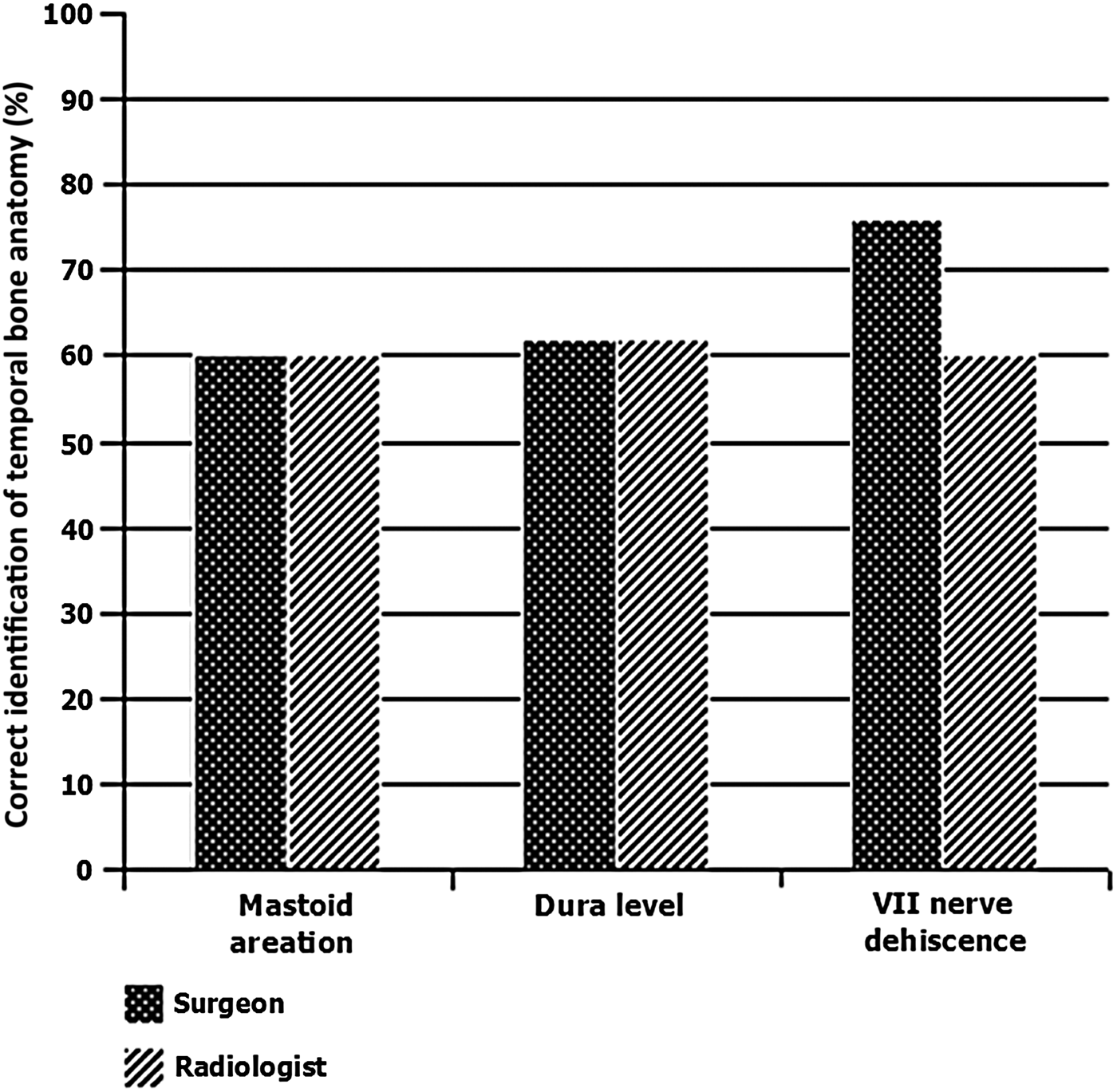
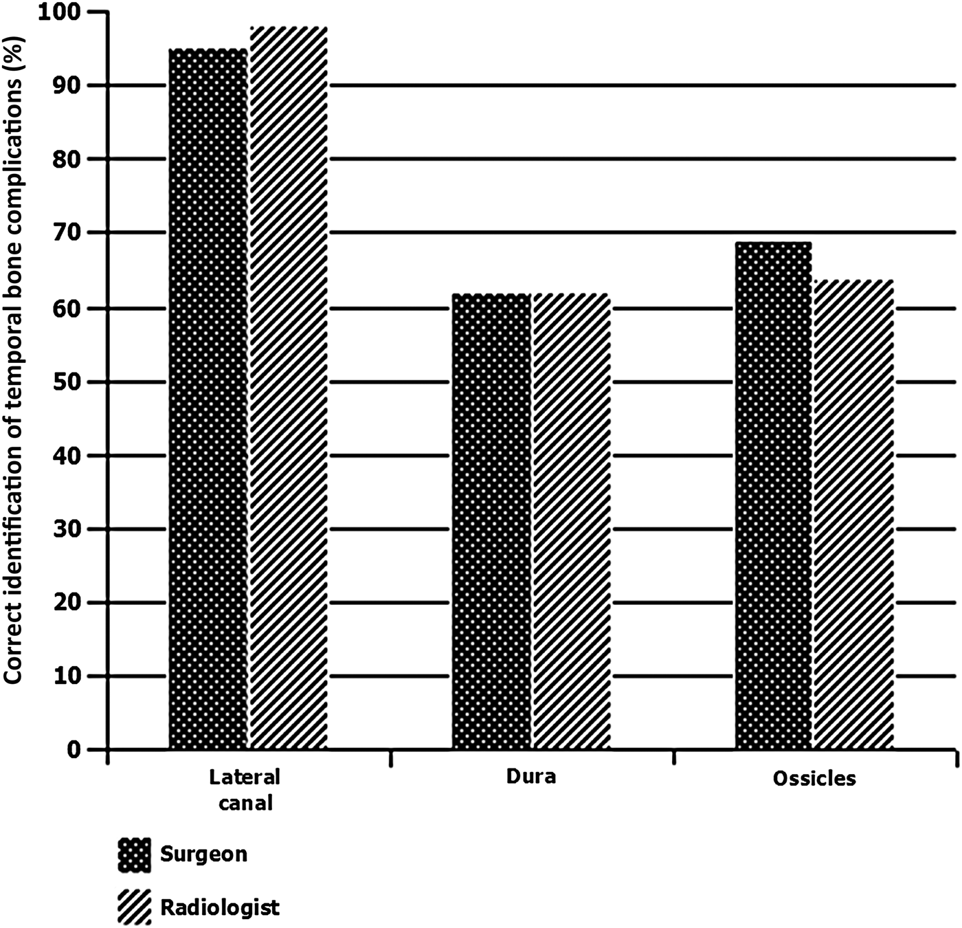
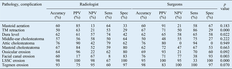
After excluding 6 patients because of incomplete operative findings, CT scans of 42 patients were independently reviewed. The average time between CT imaging and surgery was four months. In reporting the temporal bone (Figures 1–3), the percentage correct interpretations of operative and pathological findings was comparable between the two reporters, albeit with minor differences: the surgeon's performance was better regarding VII nerve dehiscence (76 vs 60 per cent; Figure 1), with higher positive (71 vs 17 per cent) and negative (77 vs 67 per cent) predictive values (Table I). Both interpreters were better at detecting lateral canal dehiscence (Figure III).
Graph showing the percentage accuracy of computed tomography interpretations of temporal bone anatomy by the two reporters.

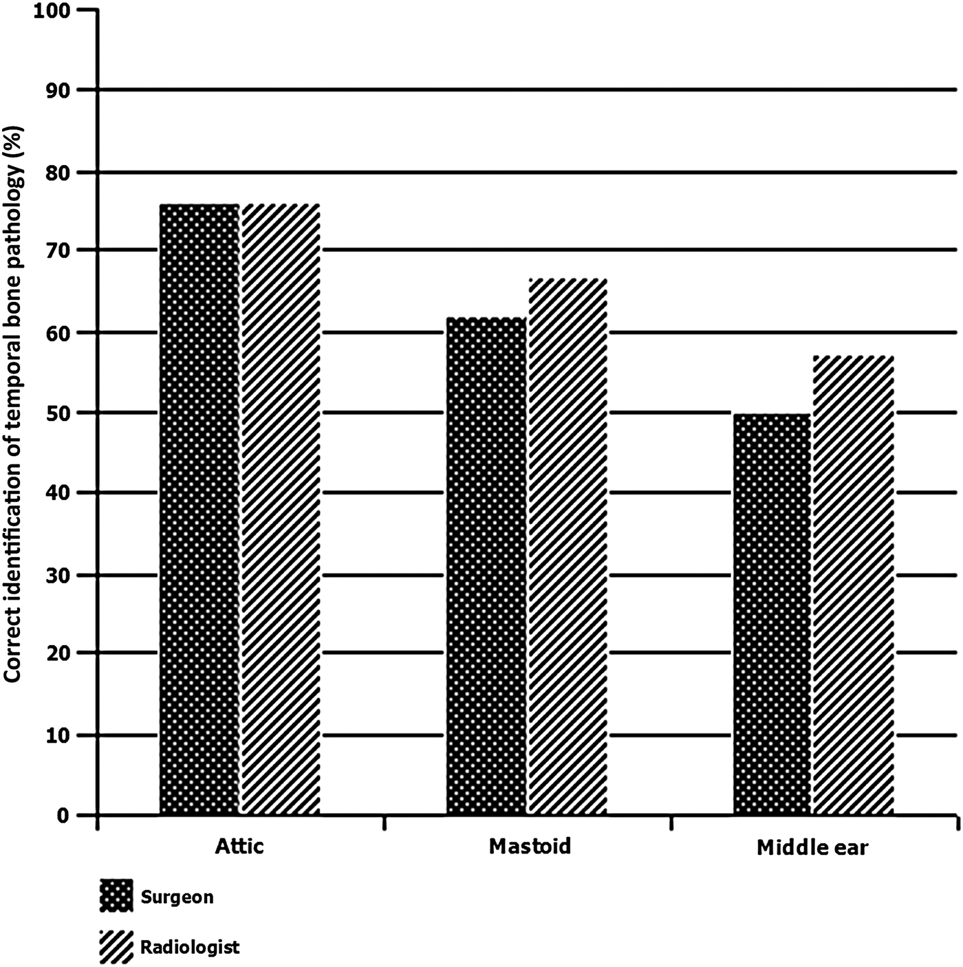
Graph showing the percentage accuracy of computed tomography interpretations of pathology affecting different parts of the temporal bone by the two reporters.
Graph showing the percentage accuracy of computed tomography interpretations of complications affecting different parts of temporal bone by the two reporters.
Performance of a surgeon and a radiologist in temporal bone CT interpretations
CT = computed tomography; PPV = positive predictive value; NPV = negative predictive value; Sens = sensitivity; Spec = specificity; TM = tympanic membrane; LSSC = lateral semicircular canal
However, the radiologist had a slightly better prediction of middle-ear disease. The radiologists had a higher accuracy and higher positive and negative predictive values for detecting cholesteatoma in the middle ear (57 vs 50 per cent, 56 vs 48 per cent and 58 vs 55 per cent, respectively), attic (76 vs 76 per cent, 90 vs 80 per cent and 42 vs 0 per cent, respectively) and mastoid (67 vs 62 per cent, 84 vs 72 per cent and 52 vs 47 per cent, respectively; Table I).
Overall, the there was no significant difference between the interpretations of temporal bone CT scans of the radiologist and otologist. However, the radiologist was significantly better at predicting lateral semicircular canal erosion, whereas the otologist was better at predicting facial canal erosion and tympanic membrane retraction. (Table I)
Discussion
This unique study evaluates the ability of an operating surgeon to report his own pre-operative CT images and compares his interpretations to those of the radiologist. Results were comparable between the two reporters, although some differences were found. These were welcomed by both reporters as providing a constructive learning experience. The methods were robust because all patients were operated on by the same surgeon and all images were evaluated by the same reporters. Use of the international otology database provided reliable information about intra-operative findings, and the use of a structured proforma added further credibility to our results. Blinding was regarded as unbiased owing to the time lag between the original reporting and/or surgery and this study.
Temporal bone CT has not gained wide acceptance as an essential aid to planning surgery for cholesteatoma, and many otologists reserve this procedure for selected patients that have complications of chronic suppurative otitis media, suspected congenital abnormalities or loss of landmarks owing to previous surgery.Reference Yates, Flood, Banerjee and Clifford2 Some authors, however, recommend routine scanning prior to all mastoid surgery because the disease process may not be apparent in clinical findings alone.Reference Blevins and Carter3
There is general consensus in the literature that mastoid CT cannot be relied upon to distinguish cholesteatoma from mucosal disease.Reference O'Donoghue, Bates, Anslow and Rothera4–Reference Garber and Dort7 Identifying the disease before surgery remains a topical issue because a higher complication risk is associated with cholesteatoma.Reference O'Donoghue, Bates, Anslow and Rothera4 Some authors have even recommended the use of magnetic resonance imaging to distinguish the two conditions.Reference Teschner, Hinz, Stöver, Lenarz and Becker12, Reference Vercruysse, De Foer, Somers, Casselman and Offeciers13 A higher accuracy of disease prediction in the middle ear, attic and mastoid by radiologists was noted in this study. This is because they are more proactive in considering bone density and erosion when making a diagnosis. In contrast, surgeons tend to concentrate more on anatomical features such as facial canal erosion and tympanic membrane retraction because these can have detrimental effects on surgical outcome.
Computed tomography was previously found to be helpful in determining middle-ear and mastoid cell anatomy.Reference Walshe, McConn Walsh, Brennan and Walsh1 This is an extremely important technique for surgeons to determine the best approach and avoid complications. One study showed that the mastoid air cell complex and sigmoid sinus are easier areas to assess radiologically compared with specific structures in the middle ear such as the oval window and the round window niche.Reference Vlastarakos, Kiprouli, Pappas, Xenelis, Maragoudakis and Troupis8 This suggests that the ability to identify different structures in the temporal bone varies according to their size, location and complexity.
Chronic otitis media usually erodes the long process of the incus, which is not normally seen on CT. The level of correct identification of this complication was moderate for both reporters. The negative predictive value for detecting ossicle erosion is reported to be no more than 27.8 per cent, which is very close to our findings (22 per cent for the radiologist and 21 per cent for the surgeon).Reference Vlastarakos, Kiprouli, Pappas, Xenelis, Maragoudakis and Troupis8 This means that visualisation of the incudomalleolar complex does not guarantee that this structure has not been eroded. O'Reilly et al. predicted an intact ossicular chain in only 50 per cent of cases, but our figures were better (69 per cent for the surgeon and 64 per cent for the radiologist).Reference O'Reilly, Chevretton, Wylie, Thakkar, Butler and Sathanathan11 Cholesteatoma can also erode the lateral semicircular canal; if the surgeon had prior knowledge of this, he would probably be more careful during dissection of diseased tissue to avoid creating a dead ear. O'Reilly et al. detected six out of eight cases and had a false positive rate of 3.5 per cent.Reference O'Reilly, Chevretton, Wylie, Thakkar, Butler and Sathanathan11 Jackler et al. had an even higher false positive rate and warned that mastoid CT gave an erroneous impression of lateral semicircular canal erosion.Reference Jackler, Dillon and Schindler14 In our series, neither the surgeon nor the radiologist had any false positives; both had 100 per cent positive predictive values.
• Pre-operative temporal bone imaging can predict intra-operative findings of chronic ear disease
• Mastoid computed tomography can accurately predict tegmen and lateral semicircular canal erosion
• It is ineffective in identifying ossicular chain continuity and facial canal dehiscence
• It can poorly differentiate cholesteatoma from mucosal disease in the middle ear
• There was no significant difference between computed tomography scan interpretation between specialists
Limitations
The limitation of our study relates to the distribution of actual pathologies, as found in any similar mastoid series.Reference Vlastarakos, Kiprouli, Pappas, Xenelis, Maragoudakis and Troupis8 Lateral canal and tegmen pathology are rare, thus reducing the sensitivity because of this calculation is based on the few abnormal cases with an eroded canal or tegmen. In contrast, most of our patient had low cellular (sclerotic) mastoids and, for the reason given above, the specificity values are lower because they are based on the few normal cases with cellular mastoid. In addition, a larger sample is required to draw more robust conclusions.
Conclusion
This study shows that pre-operative CT scanning of the temporal bone in chronic otitis media is useful for predicting attic pathology and lateral canal erosion but is less effective for delineating ossicular chain continuity, detecting facial canal dehiscence and distinguishing cholesteatoma from mucosal disease in the middle ear. The radiologist was significantly better at predicting lateral semicircular canal erosion, whereas the otologist did well in predicting facial canal erosion and tympanic membrane retraction. Better coordination between clinicians and use of a structured reporting tool can enhance the quality and usefulness of the CT scan report.
Appendix I