Implications
A comparison of androstenone measurements in pork fat between laboratories shows large differences in the absolute values, whereas inter-laboratory correlations remain high. This can be explained to a large extent by differences in the preparation of samples, as demonstrated by the better results obtained with freeze-dried samples, involving a single preparation method. These results are especially relevant to determine thresholds of consumer acceptance across countries, and to any work aiming at reducing boar taint under detection levels as well as to the definition of boar taint itself. However, any of the reported methods should apply for the ranking of animals for breeding purposes.
Introduction
When cooking/heating meat from entire male pigs, an unpleasant odour/flavour (boar taint) can occur. To prevent boar taint, male piglets are usually castrated at a young age. Thus, in the European Union, around 50% of the 161 Mio slaughtered pigs are barrows. However, according to a recent European declaration on alternatives to surgical castration of pigs, released on 16 December 2010, representatives of European farmers, the meat industry, retailers, scientists, veterinarians and animal welfare non-governmental organisations committed to a plan to voluntarily end surgical castration of pigs in Europe by 1 January 2018.
Several methods have been described in the literature to measure androstenone (5α-androst-16-en-3-one) in pork fat, one of the major compounds together with skatole and indole causing boar taint in entire male pigs. Despite the fact that the different methods effectively analyse different matrices, androstenone results are usually reported indistinctly, without specifying whether the analysis was performed in adipose tissue (AT), melted fat (MF) or pure fat (PF). This situation makes it difficult to draw meaningful conclusions for the establishment of comparable threshold levels, consumer acceptance limits and finally for the production of boar taint-free pork. In fact, many studies are performed by grouping the animals in high/medium/low-taint classes based on skatole and androstenone thresholds, with the generally accepted androstenone thresholds: 1 μg/g and 0.5 μg/g of fat, using the same levels even if those concentrations are given in MF (Matthews et al., Reference Matthews, Homer, Punter, Beague, Gispert, Kempster, Agerhem, Claudi-Magnussen, Fischer, Siret, Leask, Font i Furnols and Bonneau2000; Weiler et al., Reference Weiler, Font i Furnols, Fisher, Kemmer, Oliver, Gispert, Dobrowolski and Claus2000; Babol et al., Reference Babol, Squires and Gullett2002) or in AT (Annor-Frempong et al., Reference Annor-Frempong, Nute, Whittington and Wood1997; Bañón et al., Reference Bañón, Costa, Gil and Garrido2003; Font i Furnols et al., Reference Font i Furnols, Gispert, Guerrero, Velarde, Tibau, Soler, Hortós, García-Regueiro, Pérez, Suárez and Oliver2008) and conclusions are drawn about the abundance of animals in the given classes, or about the consumers’ acceptance by classes. Thus, comparisons are very often made between breeds, breed crosses, locations and countries (Walstra et al., Reference Walstra, Claudi-Magnussen, Chevillon, von Seth, diestre, Matthews, Homer and Bonneau1999; Bonneau et al., Reference Bonneau, Kempster, Claus, Claudi-Magnussen, Diestre, Tornberg, Walstra, Chevillon, Weiler and Cook2000; Pauly et al., Reference Pauly, Spring, O'Doherty, Ampuero Kragten and Bee2009 and Reference Pauly, Spring-Staehli, O'Doherty, Ampuero Kragten, Dubois, Messadène and Bee2010).
The methods for measuring the concentration of androstenone in pork fat include immunoassays and various chromatographic methods. The chromatographic methods are used either in combination with mass spectrometry or with fluorescence detectors. The immunoassays have been described by Claus (Reference Claus1974), Andresen (Reference Andresen1975), Claus et al. (Reference Claus, Mahler and Münster1988), Tuomola et al. (Reference Tuomola, Harpio, Knuuttila, Mikola and Lövgren1997) and Squires and Lundström (Reference Squires and Lundström1997), whereas chromatographic methods in combination with mass spectrometry have been described by Claus (Reference Claus1970), Rius and García-Regueiro (Reference Rius and García-Regueiro1988), Mågård et al. (Reference Mågård, Berg, Tagesson, Jaremo, Karlsson, Mathiasson, Bonneau and Hansen-Møller1995), Tuomola et al. (Reference Tuomola, Hakala and Manninen1998), Rius et al. (Reference Rius, Hortós and García-Regueiro2005) and Verheyden et al. (Reference Verheyden, Noppe, Aluwé, Millet, Vanden Bussche and De Brabander2007). The high-pressure liquid chromatography (HPLC) method has been described by Hansen-Møller (Reference Hansen-Møller1994). Chen et al. (Reference Chen, Zamaratskaia, Andersson and Lundström2007) and Pauly et al. (Reference Pauly, Spring, O'Doherty, Ampuero Kragten and Bee2008) mentioned HPLC-modified methods based on Hansen-Møller's method.
To our knowledge, the comparison of measurements between laboratories has not been reported. This topic is also of major interest, especially with respect to the definition of consumer acceptance thresholds across countries. In this study, measurements of androstenone were compared between three laboratories applying different assays on the same samples. The laboratories were the Norwegian School of Veterinary Science (NSVS) in Norway, the Co-operative Central Laboratory (CCL-Nutricontrol) in the Netherlands and the Institut de Recerca i Tecnologia Agroalimentàries (IRTA) in Spain. The methods used were time-resolved fluoroimmunoassay (TR-FIA) and two different methods combining gas chromatography and mass spectrometry (GC-MS). In a second trial, a fourth laboratory was included in the study, the Agroscope Liebefeld-Posieux Research Station (ALP) in Switzerland, which used HPLC.
Material and methods
Collection, conditioning and shipping conditions of AT samples
In the first trial, AT samples were collected from the neck region of 53 entire males immediately after slaughter, on a single day, in the Netherlands. Pigs were from a Duroc-based composite line with a mean age of 180 days and a mean live weight of around 125 kg at slaughter. The samples were stored at −20°C and shipped on dry ice for quantification of androstenone to NSVS in Oslo, Norway, CCL in Veghel, The Netherlands and IRTA in Girona, Spain. In all laboratories, samples were kept frozen at −20°C until analyses were performed, between 1 and 6 months, upon receipt of the samples. Under these storage conditions, at −20°C, no androstenone degradation was observed within the first years of storage as demonstrated by the stable results of several samples repeatedly analysed over 2 to 3 years after sampling (data not shown).
The second trial was carried out with freeze-dried samples. The main purpose was to circumvent the observed differences in sample preparation as routinely performed in the different laboratories, that is, analyte extraction from raw AT sample, from MF or from water-poor ‘PF’. For the second trial, back fat from 10 entire males and one castrated pig were used, and 50 g of AT from each carcass was freeze-dried at ALP, Switzerland. During the freeze-drying procedure, the AT samples were cut into pieces, cooled briefly to −20°C in a freezer and placed into closed recipients. These recipients were then immersed into liquid nitrogen. Immediately thereafter, the samples were homogenised (Vertec, Buhler) and freeze-dried (Christ Delta 1 to 24 K). Each sample was then mixed and portioned in aluminium bags, which were in turn sealed under vacuum and stored at −20°C. These lyophilised samples were then distributed to the four laboratories, including ALP in Switzerland, for androstenone analysis during the second trial. The mass (moisture) lost during the freeze-drying procedure in the 10 AT boar samples ranged from 12.6% to 23.8%. The freeze-drying process gave a fine powder, with remaining moisture of ∼3%. These samples could be readily extracted, following each laboratory's procedure as described below, in the ‘as received’ form without any further sample preparation. The sample from a castrate was also used for calibration.
Analysis of androstenone
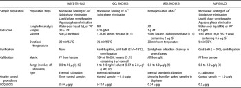
The original methods used by the four laboratories differed considerably not only in the analytical technology but also in the sample preparation (Figure 1), extraction protocol, preparation of calibration standards, calibration method and quality control procedures. A short description of the method used in each laboratory is given below and a summary of the main differences between methods, other than the detection principle, is given in Table 1.
Schema of different sample preparation procedures during routinely applied methods for the quantification of androstenone at different laboratories. 1Melted fat readily forms two phases (lipid/aqueous) after a short period of rest, especially when the aqueous fraction is important (IRTA = Institut de Recerca i Tecnologia Agroalimentàries; CCL = Co-operative Central Laboratory; NSVS = Norwegian School of Veterinary Science; ALP = Agroscope Liebefeld-Posieux Research Station).

Summary of main differences among current methods used for the determination of androstenone content in pork fat

NSVS = Norwegian School of Veterinary Science; TR-FIA = time-resolved fluoroimmunoassay; CCL = Co-operative Central Laboratory; GC-MS = gas chromatography and mass spectrometry; IRTA = Institut de Recerca i Tecnologia Agroalimentàries; ALP = Agroscope Liebefeld-Posieux Research Station; HPLC = high-pressure liquid chromatography; AT = adipose tissue; PF = pure fat; MF = melted fat; IS = internal standard; LOQ = limit of quantification; LOD = limit of determination.
1AT as sampled from a carcasse.
2IS (5α-androstan-3-one).
TR-FIA at NSVS
At NSVS, samples were analysed as described by Tuomola et al. (Reference Tuomola, Harpio, Knuuttila, Mikola and Lövgren1997). In brief, AT samples were melted in a microwave oven at 350 W for 4 min. The MF thus obtained was transferred to eppendorf tubes and centrifuged for 15 s at 6000 r.p.m., and after separation of phases, the water phase was discarded (Figure 1). A volume of 30 μl of water-poor ‘PF’ (55°C) was transferred to a glass vial containing 500 μl of methanol and extracted for 30 min at 55°C (vortexed at 0 and 15 min). The vials were then allowed to cool to room temperature for 10 min and a 50 μl aliquot of the methanol extract was diluted 1 : 9 with assay buffer. All samples were analysed in duplicate, with 2.1% variation between duplicates. Volumes of the 50 μl sample, 50 μl Eu-labelled androstenone (PerkinElmer Life and Analytical Sciences Wallac Oy, Turku, Finland) and 50 μl anti-androstenone (primary antibody, final dilution 1/45 000; Andresen, Reference Andresen1974) were pipetted into anti-rabbit coated (secondary antibody) micro-titration wells (Goat anti-rabbit IgG affinity purified antibody lot no. 021199, LabAs Ltd. Tartu, Estland). After 1.5 h incubation at room temperature, the wells were washed six times with wash solution DELFIA B117-100 at pH 7.75 (219.2 g/l of NaCl, 1.25 ml/l of Tween 20, 19.7 g/l of Tris HCl, adjusted to pH 7.7.5 with NaOH). Then, 200 μl of enhancement solution (DELFIA/autoDELFIA enhancement solution product no. B118-100 PerkinElmer, Wallac Oy, Turku, Finland) were added, and the wells shaken for 5 min at room temperature before the enhanced fluorescence was measured in a time-resolved fluorometer (Victor3 1420 multilabel counter, PerkinElmer, Turku, Finland).
Calibration standards were prepared by spiking each time 50 g of PF from a barrow with 0 to 500 μl solution of androstenone (Sigma-Aldrich A-8008, St. Louis, MO, USA) in ethanol (1 mg/ml). Thus, the calibration standards were as follows: 0.0, 0.2, 1.0, 2.0, 5.0 and 10 μg/g of androstenone in ‘PF’. These standards were then extracted and analysed in the same way as the samples.
The samples of the second trial were analysed following the normal procedure, except for the extraction step. Thus, 0.5 g of a freeze-dried sample were extracted with 1 ml methanol : water (95 : 5 v/v), instead of the 30 μl sample extracted with 500 μl methanol in the normal procedure. In this case, fresh calibration standards were prepared with 0.5 g of a freeze-dried barrow sample with 1 ml solutions of androstenone in MeOH : H2O (95 : 5 v/v), ranging from 0.1 to 5 μg/ml. Standards and samples were incubated for 30 min at 55°C, whirlmixed every 10 min and centrifuged at 13 000 r.p.m. Samples were frozen and centrifuged once more before further analysis.
GC-MS at CCL
At CCL, samples were analysed according to Verheyden et al. (Reference Verheyden, Noppe, Aluwé, Millet, Vanden Bussche and De Brabander2007). The AT samples were first prepared by removing meat residues and skin. The cleaned samples were cut into small pieces and placed in 70 ml plastic sample containers. The samples were then heated in a microwave oven at 600 W for 1 min (Figure 1). Then, 0.15 g of liquid fat was transferred to centrifuge tubes (2 ml) and 1.75 ml of extraction solvent methanol : hexane (9 : 1) was added. The tubes were placed in an ultrasonic bath at 50°C for 5 min, shaken vigorously and placed back in the ultrasonic bath for additional 30 min. They were shaken every 5 min during this period. The tubes were then centrifuged for 5 min at 10 000 r.p.m. while they were still hot and then placed on a cooled aluminium block. Around 1.5 ml of the extract was then placed into an injection vial and allowed to get to room temperature. Finally, 2 μl of the extract were injected into a GC-MS, 260°C injection temperature, in pulsed splitless mode. The separation was performed in an Rtx-5SIL MS column, 30 m × 250 μm (0.25 μm film thickness), under a constant helium flow of 1.1 ml/min. The temperature programme was as follows: 1 min at 50°C, then from 50°C to 300°C at 10°C/min, followed by 9 min at 300°C. Detection was performed in SIM mode with m/z 257 (target), 258 and 259 and a dwell time of 100 ms.
For calibration purposes, solutions of androstenone (>99%, Sigma A-8008) were prepared in 100 ml methanol : hexane (9 : 1) additioned with 5 μl corn oil. The calibration standards ranged from 6 to 240 ng/ml.
During the second trial, the normal procedures for extraction and analysis were followed, skipping the preparation step. Thus, 0.15 g of the freeze-dried sample was directly extracted with 1.75 ml of methanol : hexane (9 : 1).
GC-MS at IRTA
At IRTA, samples were analysed as described by Rius and García-Regueiro (Reference Rius and García-Regueiro1988) and Rius et al. (Reference Rius, Hortós and García-Regueiro2005). AT samples (1.00 ± 0.01 g) were homogenised (Figure 1) and extracted in 50 ml hexane : dichloromethane (50 : 50) containing 5α-androstan-3-one (2 μg/g) as the internal standard. A 5 ml aliquot was evaporated to dryness and dissolved in 2 ml hexane. Extracts were purified in a solid phase extraction (SPE) Diol column, which had been previously flushed with hexane, by elution with hexane : dichloromethane (40 : 60) and evaporated to dryness at room temperature. The collected fraction was resuspended in 2 ml methanol and further purified in a C18 SPE column. After evaporation at room temperature, the residues were dissolved in isooctane and injected in a gas chromatographer coupled to a mass spectrometry detector (Varian 3800 – Saturn 2200 Varian, Inc. Corporate Headquarters, Palo Alto, CA, USA). A capillary column BPX5 (SGE, Australia), 30 m × 0.25 mm (0.25 μm film thickness), was used. The injection mode was splitless at 250°C. The MS interface temperature was set at 280°C. A temperature programme was applied: 70°C for 2 min, then from 70°C to 200°C at 10°C/min followed by 5°C/min up to 280°C, and then for 9 min at 280°C. Mass spectrometry data was acquired in SIM mode with m/z: 257 and 272 (target) and 202, 274. All samples were evaluated in duplicate.
Calibration standards were prepared with a subcutaneous fat sample that contained <0.05 μg/g of androstenone. Each standard was prepared with 1.00 ± 0.01 g of homogenised AT and 50 ml of androstenone (>99%, Sigma A-8008) solutions in hexane:dichloromethane (50 : 50). The solvent also contained 5α-androstan-3-one as the internal standard. Fortified samples had 0.0, 0.5, 1, 2 and 4 μg/g of androstenone and 2 μg/g of 5α-androstan-3-one in AT.
During the second trial, the normal procedures for extraction and analysis were followed, skipping the preparation step. Thus, 1.00 ± 0.01 g of the freeze-dried sample was directly extracted with 50 ml hexane : dichloromethane (50 : 50).
HPLC at ALP
At ALP, the procedure for the analysis of androstenone described by Pauly et al. (Reference Pauly, Spring, O'Doherty, Ampuero Kragten and Bee2008) was followed. Although ALP participated in the second trial but not in the first one, the analytical procedure is described hereafter to illustrate the entire procedure for calibration standards. Around 5 g of raw sample (AT) are cleaned from the skin and muscular tissue and cut in pieces before introducing into a glass bottle (50 ml). The bottle with an untightened cap is heated in a microwave device for 2 × 2 min at 250 to 300 W. Twice (the analysis is made on replicate basis), 1 ml of liquid is transferred into a 2 ml eppendorf tube and immediately centrifuged at 11 250 × g, room temperature, for 2 min. The aqueous phase is then eliminated with a Pasteur pipette (Figure 1). In a 2 ml eppendorf tube, 0.50 ± 0.01 g of water-poor ‘PF’ is extracted at 47°C with 1.00 ml of MeOH : H2O (95 : 5 w/w) containing 0.500 mg/l of 5α-androstan-3-one as the internal standard. Samples are then mixed in a vortex and sonicated in an ultrasonic bath at 30°C for 5 min. Then, they are placed in an ice-water bath for 20 min. Finally, they are centrifuged at 11 000 × g, at 4°C, for 20 min. The liquid fraction is filtered through a 0.2 μm filter before injection into an HPLC (1200, Agilent Technologies, with FLD G1321A fluorescence detector). An in situ derivatisation is performed by the injection module with dansylhydrazine in the presence of BF3, with a programmed fixed derivatisation time before injection. The separation is performed by injecting 10 μl of the extract into an SB C18 column (50 × 4.6 mm, 1.8 μm particle size), at 40°C, and eluting with phosphate buffer (pH = 6) at 1.3 ml/min. The FLD parameters for the detection of androstenone are excitation/emission: 346/521 nm.
Calibration standards are prepared by spiking water-poor ‘PF’ from a barrow with 10 to 20 μl of androstenone (>99%, Sigma A-8008) solutions in MeOH : H2O (95 : 5 w/w) in order to get mass/mass standards ranging from 0.2 to 3 μg/g of androstenone in ‘PF’. These standards are treated as ‘PF’ samples and extracted and analysed following the normal procedure.
During the second trial, the normal procedures for extraction and analysis were followed, skipping the preparation step. Thus, 0.50 ± 0.01 g of the freeze-dried sample was extracted with 1 ml MeOH : H2O (95 : 5 w/w) containing 0.500 mg/l of the internal standard.
Results
First inter-laboratory trial: androstenone quantification in pork fat
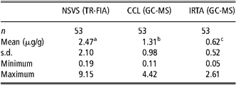
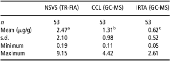
The average androstenone levels measured by three different laboratories using AT samples from the same 53 pigs in the first trial are given in Table 2. There were large differences between the average levels from different laboratories. The average values ranging from 0.62 to 2.47 μg/g. The values from NSVS ranging from as low as 0.19 to a maximum of 9.15 μg/g (in PF), whereas those from IRTA ranging from 0.05 to 2.61 μg/g (in AT). All samples were analysed in duplicate with differences between duplicates not exceeding 10% in all cases.
Characteristics of a single set of 53 AT pork samples as a result of the androstenone analysis performed independently by three different laboratories. First trial
AT = adipose tissue; NSVS = Norwegian School of Veterinary Science; TR-FIA = time-resolved fluoroimmunoassay; CCL = Co-operative Central Laboratory; GC-MS = gas chromatography and mass spectrometry; IRTA = Institut de Recerca i Tecnologia Agroalimentàries.
a,b,cLeast square means with different superscripts differ at P < 0.05.
Results are expressed in pure fat at NSVS, melted fat at CCL and AT at IRTA.
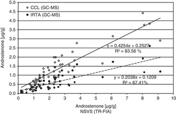
In spite of the large differences between the averages and the range of values from different laboratories, the correlations between different laboratories were high. The highest correlation was observed between CCL and IRTA (r = 0.92), and similarly, a high correlation of r = 0.91 was observed between CCL and NSVS. The correlation between IRTA and NSVS was slightly lower (r = 0.82) but still very high compared to the differences in the mean levels and range of values. These high correlations are apparent in Figure 2 showing the relationship between the androstenone concentrations measured with TR-FIA at NSVS and with GC-MS at CCL and IRTA.
Comparison of androstenone concentrations measured with different methods (time-resolved fluoroimmunoassay (TR-FIA) and gas chromatography and mass spectrometry (GC-MS)) in different laboratories. First trial. Results are expressed: in pure fat at Norwegian School of Veterinary Science (NSVS), melted fat at Co-operative Central Laboratory (CCL) and adipose tissue at Institut de Recerca i Tecnologia Agroalimentàries (IRTA).

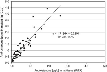
The androstenone concentrations at CCL in the Netherlands and IRTA in Spain were both estimated using a combination of GC-MS assays. However, CCL used MF samples, whereas IRTA used homogenised AT for the analysis. A comparison between the GC-MS assays using AT or MF is shown in Figure 3. The concentration estimated from MF was about 1.7 times higher than the one measured in homogenised AT.
Relationship between androstenone concentrations using homogenised adipose tissue Institut de Recerca i Tecnologia Agroalimentàries (IRTA) and melted fat Co-operative Central Laboratory (CCL). Both laboratories used a gas chromatography and mass spectrometry assay. First trial.

Second inter-laboratory trial: androstenone quantification in freeze-dried AT
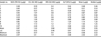
During the second inter-laboratory trial, the different laboratories performed the determinations following their own protocols except for the sample preparation. The extraction step was performed directly on the freeze-dried AT (fine powder), skipping the different melting/homogenising procedures of the sample preparation step. The results from all four laboratories of androstenone quantification in the 10 freeze-dried fat samples are given in Table 3. Correlation coefficients for pairwise comparisons were somehow higher than those from the first trial, now ranging from 0.92 between NSVS and ALP to 0.97 between IRTA or CCL and ALP. It was more interesting to note that the range of concentrations was then similar for all four laboratories.
Concentrations of androstenone in 10 freeze-dried AT samples determined independently by four laboratories. Second trial

AT = adipose tissue; NSVS = Norwegian School of Veterinary Science; TR-FIA = time-resolved fluoroimmunoassay; CCL = Co-operative Central Laboratory; GC-MS = gas chromatography and mass spectrometry; IRTA = Institut de Recerca i Tecnologia Agroalimentàries; ALP = Agroscope Liebefeld-Posieux Research Station; HPLC = high-pressure liquid chromatography.
All results are expressed in freeze-dried adipose tissue.
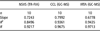
Indeed, by avoiding the different preparation steps which actually resulted in different types of matrices (homogenised AT, liquid fat or water-poor ‘PF’), the results from the different laboratories were comparable as illustrated by the slopes of pairwise comparisons, ranging from 0.2 to 0.4 in the first trial to 0.7 to 0.8 in the second trial (Table 4).
Inter laboratories relationship of androstenone analysis in a single set of freeze-dried AT samples, against ALP. Second trial
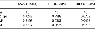
AT = AT = adipose tissue; ALP = Agroscope Liebefeld-Posieux Research Station; NSVS = Norwegian School of Veterinary Science; TR-FIA = time-resolved fluoroimmunoassay; CCL = Co-operative Central Laboratory; GC-MS = gas chromatography and mass spectrometry; IRTA = Institut de Recerca i Tecnologia Agroalimentàries.
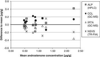
However, some systematic error seems to remain as suggested by Figure 4. In this figure, the differences between individual measurements and the mean value per sample are shown. These differences seem to spread apart with higher concentration.
Differences to mean of androstenone concentrations measured by four different laboratories in the freeze-dried adipose tissue samples. Second trial.

Discussion
Within-laboratory comparisons of chromatographic methods with immunochemical androstenone analyses have usually shown good agreement. Hansen-Møller (Reference Hansen-Møller1994) found comparable levels between HPLC and radioimmunoassay (RIA). In addition, Claus et al. (Reference Claus, Herbert and Dehnhard1997) found good agreement when comparing HPLC with enzyme immunoassay. In the study of Tuomola et al. (Reference Tuomola, Hakala and Manninen1998), supercritical fluid chromatography – mass spectrometry has shown comparable results with time-resolved fluoroimmunoassay, but the immunoassay gave slightly higher results. Although Chen et al. (Reference Chen, Zamaratskaia, Andersson and Lundström2007) found good correlation (r = 0.9), they report an overestimation of the ELISA method compared to the HPLC assay. However, they showed comparable results between HPLC and GC-MS.
In this study, large differences of androstenone determinations have been revealed between laboratories using different (NSVS v. IRTA, CCL) or comparable technologies (IRTA and CCL). The fact that individual laboratory values spread over very different ranges, Figure 2, together with high inter-laboratory correlation coefficients, shows the presence of systematic errors. An important part of the differences between the results from IRTA and CCL laboratories, both using a GC-MS method, was caused by differences in the sample type: MF at CCL and AT at IRTA. Furthermore, the differences in level between NSVS and CCL are smaller than those between NSVS and IRTA (TR-FIA v. GC-MS in both cases); again, this seems to be related to sample type: NSVS uses ‘PF’, whereas CCL uses MF and IRTA AT. This was further investigated during the second trial. In the second trial, a great part of the systematic error could be avoided by using the same preparation step, that is, freeze-drying performed in a single laboratory (ALP). Since the freeze-dried product was a readily dissolvable fine powder, no further preparation before extraction was needed and the matrix in which androstenone was quantified was the same in all cases. This was in contrast to the first trial where the matrix for extraction was AT, MF or water-poor ‘PF’.
During the second trial and for all laboratories, the differences from the mean spread mainly around the uncertainty limits of most methods (±0.2 μg/g), up to around 1.2 μg/g of androstenone in AT, Figure 4. This is a crucial range as several studies situate threshold values between 1 and 0.5 μg/g of androstenone in AT. A higher spread is observed for the one sample with the highest concentration (2.26 μg/g), which is probably an indication of systematic error due to matrix-matched calibration effects.
Besides the sample type and matrix-matched calibration effects, other parameters might be important for the harmonisation of the analysis of androstenone in pork ‘fat’. Thus, different parameters might affect accuracy to different extents and are likely to be worth studying, including parameters that are related to individual technologies. Owing to the good correlations and systematic deviations between labs during the first trial (Figures 2 and 3) together with mostly random deviations of the difference to mean during the second trial (Figure 4), minor, random effects might be expected, under the conditions of this study, from the heating and purification protocols in use (which might cause sample losses or specific evaporation of androstenone), insufficient extraction (related to solvent type or to sample/solvent ratio) and internal v. external calibrations.
The varying amount of humidity (12.6% to 23.8%) evaporated from AT samples during freeze-drying in the second trial (results not shown) indicates that the conversion between analyses in MF and in ‘PF’ can only be approximate. In other words, any conversion factor will have uncertainties over 10% because of the water content variability between samples. A similar situation can be expected concerning the amount of solid residues and the conversion between analyses in AT and in MF. These variations will add up when comparing between analyses in AT and in ‘PF’. Furthermore, the effect of fat-melting temperature on androstenone content is not clear as the composition of the liquid fraction depends on the temperature, for example, shorter fatty acid chains liquefy first and unsaturated fatty acids liquefy before saturated fatty acids (this is the reason for using quotation marks with ‘PF’ in this study). Besides, the temperature is not constant through repeated heating cycles in a microwave oven, even if the heating power remains the same (convection effect), adding to the overall variability between samples. The implication of this variability appears even more complex when taking into consideration that the analysis of androstenone is mostly correlated with consumer acceptance. However, what consumers eat is not AT or MF, and even less ‘PF’, but meat with varying amounts of ‘fat’. Indeed, sampling should be related to the tissue that is presented to the consumer or panellist.
The correlations between estimates from different laboratories using different methods are high. The range of values and standard deviations was different from one laboratory to the other but they were proportional to the means. Therefore, although the androstenone concentrations differ in absolute value when compared across the laboratories, the relative ranking between animals is not very different and for breeding purposes any of the laboratories can deliver useful results.
Conclusions
Systematic differences were observed in the analysis of androstenone in pork fat by different laboratories using different methodologies. These differences could be drastically lowered by using a single preparation method, that is, freeze-drying. The remaining systematic error could be due to matrix-matched calibrations as the differences tend to increase with concentration. Although there was no evidence here of a clear effect of the different extraction and purification protocols, they need to be specifically evaluated in order to achieve a harmonised quantification of androstenone across different laboratories and with different methods. The production of a reference material that could be used to standardise any analytical method would also be very helpful. Finally, it would be more than convenient to arrive at a convention about the sample preparation protocol, in order to universally express androstenone contents on the same basis: AT, MF, water-poor ‘PF’ or others (e.g. freeze-dried) In any case, the type of matrix should peremptorily be clearly expressed with every analytical result.
Although the ranking of animals seems not to be affected by the discrepancy in androstenone determinations, the harmonisation of the analysis of androstenone, as well as that of skatol and indole, is necessary for the study of sensory thresholds, the determination of consumer acceptance levels, the definition of clear criteria to sort out boar-tainted carcasses as well as for the development of non-specific online detection methods for boar taint.
Acknowledgements
The authors would like to thank TOPIGS for providing the data of entire boars and tissue sampling as well as S. Dubois for the preparation and analysis of the freeze-dried samples at the chemical department of ALP. This research project has been co-financed by the European Commission, within the 6th Framework Programme, contract no. FOOD-CT-2006-016250 (‘SABRE’). The study represents the authors’ views and does not necessarily represent the position of the Commission, which will not be liable for the use made of such information.