


Introduction
In many cultures around the world, dogs play a central role as pets, working animals, and guard dogs (Charlotte et al. Reference Charlotte, Claude, Stephanie, Fecteau, Marcel and Noel2022). In addition to their traditional roles as protectors and companions, dogs are increasingly recognized for the many benefits they provide to their owners. However, just like humans, dogs are susceptible to numerous diseases, including those of parasitic, bacterial, or viral origin, which can seriously impair their health and well-being. Among these diseases, canine hookworm disease caused by the parasite Ancylostoma caninum represents a significant veterinary and public health challenge worldwide. This blood-feeding intestinal worm primarily affects dogs and other carnivores but can also be transmitted to humans, posing a major health risk (Charlotte et al. Reference Charlotte, Claude, Stephanie, Fecteau, Marcel and Noel2022). In dogs, hookworm infection can lead to severe symptoms such as anaemia, digestive disorders, and even death in the most severe cases. Canine hookworm disease has a major impact on the health of dogs, their owners, and the surrounding human community. Transmission to humans occurs primarily through contact with soil contaminated by infective larvae, particularly in public environments such as parks and beaches, where infected dogs defecate, releasing eggs that contaminate the soil (Alcain et al. Reference Alcain, Waimann, Padin, Green and Chiramberro2018). In humans, infection with Ancylostoma caninum causes anaemia and skin lesions: cutaneous larval migration, a dermatosis of public health significance (Alcain et al. Reference Alcain, Waimann, Padin, Green and Chiramberro2018). To combat canine hookworm infection, dog owners use synthetic anthelminthics. However, the current economic downturn in developing countries, reduced public funding for veterinary care, the scarcity of veterinary clinics, and the high costs associated with care and the importation of synthetic anthelminthics place a heavy financial burden on dog owners and breeders. Even when these drugs are available, they cannot be used for mass treatment. Furthermore, the substances used are not always safe for animals, humans, and the environment. Moreover, the inappropriate use of synthetic drugs has contributed to the selection of organisms resistant to many anthelmintics in several developed countries. Benzimidazole resistance has been widely documented in canine hookworm populations. Molecular studies have identified several mutations in the β-tubulin gene associated with reduced drug sensitivity. For example, Venkatesan et al. (Reference Venkatesan, Jimenez Castro, Morosetti, Horvath, Chen and Redman2023) reported high frequencies of resistance-associated mutations, including F167Y and Q134H, in A. caninum populations from domestic dogs in the United States. More recently, a study by Dini et al (Reference Dini, Chen, Redman, Leutenegger, Tereski, Loo, Savard, Jimenez Castro, Miyashiro and Gilleard2026) further confirmed the spread of a novel β-tubulin allele associated with benzimidazole resistance, reinforcing concerns about the rapid evolution of drug resistance in this parasite. Consequently, strategies for the control and prevention of gastrointestinal nematodes in dogs are becoming less effective. Given the absence of new molecules on the market for several decades, alternative control solutions must be considered. Among these is the use of traditional medicine. According to the WHO (2013), in Africa, approximately 80% of the population uses traditional medicine based on herbal preparations to treat or prevent diseases. The growing use of these natural compounds is attributable to their efficacy, safety, and accessibility. These natural resources can be cultivated and contain multiple active compounds that may help slow the development of resistance. Setaria barbata (Lam.) Kunth, of the Poaceae family, is a common grass native to tropical Africa and tropical Asia, but is now cultivated in some parts of the world as an ornamental plant for its decorative inflorescences. Duarte et al. (Reference Duarte, Gomes, Catarino, Brilhante, Gomes, Rendall, Moreno, Fortes, Ferreira, Baptista, Dinis and Romeiras2022) reported that in some countries, S. barbata is used as a vegetable during times of food scarcity and as fodder for livestock. Few pharmacological studies have been conducted on S. barbata. The study by Fogue et al. (Reference Fogue Totzo, Mafo Fokam, Ngoupayo, Mfifen, Youté, Koul and Fotsing Kwetche2023) revealed the presence of polyphenols, flavonoids, anthocyanins, saponins, tannins, triterpenes, steroids, quinones, and reducing sugars in the hydroethanolic extract of S. barbata leaves. Their research also scientifically demonstrated the plant’s antibacterial and antifungal activities. To the best of our knowledge, no study has investigated the anthelmintic activity of Setaria barbata against animal gastrointestinal nematodes, and no experimental evidence has been reported regarding its activity against soil or plant nematodes. This lack of scientific evidence highlights a clear research gap and supports the rationale for evaluating its potential anthelmintic activity against Ancylostoma caninum in the present study.
Material and methods
Plant collection and processing
A preliminary study conducted among traditional healers in the village of Batoufam led us to select Setaria barbata, the whole plant of which is traditionally used in the treatment of helminthiasis. The plant material consisted of aerial parts, mainly leaves and stems. The plant was harvested in the rainy season (the month of April), in the locality of Batoufam, a rural area located in the Bayangam district, Koung-Khi division, West Region of Cameroon. A sample was packaged and sent to the National Herbarium of Cameroon for identification and was registered under reference number 488533/HNC. The plant material was dried in the shade under ambient conditions and stirred regularly to minimize visible contamination and prevent mould formation during drying. After complete drying, the material was ground into powder and packaged in plastic bags for the subsequent preparation of various extracts. No microbiological sterility testing (bacterial or fungal culture) was performed on the plant material prior to extraction. This represents a methodological limitation, as the presence of microbial contaminants cannot be fully excluded.
Preparation and dilution of plant extracts
Three (03) types of extracts were prepared: an aqueous extract by maceration, an aqueous extract by infusion, and an ethanol extract. One hundred (100) grams of plant powder were mixed with 1.5 litres of cold distilled water, 1.5 litres of distilled water brought to a boil at 100 °C, and 1.5 litres of 95% ethanol for the macerated aqueous, infused aqueous, and ethanol-macerated extracts, respectively. The mixtures were shaken vigorously and then left to stand for 48 hours and 3 hours for the macerated and infused extracts, respectively. After this period, each solution was filtered through a sieve, then through a 2.5 μm pore size filter paper placed in a funnel. The filtrates of the aqueous extracts were collected in a one-litre glass beaker, then distributed into stainless steel trays and placed in an oven at 40 °C for 3 to 5 days to evaporate the water. The dry powders of the aqueous extracts were thus obtained. The filtrate from the ethanolic extract was divided into portions of approximately 250 mL, then introduced into a flask of a rotary evaporator (Bucher-R-124 model) heated in a water bath at 45 °C to obtain the wet ethanolic extract. This wet extract was poured into wide-mouth Petri dishes (16.5 cm) and placed in a ventilated oven at 40 °C to allow complete evaporation of the remaining solvent. Forty-eight hours later, the remaining solvent had evaporated, and the dry ethanol extract was obtained. Each dry extract was weighed for yield calculation using the following formula:

The extraction yields obtained were 7.50% for the infused aqueous extract, 6.15% for the macerated aqueous extract, and 9.45% for the ethanolic extract.
Each dry extract was used to prepare a stock solution, which was then diluted with distilled water to obtain five different solutions at concentrations of 0.63, 1.25, 2.5, 5, and 7.5 mg/mL. The final concentrations tested were 0.31, 0.63, 1.25, 2.5, and 3.75 mg/mL. It should be noted that for the dilution of the ethanol extract, the dry extract was first mixed with 0.2 mL of DMSO (dimethyl sulfoxide) to ensure complete dissolution of the extract in distilled water.
Detection tests for phytochemical compounds
The methodology used for the phytochemical analyses was that of Dohou et al. (Reference Dohou, Yamni, Tahrouch, Idrissi Hassani, Badoc and Gmira2003). These analyses were conducted to identify the classes of metabolites likely responsible for the ovicidal and larvicidal activities of the plants studied. Identification was based on colorimetric and precipitation reactions depending on the metabolites being sought, notably flavonoids, phenolic compounds, sugars, alkaloids, triterpenes, and steroids.
Dog donor of Ancylostoma caninum eggs
Following coprological analysis of the faeces of several domestic dogs using the Willis flotation technique (Thienpont et al. Reference Thienpont, Rochette and Vanparijs2003), the dog selected as the parasite donor was the one whose coprological analysis, according to the Willis flotation technique, revealed the presence of Ancylostoma caninum eggs identified based on the morphological characteristics described by Tienpont et al. (Reference Thienpont, Rochette and Vanparijs2003), with an intensity of 2,000 EPG (eggs per gram of faeces), determined using the McMaster method.
Collection of Ancylostoma caninum eggs
Each morning, 3 grams of faeces were collected directly from the rectum of the donor dog and placed in a faecal collection container. Once in the laboratory, the faeces were placed in a mortar and ground, then 90 mL of a 40% saturated saline solution was added. After homogenization, the suspension was filtered using a 250 μm sieve. The filtrate was collected in a 100 mL beaker, and then distributed into 60 mm diameter test tubes until a meniscus formed. A coverslip was carefully placed on top of each test tube. Five minutes later, the coverslips were gently removed from the test tubes and placed on glass slides to be examined under a 4× microscope objective to confirm the presence of Ancylostoma caninum eggs. Once the presence of eggs was confirmed, all other coverslips were removed and placed in a beaker containing distilled water. After washing through a series of three decantations followed by siphoning, a suspension of A. caninum eggs was obtained.
Obtaining Ancylostoma caninum larvae
To obtain the larvae, we allowed the egg suspension to incubate for two days at room temperature to produce L1 larvae. Once the L1 larvae were obtained, 5 mL of Ringer’s solution was added to optimize larval survival. One to two days later, L2 larvae were obtained.
Evaluation of the effect of extracts on egg embryonation
A 24-well microtiter plate (4 × 6 layout) was used for all assays. The first row contained the primary experimental setup (one control and five treatment concentrations), while the remaining three rows served as biological replicates, providing triplicate measurements for each condition. Half a millilitre (0.5 mL) of a suspension of fresh eggs (30 to 40 eggs) was distributed among the 24 wells of a microtiter plate, and then 0.5 mL of various extracts at different concentrations was added to each well. The first well of each line of the plate served as a negative control and received 0.5 mL of 0.5% DMSO for the organic extracts and 0.5 mL of distilled water for the aqueous extracts. The microtiter wells, with a total volume of 1 mL, were incubated for 24 hours at room temperature. After this period, the contents of each well were pipetted after homogenization and placed in the two chambers of a McMaster cell, then examined under a 4× objective to count the number of embryonated eggs. Reading began with the control wells to ensure that at least 95% of the eggs were embryonated. The egg embryonation inhibition (EIn) rate was calculated using the following formula:

Evaluation of the effect of extracts on egg hatching
The effect of the various extracts on hatching was evaluated in the same manner as for the embryonation test, with the exception that, in this case, embryonated eggs (eggs containing motile larvae) were used and incubation lasted 6 hours. If, at the end of this period, a 90% hatching rate was achieved in the negative control wells, hatching of the eggs was halted in the other treated wells by adding one to two drops of Lugol’s solution. The number of L1 larvae released after hatching was counted using the McMaster method, and the mean percentage of hatching inhibition (HIn) was calculated using the following formula:

Evaluation of the effect of extracts on larvae mortality
Half a millilitre (0.5 mL) of suspension containing 20 to 30 L1- or L2-stage larvae was distributed among the 24 wells of the microtiter plate and exposed to different concentrations of extracts, then incubated at room temperature for 24 hours. At the end of this incubation period, the number of dead larvae was counted using a 4× microscope objective. Larvae were considered dead if they exhibited the following characteristics: immobility for at least 5 minutes, degraded cuticle, and an elongated, crenulated, or swollen appearance (Wabo Poné Reference Wabo2007). The mortality percentage was determined using the following formula:

All incubation steps were conducted under ambient laboratory conditions (22–23 °C), ensuring uniform experimental conditions across all assays.
Determination of the 50% inhibitory concentration (Ic50) and 50% larvicidal concentration (Lc50)
The egg inhibition rates and larval mortality rates obtained with the various extracts were converted to probit values using a probit table. Regression lines (D) between the probit value and the decimal logarithm of the extract concentration were plotted, and the equations of these lines were used to calculate the IC₅₀ and LC₅₀.
Statistical analyses
The results were entered into Excel 2013 and then transferred to SPSS version 24.0. The results of the in vitro tests are expressed as mean ± standard deviation. The mean percentages of inhibition of embryonation, hatching, and larval mortality for the different extracts at various concentrations were compared using a one-way analysis of variance (ANOVA). When significant differences were observed, Duncan’s test was applied to separate the means at the 0.05 significance level.
Results
Phytochemical composition of the plant extracts in the study
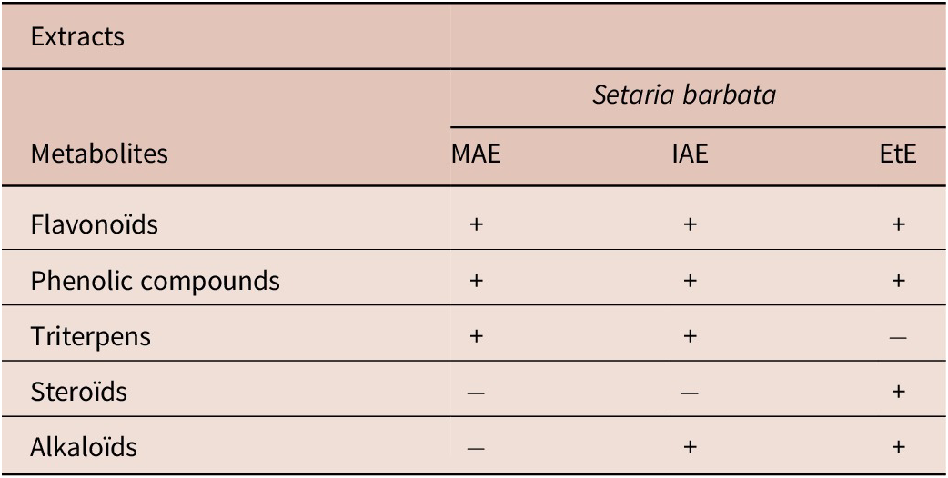
The secondary metabolites present in the various extracts of Setaria barbata are listed in Table 1. It appears that flavonoids and phenolic compounds are present in all plant extracts. The macerated aqueous extract of S. barbata contains the fewest secondary metabolites, with no steroids or alkaloids present.
Phytochemical constituents of Setaria barbata extracts

Table 1. Long description
The table consists of four columns. The first column lists the Metabolites, while the subsequent three columns represent the extracts: M A E (Macerated aqueous extract), I A E (Infused aqueous extract), and EtE (Ethanol extract). Presence is indicated by a plus sign and absence by a minus sign.
* Flavonoids: Present in M A E, I A E, and EtE.
* Phenolic compounds: Present in M A E, I A E, and EtE.
* Triterpens: Present in M A E and I A E, but absent in EtE.
* Steroids: Absent in M A E and I A E, but present in EtE.
* Alkaloids: Absent in M A E, but present in I A E and EtE.
Legend: MAE, Macerated aqueous extract; IAE, infused aqueous extract; EtE, ethanol extract; +, present; −, absent.
Ovicidal effect of Setaria barbata
The ovicidal effect of the plant extracts was evaluated on the embryonation and hatching of Ancylostoma caninum eggs.
Table 2 presents the effect of different concentrations of S. barbata extract on the embryonation of fresh eggs 24 hours after treatment. This table shows that distilled water and 0.5% DMSO do not influence the egg embryonation process, as evidenced by the average inhibition percentages of 0%. The S. barbata extracts exhibited concentration-dependent activity. Complete inhibition of egg embryonation was achieved at a concentration of 2.5 mg/mL with all extracts. The lowest IC50 (0.74 mg/mL) was obtained with the infused aqueous extract of S. barbata, resulting in the following efficacy gradient: infused aqueous extract (IAE) > macerated aqueous extract (MAE) > ethanolic extract (EtE).
Mean percentage (mean ± standard deviation) of inhibition of Ancylostoma caninum egg embryonation and IC50 of aqueous and ethanol extracts of Setaria barbata

Table 2. Long description
The table consists of 10 columns. The headers are: Plant, Extracts, Negative controls (divided into D W and D M S O), Concentrations in mg/mL (0.31, 0.63, 1.25, 2.5, 3.75), and I C sub 50 in mg/mL.
Data for Setaria barbata extracts:
* M A E (macerated aqueous extract): Negative control D W is 0 plus or minus 0. Concentrations: 0.31 is 0 plus or minus 0; 0.63 is 51.3 plus or minus 7.2; 1.25, 2.5, and 3.75 are all 100 plus or minus 0. I C sub 50 is 0.79.
* I A E (infused aqueous extract): Negative control D W is 0 plus or minus 0. Concentrations: 0.31 is 0 plus or minus 0; 0.63 is 12.9 plus or minus 5.8; 1.25 is 94.9 plus or minus 3.9; 2.5 and 3.75 are 100 plus or minus 0. I C sub 50 is 0.74.
* EtE (ethanol extract): Negative control D M S O is 0 plus or minus 0. Concentrations: 0.31 is 1.5 plus or minus 0.5; 0.63 is 6.2 plus or minus 1.7; 1.25 is 76.0 plus or minus 4.6; 2.5 and 3.75 are 100 plus or minus 0. I C sub 50 is 0.87.
Statistical significance is indicated by capital letters A through D, where different letters in a row denote p less than 0.05.
The capital letters compare the means within the same row. Different letters indicate a significant difference (p < 0.05).
Legend: DW, Distilled water; DMSO, dimethyl sulfoxide; MAE, macerated aqueous extract; IAE, infused aqueous extract; EtE, ethanol extract.
Table 3 highlights the activity of S. barbata extracts on the hatching of Ancylostoma caninum eggs at different concentrations. It shows concentration-dependent inhibition of A. caninum egg hatching for all plant extracts tested. The various negative controls had no significant effect on egg hatching. All S. barbata extracts proved effective, particularly the macerated aqueous extract, which caused a 100 ± 0% inhibition of the hatching of embryonated eggs starting at a concentration of 0.63 mg/mL. The macerated aqueous extract of S. barbata had the lowest IC50 (0.31 mg/mL). The IC50 values indicate the following order of efficacy: MAE > EtE > IAE.
Mean percentage (mean ± standard deviation) of inhibition of Ancylostoma caninum egg hatch and IC50 of aqueous and ethanol extracts of Setaria barbata

Table 3. Long description
The table consists of 10 columns and 5 rows including headers.
Column headers from left to right: Plant, Extracts, Negative controls (divided into D W and D M S O), Concentrations in mg/mL (0.31, 0.63, 1.25, 2.50, 3.75), and I C sub 50 in mg/mL.
Data rows for Setaria barbata:
1. M A E (macerated aqueous extract): D W control is 0 plus or minus 0. Inhibition increases from 1.1 plus or minus 2.2 at 0.31 mg/mL to 100 plus or minus 0 at all concentrations from 0.63 to 3.75 mg/mL. I C sub 50 is 0.31.
2. I A E (infused aqueous extract): D W control is 0 plus or minus 0. Inhibition remains at 0 until 1.25 mg/mL (11.5 plus or minus 0.5), reaching 100 plus or minus 0 at 3.75 mg/mL. I C sub 50 is 0.75.
3. EtE (ethanol extract): D M S O control is 0 plus or minus 0. Inhibition starts at 14.4 plus or minus 3.8 at 0.31 mg/mL and reaches 100 plus or minus 0 at 2.50 mg/mL and above. I C sub 50 is 0.66.
Statistical significance is indicated by capital letter superscripts (A through E) where different letters within a row indicate a significant difference (p < 0.05).
The capital letters compare the means within the same row. Different letters indicate a significant difference (p < 0.05).
Legend: DW, Distilled water; DMSO, dimethyl sulfoxide; MAE, macerated aqueous extract; IAE, infused aqueous extract; EtE, ethanol extract.
Larvicidal effect of Setaria barbata extracts
The larvicidal effect of S. barbata extracts was evaluated on first- and second-generation Ancylostoma caninum larvae. The lethal activity of the plant extracts on A. caninum L1 larvae after 24 hours of contact is presented in Table 4. As with fresh and embryonated eggs, the negative controls (distilled water, 0.5% DMSO) had no adverse effect on the survival of L1 larvae. However, in the treated wells, a progressive increase in mortality rates was observed as the concentrations of Setaria barbata extracts increased. The infused extract proved more effective against these larvae, inducing 100 ± 0% mortality starting at a concentration of 1.25 mg/mL, with an LC₅₀ of 0.42 mg/mL. The efficacy gradient is as follows: IAE > MAE > EtE.
Mean percentage (mean ± standard deviation) of L1 larval mortality of Ancylostoma caninum and IC50 of aqueous and ethanol extracts of Setaria barbata

Table 4. Long description
The table presents the mean percentage of L sub 1 larval mortality plus or minus standard deviation.
Columns include: Plant, Extracts, Negative controls (D W and D M S O), Concentration in mg/mL (0.31, 0.63, 1.25, 2.50, 3.75), and L C sub 50 in mg/mL.
Data rows for S. barbata:
- M A E (macerated aqueous extract): Mortality increases from 6.2 plus or minus 1.2 at 0.31 mg/mL to 89.8 plus or minus 3.5 at 3.75 mg/mL. L C sub 50 is 1.25.
- I A E (infused aqueous extract): Mortality increases from 18.6 plus or minus 5.0 at 0.31 mg/mL to 100 plus or minus 0 at 1.25 mg/mL and above. L C sub 50 is 0.42.
- EtE (ethanol extract): Mortality is 0 until 1.25 mg/mL, then increases to 82.0 plus or minus 8.2 at 3.75 mg/mL. L C sub 50 is 2.23.
Negative controls for D W and D M S O all show 0 plus or minus 0 mortality. Capital letters A through E indicate significant differences (p < 0.05) within rows.
The capital letters compare the means within the same row. Different letters indicate a significant difference (p < 0.05).
Legend: DW, Distilled water; DMSO, dimethyl sulfoxide; MAE, macerated aqueous extract; IAE, infused aqueous extract; EtE, ethanol extract.
Table 5 illustrates the average mortality rates of Ancylostoma caninum L2 larvae as a function of the concentrations of aqueous and ethanolic extracts of S. barbata. The infused and ethanolic extracts of S. barbata appear to be more lethal to L2 larvae, inducing mortality rates of 100% at a concentration of 1.25 mg/mL. The macerated extract showed considerable activity at concentrations of 2.5 mg/mL and 3.75 mg/mL, resulting in average mortality rates of 70.5 ± 5.4% and 100 ± 0%, respectively. The lowest LC50 was obtained with the ethanolic extract of S. barbata, resulting in the following efficacy gradient: EtE > IAE > MAE.
Mean percentage (mean ± standard deviation) of L2 larval mortality of Ancylostoma caninum and IC50 of aqueous and ethanol extracts of Setaria barbata

Table 5. Long description
The table presents mean percentage mortality plus or minus standard deviation for three types of S. barbata extracts across five concentrations (0.31, 0.63, 1.25, 2.50, and 3.75 mg/mL) and two negative controls (D W and D M S O).
* M A E (macerated aqueous extract): Negative control D W is 0 plus or minus 0. Mortality increases from 5.1 plus or minus 2.5 at 0.31 mg/mL to 100 plus or minus 0 at 3.75 mg/mL. L C sub 50 is 1.02 mg/mL.
* I A E (infused aqueous extract): Negative control D W is 0 plus or minus 0. Mortality increases from 5 plus or minus 0.2 at 0.31 mg/mL to 100 plus or minus 0 at 1.25 mg/mL and above. L C sub 50 is 0.50 mg/mL.
* EtE (ethanol extract): Negative control D M S O is 0 plus or minus 0. Mortality increases from 9.7 plus or minus 2.7 at 0.31 mg/mL to 100 plus or minus 0 at 1.25 mg/mL and above. L C sub 50 is 0.48 mg/mL.
Capital letter superscripts (A, B, C, D, E) indicate significant differences (p < 0.05) within each row.
The capital letters compare the means within the same row. Different letters indicate a significant difference (p < 0.05).
Legend: DW, Distilled water; DMSO: dimethyl sulfoxide; MAE, macerated aqueous extract; IAE, infused aqueous extract; EtE, ethanol extract.
Discussion
In developing countries such as Cameroon, the identification of medicinal plants with anthelmintic properties could contribute to the establishment of a sustainable approach to combating gastrointestinal nematodes. With this in mind, we sought to evaluate the in vitro anthelmintic potential of aqueous and ethanolic extracts of the Cameroonian medicinal plant Setaria barbata against Ancylostoma caninum.
The negative controls, namely distilled water and 0.5% DMSO, had no effect on the development of fresh eggs, the hatching of embryonated eggs, or the survival of Ancylostoma caninum larvae. These same observations were made by Mbogning Tayo et al. (Reference Mbogning Tayo, Payne, Wabo, Komtangi, Yondo, Ngangout, Mpoame and Bilong Bilong2012) and Payne et al. (Reference Payne, Wabo, Mbogning Tayo, Fidelia and Joseph2014). These results show that distilled water and 0.5% DMSO have no adverse effect on the development of Ancylostoma caninum eggs. This confirms that the variations in embryonation inhibition rates, hatching rates, and larval mortality observed in the microtiter wells treated with the various S. barbata extracts result from the bio-ovicidal and bio-larvicidal action of these products. Chagas (Reference Chagas2015) emphasized the importance of using appropriate solvents during the in vitro evaluation of plant extracts, suggesting that an unsuitable solvent could be toxic to the eggs, leading to false positives and masking the extract’s actual effect. Furthermore, D’Angelo et al. (Reference D’Angelo, Poné, Yondo, Komtangi, Vittori and Mbida2014) reported that the potential effects of plant extracts could be underestimated or overlooked due to external factors such as improper dilution in unsuitable solvents, which could lead to the rejection of potential alternative sources of anthelmintic agents.
In this study, the S. barbata extracts showed concentration-dependent activity on the eggs and larvae of Ancylostoma caninum. The concentration-dependent activity of the extracts could be explained by the fact that an increase in concentration corresponds to the addition of active compounds. It may also be due to the progressive saturation of target receptors as the concentration increases. At a concentration of 0.63 mg/mL, the macerated aqueous extract of S. barbata caused total inhibition of hatching. It is likely that, at this concentration, the active compounds induced a disruption of the ionic balance of the membrane potential, leading to lethal cellular disorganization for the embryo (Doligalska et al. Reference Doligalska, Jozwisole, Kiernsowska, Mroczeck, Packzkowski and Janiszowska2011). Overall, the S. barbata extracts were more effective against fresh eggs than embryonated eggs, as confirmed by the IC50 values. This observation aligns with the sensitivity scale described by Soulsby (Reference Soulsby1982). This could be due to the easier diffusion of active substances through the membranes of fresh eggs. According to Botuma et al. (Reference Botura, Dos Santos, Da Silva, De Lima, De Oliveira, De Almeida, Batatinha and Branco2013), the hatching of nematode eggs is triggered by environmental stimuli. These stimuli cause the release of enzymes such as proteases, lipases, and chitinases by the larvae, which degrade the egg membrane. Therefore, the active compounds in the extracts likely had a minimal effect on inhibiting the activity of these enzymes.
In general, the S. barbata extracts were effective on both eggs and larvae. In traditional medicine, Setaria barbata is used in infusion to treat gastrointestinal helminthiasis. The infused extract of this plant induced significant inhibition rates for embryonation, hatching, and larval mortality, resulting in the lowest IC50 values, particularly for egg embryonation inhibition and L1 larval mortality.
Phytochemical screening revealed the absence of steroids in the aqueous extracts of S. barbata, which were present in the ethanol extract. This could be explained by the fact that water is a polar solvent, while steroids are non-polar compounds. The variability in the chemical composition of the extracts is likely due to the type of solvent used for extract preparation. Water is more polar than ethanol, thus allowing better extraction of the plant’s polar bioactive compounds (Nawaz et al. Reference Nawaz, Aslam Shad, Najiha, Hina and Najeeb2020).
Conclusion
This study was conducted to evaluate the anthelmintic activity of aqueous and ethanolic extracts of Setaria barbata against Ancylostoma caninum in order to determine their potential use for the control of canine hookworm disease. The results showed that the Setaria barbata extracts studied exhibited concentration-dependent in vitro anthelmintic activity against the free-living life stages (eggs and larvae) of A. caninum. The application of these extracts to the environment could therefore disrupt the life cycle of these nematodes and consequently reduce the rates of hookworm infestations. However, microbiological sterility assessments of the plant material, chemical characterization of bioactive compounds using chromatographic techniques, and evaluation of toxicity on relevant biological models are required to better understand and validate the anthelmintic activity of the plant extracts.
Acknowledgements
The authors would like to express their sincere gratitude to the Applied Biology and Ecology Research Unit (URBEA) of the Faculty of Sciences at the University of Dschang, where this study was conducted. They also thank Mr. Jean-Pierre Toukam, a traditional healer in Batoufam, who provided them with information on the plant specimen used in this study.
Financial support
This research received no specific grant from any funding agency.
Competing interest
The authors declare none.


 Open access

