Introduction
Candelariaceae Hakul. is characterized within Lecanoromycetes primarily by the foliose or crustose thallus with pulvinic acid and related pigments which give the Candelariaceae its distinctive yellow to orange-yellow colour. The family has a worldwide distribution and consists of four genera with the largest, Candelariella Müll. Arg., comprising c. 50 accepted species. Among the species of Candelariella, a small group can be recognized that share the following features: a grey thallus, lacking lichen substances detected by high performance liquid chromatography (HPLC) and thin-layer chromatography (TLC), 8-spored asci and mostly lecanorine apothecia (Westberg Reference Westberg2005). The representatives of this group are predominantly found in open woodlands, steppes and prairies growing on bark, wood, plant debris or calcareous rocks, more often in dry and sun-exposed habitats. Such Candelariella species with a grey thallus are now frequently reported by many researchers (e.g. Malíček et al. Reference Malíček, Palice, Bouda, Czarnota, Halda, Liška, Müller, Peksa, Svoboda and Syrovátková2008; Vondrák et al. Reference Vondrák, Guttová and Mayrhofer2008; Aptroot Reference Aptroot2011; Westberg & Clerc Reference Westberg and Clerc2012; Westberg & Sohrabi Reference Westberg and Sohrabi2012; Yakovchenko et al. Reference Yakovchenko, Ismailov and Westberg2012; Gasparyan et al. Reference Gasparyan, Sipman and Brackel2014; Urbanavichus & Urbanavichene Reference Urbanavichus and Urbanavichene2014; Kocakaya & Halici Reference Kocakaya and Halici2015).
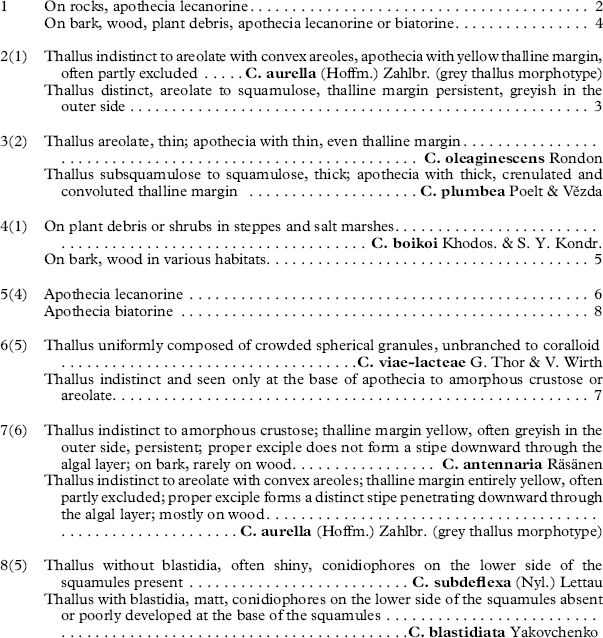
Among the species with a grey thallus, Candelariella subdeflexa (Nyl.) Lettau is distinct in its biatorine apothecia and well-developed thallus formed by squamules with occasional conidiophores producing globose conidia on the lower side of the thallus (Westberg Reference Westberg2007). During a revision of Candelariaceae in the herbaria H, LE and TNS, the first author found several specimens identified as Candelariella subdeflexa with blastidiate squamules. The aim of this study is to compare these individuals with other related taxa employing morphological, chemical and DNA sequence data. A worldwide key to grey thalline Candelariella is also presented.
Materials and Methods
Specimens and phenotype studies
The specimens examined are housed in ALTB, G, GZU, H, LE, PRA, SMR and TNS. Morphological observations were made using a dissecting microscope. Hand-cut sections of apothecia and thalli were observed after mounting in water. Measurements are presented as the mean
 $(\bar{x})$
and range including 85% of the variation, bounded by the smallest and largest observed values, followed by the sample size (n) following Westberg (Reference Westberg2005).
Lichen substances were examined using thin-layer chromatography (TLC) with solvent B' (hexane: methyl tert-butyl ether: formic acid, 140 : 72 : 18) and solvent C (toluene: acetic acid, 170 : 30) (Culberson & Kristinsson Reference Culberson and Kristinsson1970; Culberson & Johnson Reference Culberson and Johnson1982) for the specimen in TNS.
Sequences and phylogenetic reconstructions
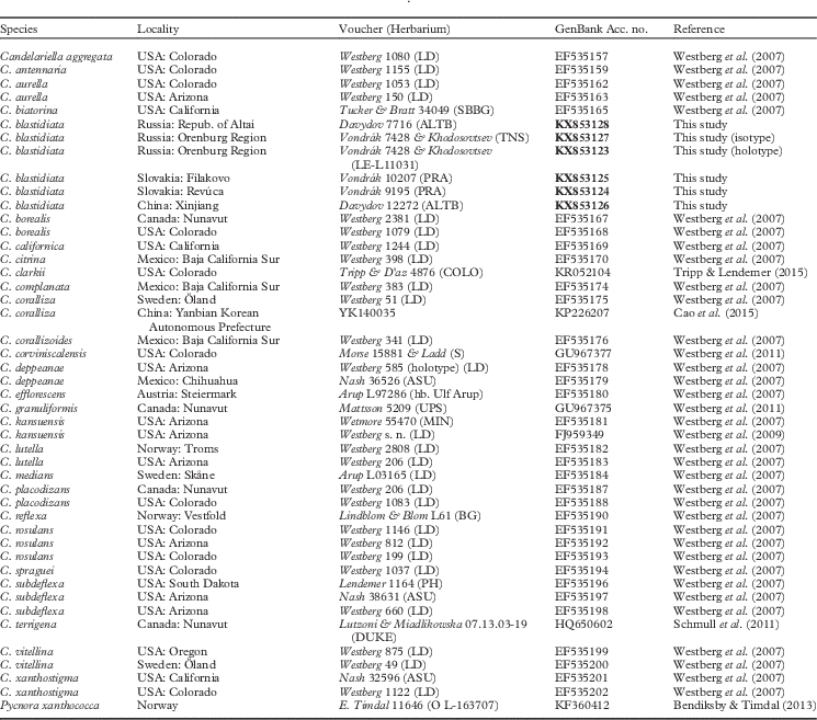
To test the monophyly of Candelariella blastidiata and its relationship with other species, ITS nrDNA sequences of fresh material and those retrieved from the NCBI database (GenBank) were used for molecular phylogenetic analysis. ITS nrDNA sequence data were used because GenBank includes a number of ITS sequences of Candelariella, whereas other loci are poorly represented. Furthermore, ITS single-locus genealogies are usually consistent with phenotypic data (Westberg et al. Reference Westberg, Arup and Kärnefelt2007, Reference Westberg, Frödén and Wedin2009; Westberg & Arup Reference Westberg and Arup2011; Tripp & Lendemer Reference Tripp and Lendemer2015). One or more different sequences of appropriate length (and preferably lacking uncertain nucleotides) were selected from every species of Candelariella. Our sampling comprised 26 species representing morphological variability within Candelariella, 10 of which were represented by two or more accessions. Information on the samples with GenBank Accession numbers is shown in Table 1.
Locality, voucher information and GenBank Accession numbers for the species used in this study for phylogenetic analysis. New sequences are in bold

Two apothecia or fragments of thallus (for sterile specimens) were used for DNA extraction. Samples were frozen in liquid nitrogen and made into a powder in tubes using sterile pestles. The DNeasy Plant Mini Kit (Qiagen, Germany) was used for DNA extraction following the manufacturer’s instructions. The primer set was ITS1F (Gardes & Bruns Reference Gardes and Bruns1993) and LR1 (Vilgalys & Hester Reference Vilgalys and Hester1990). PCR settings were as follows: 95°C for 2 min, 35 cycles of 94°C for 20 s, 53°C for 60 s, 72°C for 2 min, followed by 72°C for 15 min and then maintained at 14°C. The sequences obtained in this study and the reference sequences from GenBank were aligned by Geneious 6.0 (Biomatters Ltd., New Zealand) and manually optimized. The sequence of Pycnora xanthococca (Sommerf.) Hafellner was used as outgroup since Pycnora has been identified as a possible sister clade to Candelariaceae (Bendiksby & Timdal Reference Bendiksby and Timdal2013). Total alignment of 461 bp was used for the maximum likelihood (ML) and Bayesian analyses. Optimal substitution models were inferred separately for ITS1, 5.8S and ITS2 using PartitionFinder version 1.1.1 (Lanfear et al. Reference Lanfear, Calcott, Ho and Guindon2012). The symmetrical substitution model with gamma-distribution (SYM+G) was selected for the 5.8S partition and Kimura 2-parameter with proportion of invariable sites (K80+I) for ITS1+ITS2. Bayesian inference with the Markov chain Monte Carlo (BMCMC) method (Larget & Shimon Reference Larget and Shimon1999) was performed using MrBayes 3.2.3 (Ronquist et al. Reference Ronquist, Teslenko, van der Mark, Ayres, Darling, Höhna, Larget, Liu, Suchard and Huelsenbeck2012). Three parallel Bayesian analyses were run for 20 000 000 generations in six chains and every 200th generation was sampled. The first 50% of trees were discarded as burn-in and a 50% majority-rule consensus tree calculated from the remaining trees of three runs with the sumt command implemented in MrBayes 3.2.3. Bootstrap support values and BMCMC posterior probability were noted on the best scoring tree (numbers are shown only when≥50% in both). The ML tree and 1000 bootstrap replicates were calculated using RAxML 8.0.26 (Stamatakis Reference Stamatakis2014) by raxmlGUI software version 1.3.1 (Silvestro & Michalak Reference Silvestro and Michalak2012), applying the GTRGAMMA model of substitution because RAxML does not support SYM and K80 models.
Results
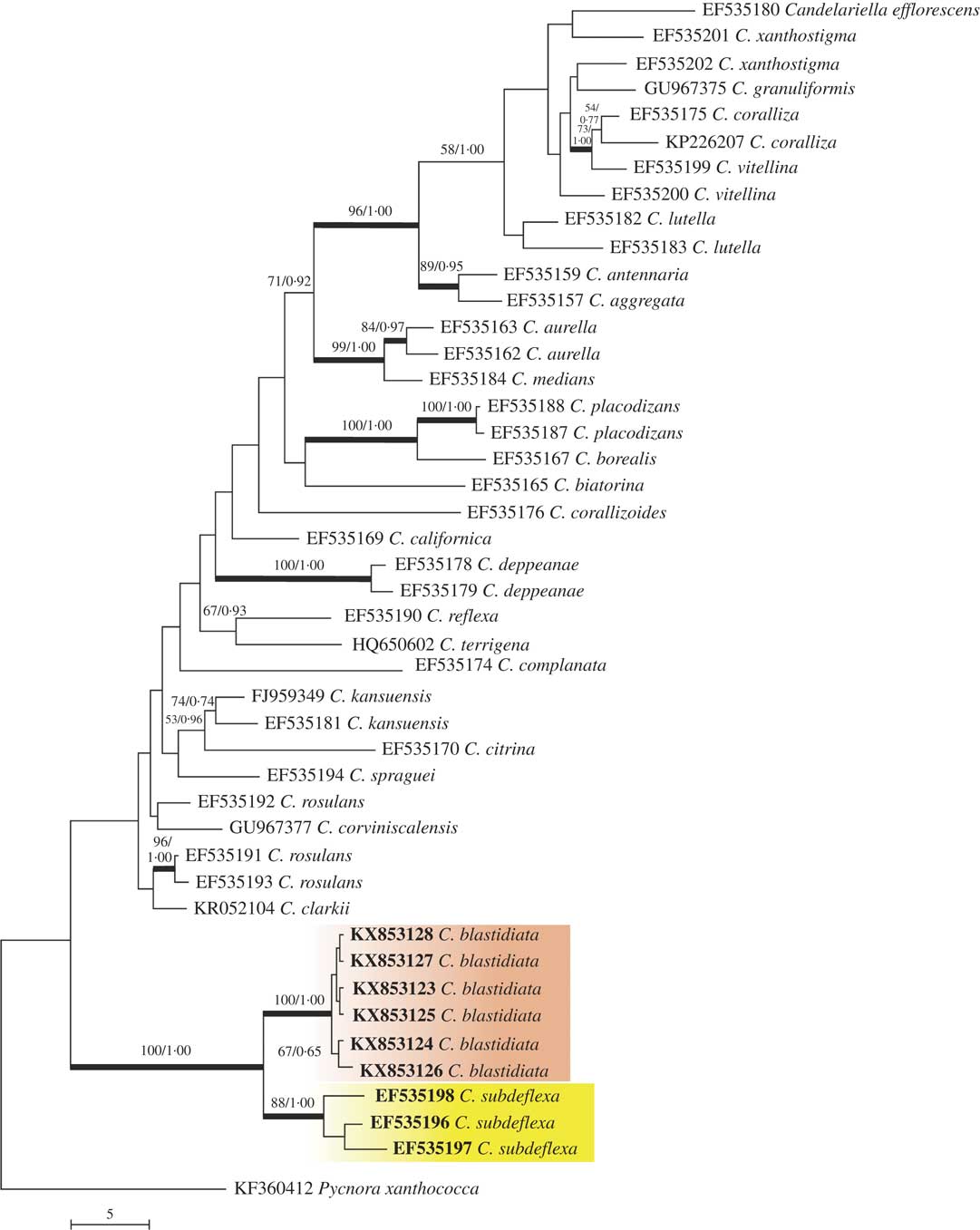
Six ITS sequences of blastidiate specimens of Candelariella were successfully obtained. They formed a monophyletic clade in the ITS phylogeny of Candelariella with support values of 100/1·00 (bootstrap/posterior probability) in the phylogenetic tree (Fig. 1). Variability in ITS sequences among four of the European and Russian specimens is only 1 residue, but the Chinese sequence differs from others by 3 residues. A supported sister clade (88/1·00) includes three sequences of C. subdeflexa. Four constant residues differentiate C. subdeflexa from blastidiate specimens. Both species form a group well-separated from all other included species.
Reconstruction of the Candelariella ITS phylogeny showing positions of C. blastidiata and C. subdeflexa. The tree was constructed by Maximum Liklihood (ML), and the reliability of each branch was tested by ML and Bayesian methods. Numbers at tree nodes indicate bootstrap values of ML (left) and BMCMC posterior probabilities (right) (only ML values ≥50% and PP values ≥0·50 are depicted). Thicker branches indicate where both the ML bootstrap value is ≥70% and the BMCMC posterior probability is ≥0·95. GenBank Accession numbers are given as OTU names (see Table 1). Pycnora xanthococca is the outgroup.

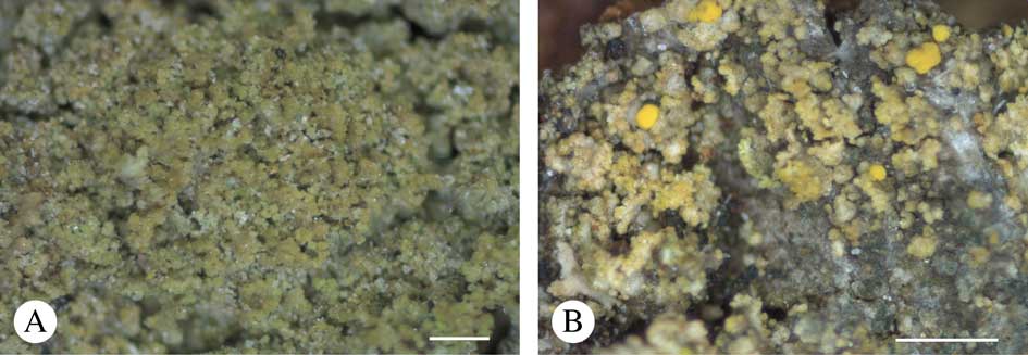
The close relationship of blastidiate specimens with C. subdeflexa is well explained by the close morphology (i.e. grey thallus with biatorine apothecia) as well as growing in similar habitats. However, we consider the two clades as distinct and morphologically recognizable species and one of them is described here as Candelariella blastidiata Yakovchenko. Specimens of C. blastidiata studied include samples without conidiophores (European specimens) and samples with a poorly developed layer of conidiophores at the base of the squamules (Altaian specimens: Russia, ED 7716; China, ED 12272). The Chinese specimen also shows slight morphological differences, with larger squamules and more abundant blastidia (Fig. 2A).
Candelariella blastidiata. A, sterile specimen with abundant blastidia, China, Altai, ED 12272 (ALTB); B, isotype (ALTB). Scales: A=200 µm; B=500 µm. In colour online.

Candelariella blastidiata Yakovchenko sp. nov.
MycoBank No.: MB 817756
Thallus corticolous, grey, squamulose with blastidia on margins and/or the lower side of squamules 25–34(–60) µm wide, apothecia biatorine, convex, yellow, (0·3–)0·5(–0·9) mm in diam; apothecial margin indistinct. Asci 8-spored, ascospores narrowly-ellipsoid, (7·5–)10·0–14·5(−15·0)×(3·5–)4·0–5·0 µm, conidiophores absent or partly-developed on the lower side of the squamules.
Type: Russia, Orenburg Region, Buzuluk District, National Park ‘Buzuluksky bor’, Partizanskiy Village, bank of the Borovka River, 52°59'54''N, 52°07'42''E, 80 m, on bark of Salix, 2009, J. Vondrák JV7428 & A. Khodosovtsev [Obermayer, Lichenotheca Graecensis no. 405] (LE–L11031—holotype; ALTB, ASU, B, C, CANB, CANL, E, G, GZU, H, HAL, HMAS, M, MAF, MIN, O, PRA, TNS, UPS—isotypes labelled “C. subdeflexa”).
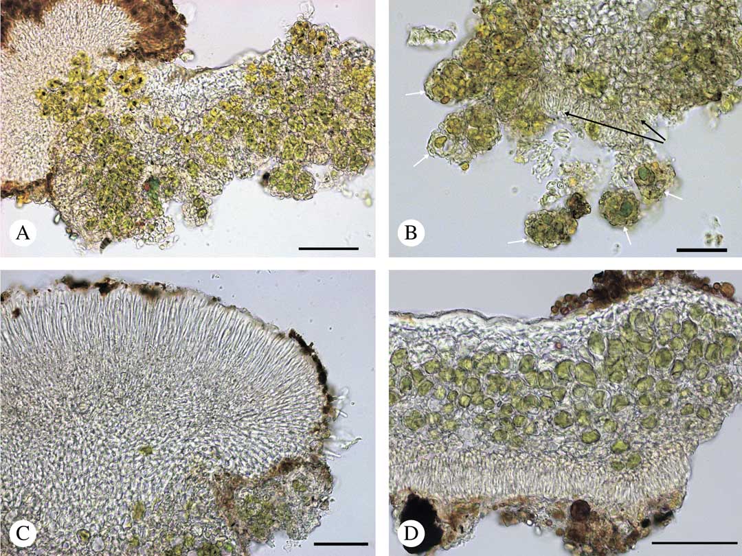
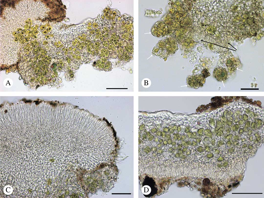
A–C, Candelariella blastidiata, vertical sections through thallus; A, thallus with blastidia; B, lower part of thallus (white arrows, blastidia; black arrows, conidiophores); C, apothecium. D, Candelariella subdeflexa, vertical section through thallus with conidiophores on the lower side, lectotype (H–9506465). Scales: A=100 µm; B–D=50 µm. In colour online.
Thallus squamulose. Squamules first prostrate to soon ascending and erect, scattered to crowded and somewhat imbricate,±rounded, (0·2–)0·3–0·4–0·5(–0·7)mm long (n=29) with irregularly incised margin; squamules (75–)103–122–141(–163)µm thick (n=19); epinecral layer absent; cortex alveolate, (0–)4–8–13(–25) µm thick (n=35), consisting of gelatinized hyphae with±isodiametric cells, (2·5–)4·0–5·5–7·0(–10·0)µm wide (n=30); algae chlorococcoid, (18–)19–20–21(–25)µm diam. (n=20), lower surface covered by blastidia and sometimes also with a layer of conidiophores at the base of the squamules; conidiophores (15–)23–32–41(–50)µm tall (n=11). Surface pale grey to pale brownish grey, smooth and matt. Vegetative propagules: blastidia, (25–)27–34–41(–60)µm wide (n=36), same colour as thallus or with greenish tint, marginal to spreading on the lower side of the squamules.
Apothecia biatorine, yellow, scattered to crowded and coalescing, (0·3–)0·4–0·5–0·5(–0·9) mm diam. (n=32); disc first rounded and weakly convex, becoming irregular in outline and strongly convex with age; proper margin not visible, hidden below margin of the convex disc, of branched and anastomosing hyphae radiating towards the margin, consisting of ±rectangular cells c. 4·5–10·0×1·5–2·5 µm (n=10), ±similar throughout; epihymenium yellow-brown; hymenium colourless, c. 45·0–62·5 µm tall (n=12); subhymenial layers colourless with oil droplets; paraphyses simple to branched, in midhymenium 1·5–2·5 µm wide (n=15); tips cylindrical or weakly tapering, rarely narrowly clavate, up to 3 µm wide; asci clavate to widely-clavate, 8-spored, c. 30–50×12·5–15·0 µm (n=17); ascospores narrowly ellipsoid, with thin septa or rarely simple, straight to slightly curved, (7·5–)10·0–12·0–14·5(–15·0)×(3·5–)4·0–5·0–5·0 µm (n=42).
Conidiogenous cells cylindrical to flask-shaped; conidia globose to widely teardrop-shaped, (2·5–)4·0–5·5–6·5(–7·5) µm diam. (n=41). Pycnidia not seen.
Chemistry. Spot tests: apothecia K+ reddish, KC−, C−; thallus K−, KC−, C−. TLC: thallus, no substances; apothecia containing calycin, pulvinic acid, and pulvinic dilactone.
Etymology. The species is named with reference to the blastidia.
Ecology. Candelariella blastidiata was found on the bark of deciduous trees. It grows in open woodland or on solitary trees at elevations of 49–1050 m near rivers on trunks of Acer, Cercidium, Populus, Tilia and Salix. It often grows together with Candelariella antennaria Räsänen.
Distribution. Candelariella blastidiata is currently known from a small number of localities in Eurasia and North America: Central Europe, the European part of Russia (Orenburg, Samara Region), Siberia and the adjacent territory of China (Altai Mountains), and western North America (North Dakota and Arizona).
Additional specimens examined. Slovakia: Banská Bystrica Region: Revúca, Muráň, in village of Predná Hora, Muránská planina Mts, 48°46'21"N, 20°6'13"E, 760 m, on bark of Tilia cordata, 2011, J. Vondrák 9195 (PRA); Filakovo, Hajnáčka, Šurice, S-slope of hill Soví hrad, Cerová vrchovina upland, 48°13'34"N, 19°54'45"E, 250 m, on bark of solitary Acer campestre on sun-exposed rock, 2012, J. Vondrák 10207 (PRA).—Russia: Orenburg Region: Buzuluk District, National Park ‘Buzuluksky bor’, Koltubanovskiy Village, square 95, 52°53'03·13''N, 52°03'16·31''E, 64 m, on bark of Populus tremula and Salix alba, 2013, V. P. Travkin 167, 168 (SMR); same National Park, Borovo-opytniy Village, square 50, 52°59'52·87''N, 52°07'35·55''E, 81 m, on bark of Salix alba, 2015, V. P. Travkin 171 (SMR). Samara Region: Kinel District, Krasnosamarskiy Village, square 81, 52°59'45·61"N, 51°04'53·82"E, 49 m, on bark of Populus tremula, 2008, E. S. Korchikov 172, 173 (SMR). Republic of Altai: Ongudaisky District, Altai Mts, Terektinsky range, right bank of Bolshoi Yaloman River, 1 km upstream from its mouth, on bark of Populus laurifolia, 50°31'27''N, 86°33'23''E, 770 m, 2009, E. A. Davydov 7716 (ALTB).—China: Xinjiang: Altai Mts, Kabinsky range, at 22 km NNE of Keldynen-Bulak settlement, the valley of Terekty River, on bark of Populus laurifolia, 48°28'25''N, 86°41'29''E, 1050 m, 2005, E. A. Davydov 12272 (ALTB).—USA: North Dakota: Mercer County, Knife River Indian Villages National Historic Site, 0·5 mi NE of headquarters (50 mi NW of Bismarck), in woods along Knife River with dead green ash and box elder and small prairie knoll, sec. 33, T 145 N, R 84 W, on boulder, 47°20'23''N, 101°23'10''W, 1998, C. M. Wetmore 79964/51-2000 (GZU). Arizona: Pinal Co., between Boyce Thompson arboretum and Picket Post Mt., upper Sonoran Desert, 33°16'N, 111°10'W, 600–800 m, at the base of trunk of Cercidium sp., NE-exp. (very shady), 1988, P. Clerc 88/394 (G00295824).
Discussion
Candelariella blastidiata is easily recognized by the grey to brownish grey squamulose thallus, composed of prostrate to ascending and erect squamules with marginal blastidia spreading on the lower side of the squamules, the presence of biatorine apothecia with 8 spores per ascus and an absent or poorly developed layer of conidiophores at the base of the squamules (Fig. 3A & B).
The presence of biatorine apothecia and blastidia distinguishes C. blastidiata from other corticolous and lignicolous species with grey thalli: Candelariella antennaria Räsänen, C. viae-lacteae G. Thor & V. Wirth, C. boikoi Khodos. & S. Y. Kondr. and forms of C. aurella (Hoffm.) Zahlbr. with an eroded non-yellow thallus. It differs from C. subdeflexa by having blastidia and the absence of an epinecral layer. In addition, the layer of conidiophores on the lower side of the squamules is absent or poorly developed in C. blastidiata, whereas it is prominent in C. subdeflexa (Fig. 3D). In North America both species occur together in North Dakota. They could be easily distinguished from each other morphologically: North American specimens of C. subdeflexa typically have a well-developed thallus composed of thick, shiny squamules forming a thick imbricate crust (Westberg & Clerc Reference Westberg and Clerc2012) while C. blastidiata is distinguished by having flattened, matt squamules with marginal or lower-side blastidia.
Candelariella blastidiata can be found sterile and then its grey thallus is not likely to be identified as Candelariella. In a sterile state, C. blastidiata could be confused with C. viae-lacteae G. Thor & V. Wirth. Candelariella antennaria with lecanorine apothecia might grow among squamules of sterile C. blastidiata and the combination of both species could be erroneously identified as C. viae-lacteae, which is characterized by lecanorine apothecia and a thallus composed of crowded granules resembling blastidia (Thor & Wirth Reference Thor and Wirth1990; Khodosovtsev et al. Reference Khodosovtsev, Kondratyuk and Kärnefelt2004; Khodosovtsev Reference Khodosovtsev2005). In such cases C. blastidiata is distinguished from C. viae-lacteae by permanent squamules while the thallus of C. viae-lacteae soon dissolves into granular blastidia. Some unrelated species (e.g. Caloplaca substerilis Vondrák et al.; Vondrák et al. Reference Vondrák, Frolov, Říha, Hrouzek, Palice, Nadyeina, Halici, Khodosovtsev and Roux2013) might also be very similar to sterile crusts of Candelariella. blastidiata.
Morphological and molecular phylogenetic data support the close relationship of C. subdeflexa and C. blastidiata representing a so-called “species pair” (Poelt Reference Poelt1970) where C. subdeflexa reproduces by sexual propagules and C. blastidiata mostly by vegetative ones. Both species also possess asexual conidia as additional propagules; however, production of conidia appears to be less effective in C. blastidiata. Divergence by switching between reproductive modes has been shown for species in different families, such as Parmeliaceae (Poelt Reference Poelt1970; Miądlikowska et al. Reference Miądlikowska, Schoch, Kageyama, Molnar, Lutzoni and McCune2011), Physciaceae (Hafellner et al. Reference Hafellner, Muggia and Obermayer2012), Teloschistaceae (Vondrák et al. Reference Vondrák, Frolov, Davydov, Urbanavichene, Chesnokov, Zhdanov, Muchnik, Konoreva, Himelbrant and Tchabanenko2016) and Umbilicariaceae (E. A. Davydov, D. Peršoh & G. Rambold, unpublished data), and often results in a reduction of sexual reproduction in species with vegetative propagules. As all our ITS sequences of C. blastidiata are from Eurasia and the two C. subdeflexa sequences are from North America, our results might be alternatively interpreted as an existence of two species that differ in geographical range and that have a blastidiate or non-blastidiate appearance. We cannot deny this possibility because of the limited availability of specimens.
Further specimens of C. blastidiata could probably be found amongst those labelled as “Candelariella subdeflexa” in herbaria. Specimens of C. subdeflexa from southern and central Europe (e.g. Austria, France, Germany, Italy, Spain and Switzerland) and from North Africa (Algeria) (Westberg & Clerc Reference Westberg and Clerc2012) should be re-evaluated. The record from Russia (Obermayer Reference Obermayer2012) is based on C. blastidiata, so C. subdeflexa should be excluded from the lichen flora of Russia.
Selected specimens of Candelariella subdeflexa examined. Algeria: Constantine: An Baumrinden am Wege gegen Balna bei Canstantine, 500 m, 1888, Flagey 1433 (H).—Afghanistan: Samangan: Kotal-i-Mirza Atbili Pass on the road from Pule-Khumri to Samangan (Aybak), N-facing slope, silt, at the base of a stump of Pistacia vera, 1250 m, 1972, P. Uotila 16702 (H).—USA: North Dakota: Ward County, 5·5 mi NNE of Sawyer (14 mi ESE of Minot), in grove of box elder (Acer negundo) and green ash (Fraxinus pennsylvanica) surrounded by cultivated fields, 48°06'N, 101°03'W, 470 m, 1979, T. D. Trana 9940 [distributed by C. M. Wetmore] 37-98 (GZU). Minnesota: Becker County, 1 mi N of Stinking Lake (18 mi NW of Detroit lakes), lone stream near farm with box elder and bur oak, sec. 18, T 140 N, R 43 W, 1978, C. M. Wetmore 35958 (H); Clay County, 8 mi NW of Hawley (18 mi NE of Moorhead), near gravel pit in grove of trees with box elder, cottonwood and willow, sec. 13, T 140 N, R 45 W, 1978, C. M. Wetmore 35984/233-81 (GZU).
We are grateful to Dr Christian Printzen for the opportunity to sequence the material; Ivan Frolov kindly sequenced specimens from Slovakia. We thank Prof. Teuvo Ahti for access to the type material of C. subdeflexa, as well as Dr Philippe Clerc and Dr Walter Obermayer for sending material from G and GZU. The study was partly financially supported by the Russian Foundation for Basic Research (grants 15–04–09245 & 15-34-50213).