

Introduction
TBI, defined as an alteration in brain function or other evidence of brain pathology caused by an external force (Menon et al., Reference Menon, Schwab, Wright and Maas2010), is a leading cause of death and long-term activity limitation globally (Hyder, Wunderlich, Puvanachandra, Gururaj, & Kobusingye, Reference Hyder, Wunderlich, Puvanachandra, Gururaj and Kobusingye2007). The reported incidence of TBI ranges from 108 to 392 per 100,000 population (Abelson-Mitchell, Reference Abelson-Mitchell2008) and although the majority of these injuries are mild (Hyder et al., Reference Hyder, Wunderlich, Puvanachandra, Gururaj and Kobusingye2007), there are still a significant number of people who sustain a serious TBI and survive. The sequelae of severe TBI varies extensively depending on the site and extent of the lesion, and include cognitive impairments (e.g., deficits in processing speed), behavioural disturbances (e.g., depression), and primary and secondary physical impairments (e.g., muscle weakness).
Reduced cardiorespiratory fitness or cardiorespiratory deconditioning is a secondary physical impairment commonly reported to affect people after TBI both in the short- and long-term. Cardiorespiratory fitness is influenced by both central and peripheral factors, and is defined as the ability of the body to transport (i.e., central factors, namely cardiac output) and utilise (i.e., peripheral factors, reflected by the arteriovenous oxygen difference) oxygen during sustained physical activity (Keteyian & Brawner, Reference Keteyian, Brawner and Kaminsky2006). Although the pulmonary system plays a vital role in blood oxygenation, it does not generally limit cardiorespiratory fitness in apparently healthy individuals (Keteyian & Brawner, Reference Keteyian, Brawner and Kaminsky2006). Maximal oxygen uptake  $\left( {{\rm \dot VO}_{{\rm 2 max }} } \right)$ is the gold standard measurement of cardiorespiratory fitness. In some clinical populations including people with TBI, the criteria for obtaining
 ${\rm \dot VO}_{{\rm 2 max }}$ (Howley, Bassett, & Welch, Reference Howley, Bassett and Welch1995) are difficult to achieve due to the impairments from their health condition (Noonan & Dean, Reference Noonan and Dean2000). For this reason,
 ${\rm \dot VO}_{{\rm 2 peak }}$ or symptom-limited peak oxygen uptake is the preferred terminology.
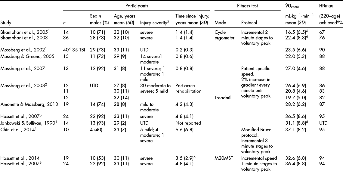
Eleven studies have measured ${\rm \dot VO}_{{\rm 2peak }}$ (i.e., using expired gas analysis) among people with TBI, and have either directly or indirectly compared cardiorespiratory fitness to age-matched healthy adults (Table 1). These studies were identified from a PubMed search combining the search terms ‘traumatic brain injury’ and ‘fitness’ conducted in March 2015, as well as studies known to the authors from their previous research. The 11 studies provide data for 234 adults who were predominantly males (64%) with an average age of 31 years, who had sustained TBI's of varying severity (at least 50% severe), and who were predominantly more than one year post injury. Only one study included participants who were, on average, less than 6 months post injury (Mossberg, Kuna, & Masel, Reference Mossberg, Kuna and Masel2002). ${\rm \dot VO}_{{\rm 2peak }}$ was measured using expired gas analysis whilst exercising on a cycle ergometer (Bhambhani, Rowland, & Farag, Reference Bhambhani, Rowland and Farag2003, Reference Bhambhani, Rowland and Farag2005), a treadmill (Amonette & Mossberg, Reference Amonette and Mossberg2013; Chin et al., Reference Chin, Chan, Woolstenhulme, Christensen, Shenouda and Keyser2014; Hassett, Harmer, Moseley, & Mackey, Reference Hassett, Harmer, Moseley and Mackey2007; Jankowski & Sullivan, Reference Jankowski and Sullivan1990; Mossberg, Ayala, Baker, Heard, & Masel, Reference Mossberg, Ayala, Baker, Heard and Masel2007; Mossberg & Greene, Reference Mossberg and Greene2005; Mossberg, Masel, Gilkison, & Urban, Reference Mossberg, Masel, Gilkison and Urban2008; Mossberg et al., Reference Mossberg, Kuna and Masel2002) or during the modified 20-metre shuttle test (Hassett, Harmer, Moseley, & Van der Ploeg, Reference Hassett, Harmer, Moseley and Van der Ploeg2014; Hassett et al., Reference Hassett, Harmer, Moseley and Mackey2007) using exercise protocols with 1-, 2- or 3-minute power increments until symptom-limited or voluntary peak was reached. The mean (SD) ${\rm \dot VO}_{{\rm 2peak }}$ of the 11 studies was 27.2 (6.7) mL·kg−1·min−1 (range 16.6 to 37.1 mL·kg−1·min−1) (or 7.6 METs); and a mean 87% (range 67 to 95%) of predicted maximum heart rate was achieved. Comparing these values to age-matched data for able-bodied males (American College of Sports Medicine, 2000), all are below the average fitness level (41 mL·kg−1·min−1); and the pooled mean ${\rm \dot VO}_{{\rm 2peak }}$ of the 11 studies and the mean ${\rm \dot VO}_{{\rm 2peak }}$ for seven of the eleven studies are below the lowest fitness level rating (i.e., below the 10th percentile fitness level; 32.5 mL·kg−1·min−1). Thus, collectively these studies provide evidence that adults with TBI have markedly lower cardiorespiratory fitness than their age-matched peers. Given that low fitness increases the risk of morbidity and all-cause mortality (Kodama et al., Reference Kodama, Saito, Tanaka, Maki, Yachi, Asumi and Sone2009; Lee, Artero, Sui, & Blair, Reference Lee, Artero, Sui and Blair2010) and that increasing fitness by 1 MET can reduce the risk of mortality by 15% (Kodama et al., Reference Kodama, Saito, Tanaka, Maki, Yachi, Asumi and Sone2009), preventing or reversing cardiorespiratory deconditioning among people with TBI should be a priority.
Studies Measuring Peak Oxygen Uptake  $\left( {{\rm \dot VO}_{{\rm 2 peak }} } \right)$ Among Adults who have Sustained a TBI. The Studies have been Grouped by Mode of Exercise and then Ranked by Time Since Injury

Key: 1Pre-intervention values for intervention studies; 2Participants were divided into three groups based on their growth hormone levels; 3The same participants completed two different tests 1 week apart; 4Participants were a mixed sample of people with various acquired brain injuries (e.g., stroke, anoxia, subarachnoid haemorrhage, TBI); 5Injury severity determined using GCS apart from Hassett (2007) and Hassett (2014) which measured injury severity by length of post traumatic amnesia and Chin (2014) which used Veteran Affair and Department of Defense Clinical Practice guidelines (Management of Concussion/m TBI Working Group, 2009). 6Value calculated from data presented in paper. UTD = unable to determine from data presented in paper. M20MST = modified 20-metre shuttle test.
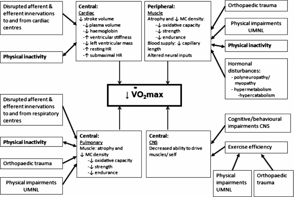
To determine how to prevent or reverse cardiorespiratory deconditioning in people with TBI, we must first understand how it develops. Unfortunately, the aetiology has not been well investigated, thus much of our current understanding is informed by studies on prolonged bed rest and studies of the acute consequences of TBI. These studies indicate that both central (cardiac, pulmonary and, in this context, central nervous system) and peripheral (muscle) factors contribute to cardiorespiratory deconditioning after TBI, and that these changes occur because of both the initial brain damage and orthopaedic trauma sustained, and the prolonged and initially extreme physical inactivity that is commonly experienced after this type of injury. From these data, we have proposed a model to explain the factors contributing to cardiorespiratory deconditioning in adults with severe TBI (Figure 1). The primary aim of this paper is to present this model to describe the physiological factors contributing to the development of cardiorespiratory deconditioning among people with severe TBI. Secondary aims are to discuss the relationship between physical inactivity and reduced cardiorespiratory fitness and the clinical and research implications of deconditioning in this population.
Proposed mechanism for the development of cardiorespiratory deconditioning in adults with severe TBI. Key: CNS: central nervous system; UMNL: upper motor neuron lesion; MC: mitochondria.

Aetiology of Reduced Cardiorespiratory Fitness in Adults with Severe TBI
After a severe TBI, it is not uncommon for the person to spend days to weeks in the acute care setting before being admitted to an inpatient rehabilitation unit. For example, a state-wide Australian study incorporating 2545 adults with severe TBI reported a median of 25 (IQR = 24) days between injury and admission to inpatient rehabilitation (Baguley et al., Reference Baguley, Nott, Howle, Simpson, Browne, King and Hodgkinson2012). Similarly, data from 21 TBI Model System Centers in North America incorporating 6975 adults with moderate to severe TBI evidenced a mean of 21 (SD = 16) days between injury and admission to inpatient rehabilitation (Dahdah et al., Reference Dahdah, Barisa, Schmidt, Barnes, Dubiel, Dunklin and Shafi2014). During this acute phase, the person with TBI is likely to spend the majority of the time confined to bed, with only short periods sitting out of bed or mobilising around the ward, depending on their impairments and the ward's capacity. Once admitted to inpatient rehabilitation, physical rehabilitation commences and the length of stay is often extensive (e.g., median length of stay = 37 (IQR = 66) days (Baguley et al., Reference Baguley, Nott, Howle, Simpson, Browne, King and Hodgkinson2012); mean length of stay = 26 (SD = 18) days (Dahdah et al., Reference Dahdah, Barisa, Schmidt, Barnes, Dubiel, Dunklin and Shafi2014)). Unfortunately, in the rehabilitation setting, extended periods of lying in bed and prolonged sitting are still prevalent. We recently investigated the time use in this setting over a 5-day period using behaviour mapping, and determined that participants spent on average 83% of the observed time (7:30 am until 7:30 pm) sitting or lying, and only 17% of time standing, walking or wheeling (Hassett et al., Reference Hassett, Wong, Sheaves, Daher, Egan, Grady and Moseley2015). This equates to approximately 10 hours per day engaged in sedentary behaviour and is higher than the sitting time reported in a large population-based cohort study (75% of 222 497 adults over 45 years reported spending less than 8 hours per day sitting) (van der Ploeg, Chey, Korda, Banks, & Bauman, Reference van der Ploeg, Chey, Korda, Banks and Bauman2012). Thus physical inactivity from prolonged and sustained bed rest is extensive and extreme in the first days to weeks after a severe TBI.
The Effects of Bed Rest on Cardiorespiratory Deconditioning Among Healthy Adults
A number of small studies and reviews have investigated the effects of bed rest on cardiorespiratory deconditioning (Bloomfield, Reference Bloomfield1997; Capelli et al., Reference Capelli, Antonutto, Kenfack, Cautero, Lador, Moia and Ferretti2006; Convertino, Bloomfield, & Greenleaf, Reference Convertino, Bloomfield and Greenleaf1997; Ferretti et al., Reference Ferretti, Antonutto, Denis, Hoppeler, Minetti, Narici and Desplanches1997; Lee, Moore, Everett, Stenger, & Platts, Reference Lee, Artero, Sui and Blair2010; Miokovic, Armbrecht, Felsenberg, & Belavy, Reference Miokovic, Armbrecht, Felsenberg and Belavy2012; Saltin et al., Reference Saltin, Blomqvist, Mitchell, Johnson, Wildenthal and Chapman1968; Wall, Dirks, & van Loon, Reference Wall, Dirks and van Loon2013), mainly to mimic and investigate the effects of prolonged anti-gravity on astronauts during spaceflight. These studies have mostly been conducted among young healthy men, and given the similar demographics of the typical TBI survivor (Hyder et al., Reference Hyder, Wunderlich, Puvanachandra, Gururaj and Kobusingye2007), may provide valuable insight into physiological mechanisms of deconditioning in TBI. In these studies of bed rest, ${\rm \dot VO}_{2{\rm max}}$ undergoes a rapid decrease over the first few days followed by a more gradual loss thereafter. ${\rm \dot VO}_{2{\rm max}}$ declines, on average, 0.8 to 0.9% per day over 30 days of bed rest (Lee et al., Reference Lee, Artero, Sui and Blair2010), although this rate is attenuated as the time of bed rest is prolonged, e.g., 0.35% per day over 90 days of bed rest (Capelli et al., Reference Capelli, Antonutto, Kenfack, Cautero, Lador, Moia and Ferretti2006).
Central cardiac changes and peripheral changes both contribute to the reduction in ${\rm \dot VO}_{2{\rm max}}$ with bed rest (Ferretti et al., Reference Ferretti, Antonutto, Denis, Hoppeler, Minetti, Narici and Desplanches1997; Lee et al., Reference Lee, Artero, Sui and Blair2010). A reduction in stroke volume (central cardiac factor) primarily due to reduced plasma volume and haemoglobin content is thought to be the main contributor to the initial rapid deconditioning (Ferretti et al., Reference Ferretti, Antonutto, Denis, Hoppeler, Minetti, Narici and Desplanches1997; Lee et al., Reference Lee, Artero, Sui and Blair2010). As bed rest continues over one to two weeks, physical cardiac adaptations become evident, including increased ventricular stiffness and reduced left ventricular mass which contribute to the further reduction in stroke volume and hence reduced ${\rm \dot VO}_{2{\rm max}}$. In addition, heart rate at rest and during submaximal exercise increases, mainly due to reduced stroke volume. Peripherally, muscle atrophy has been detected after periods of bed rest of as little as 4 days (Wall et al., Reference Wall, Dirks and van Loon2013), although it is thought that peripheral changes from more prolonged bed rest (at least 1 week) are more likely to contribute significantly to a reduction in ${\rm \dot VO}_{2{\rm max}}$ (Lee et al., Reference Lee, Artero, Sui and Blair2010). In a recent review of studies of 10 to 42 days of bed rest or immobilisation, the rate of muscle atrophy ranged from 0.5% to 0.6% per day with a concomitant loss of muscle strength ranging from 0.3% to 4.2% per day (Wall et al., Reference Wall, Dirks and van Loon2013). This peripheral muscle atrophy is not uniform, with the fastest rate of atrophy seen predominantly in type II (fast twitch) fibres and anti-gravity muscles (Bloomfield, Reference Bloomfield1997) with the calf muscles having the fastest rate of atrophy (Miokovic et al., Reference Miokovic, Armbrecht, Felsenberg and Belavy2012). In addition to the reduction in muscle fibre area (atrophy), other physiological changes within or affecting the muscle include a reduced density of mitochondria and a reduction in capillary length as well as altered neural input to muscles (Bloomfield, Reference Bloomfield1997; Ferretti et al., Reference Ferretti, Antonutto, Denis, Hoppeler, Minetti, Narici and Desplanches1997; Lee et al., Reference Lee, Artero, Sui and Blair2010). Atrophy reduces muscle strength (and endurance) and reduction in mitochondrial density reduces muscle oxidative capacity and ${\rm \dot VO}_{2{\rm max}}$. This results in an earlier rise in lactic acid accumulation in muscle and, after diffusion of hydrogen ions to blood, acidaemia. Buffering of acidaemia increases carbon dioxide production which drives ventilation; hence in incremental exercise, the ventilatory threshold is reached earlier. Thus, after bed rest, any given task requires higher ventilation and more effort because it requires a higher percentage of the person's (reduced) ${\rm \dot VO}_{2{\rm max}}$; and hence fatigue occurs earlier.
Central Cardiac Factors Contributing to Cardiorespiratory Deconditioning in TBI
Given the extensive period of bed rest after TBI, it is probable that this prolonged physical inactivity causes central cardiac changes as described above which contributes to the loss of cardiorespiratory fitness and early fatigue in this population (Figure 1). Physical inactivity appears to be the primary cause of these changes, although a lesion in the medulla oblongata where the cardiac centre is located may also contribute to cardiac limitations by disrupting afferent and efferent innervations (Bhambhani et al., Reference Bhambhani, Rowland and Farag2003).
Central Pulmonary Factors Contributing to Cardiorespiratory Deconditioning in TBI
Although pulmonary function does not usually contribute to cardiorespiratory deconditioning in healthy populations, among people with TBI, impairments in pulmonary function may contribute to the development of earlier fatigue and hence the associated deconditioning (Figure 1). Pulmonary impairments may be directly related to the orthopaedic trauma sustained (e.g., fractured ribs causing ongoing pain and reduced tidal volume which may impair gas exchange), as a result of the physical impairments from the upper motor neuron lesion (e.g., weakness or loss of coordination of the ventilatory muscles) (Wiercisiewski & McDeavitt, Reference Wiercisiewski and McDeavitt1998), as a result of damage to the medulla oblongata where the respiratory centre is located (Bhambhani et al., Reference Bhambhani, Rowland and Farag2003) or due to the initial extreme physical inactivity (i.e., sedation and mechanical ventilation in an intensive care unit). Many individuals who sustain a moderate to severe TBI may spend days to weeks in an intensive care unit on mechanical ventilation during which time the diaphragm and the accessory muscles of breathing are not active (or have reduced activity) and are therefore susceptible to significant deconditioning (Faulkner, Reference Faulkner and Roussos1995; Levine et al., Reference Levine, Nguyen, Taylor, Friscia, Budak, Rothenberg and Shrager2008). After removal of mechanical ventilation, the individual with TBI may spend many days to weeks either immobile in bed or participating in minimal physical activity due to altered levels of consciousness and physical impairments, thus likely not activating all the motor units within the respiratory muscles. The ventilatory muscles (e.g., diaphragm) are skeletal muscles and with extreme physical inactivity appear to undergo the same adaptations as other skeletal muscles (Faulkner, Reference Faulkner and Roussos1995; Levine et al., Reference Levine, Nguyen, Taylor, Friscia, Budak, Rothenberg and Shrager2008). Dormant motor units of respiratory muscles atrophy and mitochondrial density is reduced leading to reduced oxidative capacity and reduced strength and endurance; hence increasing the effort required to sustain ventilation at any workload (Amonette & Mossberg, Reference Amonette and Mossberg2013; Faulkner, Reference Faulkner and Roussos1995). Thus, amongst people with TBI, pulmonary impairments are likely to contribute to reduced cardiorespiratory fitness.
Peripheral Factors Contributing to Cardiorespiratory Deconditioning in TBI
As described above for the central cardiac factors, the prolonged period of bed rest after TBI (i.e., physical inactivity) causes peripheral changes which contribute to the loss of cardiorespiratory fitness (Figure 1). However, clinical observation of people with TBI who are admitted to rehabilitation with substantial weight loss (mean 11 (SD = 6) kg loss in body weight in the initial 38 (SD = 40) days) (Crenn et al., Reference Crenn, Hamchaoui, Bourget-Massari, Hanachi, Melchior and Azouvi2014) suggests that peripheral factors may indeed have a greater contribution to reduced fitness compared to the young healthy men in the bed rest studies described earlier. Whilst fat mass may be reduced, some of this weight loss is due to muscle atrophy. Again, as for the pulmonary factors, this is likely to be due to the extreme level of physical inactivity, orthopaedic trauma and physical impairments from the upper motor neuron lesion. But in addition to these factors, hormonal disturbances from the brain injury and acute care management are likely to also contribute to the peripheral changes causing a reduction in cardiorespiratory fitness (Lipshutz & Gropper, Reference Lipshutz and Gropper2013; Mossberg et al., Reference Mossberg, Masel, Gilkison and Urban2008; Pepe & Barba, Reference Pepe and Barba1999). A condition more recently described in the literature is intensive care unit acquired weakness (Lipshutz & Gropper, Reference Lipshutz and Gropper2013), which results from polyneuropathy and myopathy. This weakness is more extreme than that expected from bed rest alone and is thought to be due to the combination of the extreme physical inactivity combined with hormonal changes from the critical illness and drug management. In TBI, acute changes in hormone levels lead to hypermetabolism (elevated basal metabolic rate), and this is one factor that may promote hypercatabolism (elevated protein degradation in skeletal muscles). This leads to significant muscle atrophy with a loss of up to 1 kg of muscle tissue per day in the acutely ill patient with TBI (Pepe & Barba, Reference Pepe and Barba1999). Persistence of the hypercatabolic state for up to 3 months after the initial injury may therefore result in significant muscle atrophy and weakness if sufficient nutrition is not achieved (Pepe & Barba, Reference Pepe and Barba1999). Peripheral muscle atrophy due to disuse and the hypercatabolic state would reduce the oxidative capacity of the muscle and cause a reduction in muscular strength and endurance at submaximal and maximal intensities, hence leading to early fatigue at submaximal workloads and a reduction in ${\rm \dot VO}_{2{\rm max}}$.
Central CNS Factors Contributing to Cardiorespiratory Deconditioning in TBI
In addition to the central and peripheral physiological adaptations causing a decrease in cardiorespiratory fitness, other factors related to damage to the central nervous system from the brain injury may also contribute to deconditioning by affecting the individual's ability to perform submaximal exercise or to drive their muscles or themselves to reach their ${\rm \dot VO}_{2{\rm max}}$ (Figure 1). One such factor may be reduced exercise efficiency due to physical impairments from the upper motor neuron lesion and orthopaedic trauma. In a study of 14 people with TBI (Jankowski & Sullivan, Reference Jankowski and Sullivan1990), the average energy cost of walking at 1.3 m·s−1 was 161% of able-bodied values (American College of Sports Medicine, 2000). Because physiologic efficiency is an important factor in fatigability (Jankowski & Sullivan, Reference Jankowski and Sullivan1990), the findings from this study indicate that the inefficient movement pattern could significantly contribute to premature fatigue, and hence over time, deconditioning. Other contributing factors may include cognitive impairments from damage to the central nervous system such as diminished drive and poor attention, and behavioural disturbances such as apathy and depression. These cognitive impairments and behavioural disturbances may result in the individual not being able to push him or herself to exercise at moderate to vigorous levels or to reach their ${\rm \dot VO}_{2{\rm max}}$.
Relationship Between Physical Inactivity and Reduced Cardiorespiratory Fitness
Enforced physical inactivity during an often extensive hospital admission, plus orthopaedic trauma and brain injury related factors, contributes to the early development of cardiorespiratory deconditioning. As the individual with TBI starts to recover, cardiorespiratory deconditioning (muscle weakness and reduced oxidative capacity) itself then contributes to ongoing physical inactivity due to increased fatigability which discourages or limits participation in some forms of physical activity (Figure 2). In addition, environmental factors such as healthcare provider recommendations (i.e., cautious restrictions on exercise) and lack of opportunities for leisure-time physical activity (Self, Driver, Stevens, & Warren, Reference Self, Driver, Stevens and Warren2013) and personal factors such as pre-injury exercise history and age (Hassett, Tate, Moseley, & Gillett, Reference Hassett, Tate, Moseley and Gillett2011) influence participation in community-based physical activities and will contribute to deconditioning. As cardiorespiratory deconditioning worsens, the reserve capacity  ${\rm (\dot VO}_{{\rm 2peak}} - {\rm \dot VO}_{{\rm 2rest}} )$ decreases and submaximal tasks require an even greater percentage of the reserve capacity, thus necessitating higher effort and resulting in earlier fatigue and further physical inactivity. Therefore, a vicious cycle between deconditioning and physical inactivity is established.
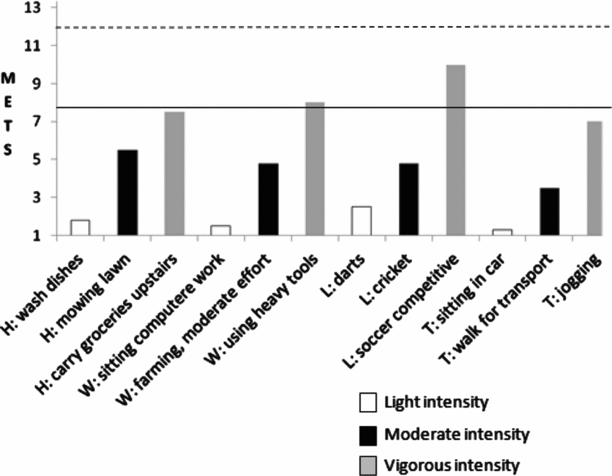
Examples of various intensities (light >1.0 to 3 METs; moderate 3 to 6 METs and vigorous >6 METs) of physical activities within the domains of household (H), work-related (W), leisure (L) and transport (T). The metabolic equivalents (METs) are taken from the compendium of physical activities (Ainsworth et al., Reference Ainsworth, Haskell, Herrmann, Meckes, Bassett, Tudor-Locke and Leon2011). The solid horizontal line represents the average ${\rm \dot VO}_{{\rm 2peak}}$ of adults with TBI from the studies presented in Table 1 (7.6 METs or 27.2 mL·kg−1·min−1) and demonstrates a reduced reserve capacity  ${\rm (\dot VO}_{{\rm 2peak}} - {\rm \dot VO}_{{\rm 2rest}} )$ to participate in many moderate intensity physical activities (e.g., mowing the lawn) and insufficient aerobic capacity to participate in some vigorous physical activities (e.g., soccer). The dashed line is the average ${\rm \dot VO}_{{\rm 2peak}}$ of males aged 30–39 years (American College of Sports Medicine, 2000).
Reduced cardiorespiratory fitness can directly restrict return to and participation in pre-injury physical activities (Jankowski & Sullivan, Reference Jankowski and Sullivan1990) because the individual may no longer have the aerobic capacity to meet the metabolic demands of various work and leisure activities (Figure 2). For example, if an individual with TBI has a ${\rm \dot VO}_{{\rm 2peak}}$ of 27.2 mL·kg−1·min−1 (the average ${\rm \dot VO}_{{\rm 2peak}}$ of studies included in Table 1), they would have insufficient aerobic capacity to participate in a pre-injury work activity of using heavy tools or a pre-injury leisure activity of soccer (Figure 2). Both of these activities are estimated to require ≥ 8 times the metabolic energy expenditure of sitting quietly (Ainsworth et al., Reference Ainsworth, Haskell, Herrmann, Meckes, Bassett, Tudor-Locke and Leon2011), which equates to working at an aerobic capacity ≥ 28 mL·kg−1·min−1. Thus, an individual with a ${\rm \dot VO}_{{\rm 2peak}}$ of 27.2 mL·kg−1·min−1 could only perform these (now supra-maximal) activities for a very brief period before fatigue. Thus, cardiorespiratory deconditioning can directly restrict participation in physical activity, and more broadly, reintegration back into their previous roles within their family, work and community.
Among people with TBI, low cardiorespiratory fitness and physical inactivity likely contribute to increased morbidity and the more than doubled risk of cardiac-related mortality compared with the general population (Baguley et al., Reference Baguley, Nott, Howle, Simpson, Browne, King and Hodgkinson2012; Selassie, Cao, Church, Saunders, & Krause, Reference Selassie, Cao, Church, Saunders and Krause2014). In the general population, physical inactivity has been identified as one of the major causes of mortality and contributes to the development of secondary co-morbid conditions including coronary heart disease, type 2 diabetes, colon cancer, breast cancer, stroke and depression (Lee et al., Reference Lee, Shiroma, Lobelo, Puska, Blair and Katzmarzyk2012). Similarly, a low fitness level accounted for 16% of deaths in a large cohort study of more than 50,000 people and was a substantially greater risk factor for mortality than other more commonly reported risk factors such as smoking and obesity (Blair, Reference Blair2009). The development of secondary co-morbidities due to physical inactivity has been shown to have significant economic costs in the general population, including direct (i.e., increase health sector costs), indirect (i.e., lost productivity due to illness or premature death) and intangible costs (i.e., quality of life of the individual and their family) (Stephenson, Bauman, Armstrong, Smith, & Bellew, Reference Stephenson, Bauman, Armstrong, Smith and Bellew2000). Therefore, it is likely that physical inactivity and low cardiorespiratory fitness will considerably increase the already major economic burden of TBI (Ma, Chan, & Carruthers, Reference Ma, Chan and Carruthers2014).
Clinical and Research Implications
Given the paucity of data about the aetiology of cardiorespiratory deconditioning in TBI, further research should be conducted to determine the amount and time-course of the development of peripheral and central changes and their relationship to change in fitness over time. From there, interventions to address the specific impairments as well as overall fitness need to be evaluated. For example, a randomised controlled trial among people with chronic heart failure evaluated aerobic exercise combined with inspiratory muscle training and peripheral muscle resistance training compared to aerobic training alone (Laoutaris et al., Reference Laoutaris, Adamopoulos, Manginas, Panagiotakos, Kallistratos, Doulaptsis and Dritsas2013). The combined exercise group showed greater improvements in some aspects of fitness as well as in respiratory and peripheral impairments compared to the aerobic training alone group. Respiratory muscle training has not been evaluated among people with TBI. While ballistic (Williams, Clark, Hansson, & Paterson, Reference Williams, Clark, Hansson and Paterson2014) and isokinetic (Killington, Mackintosh, & Ayres, Reference Killington, Mackintosh and Ayres2010) strength training has been investigated in this population, changes in cardiorespiratory fitness were not evaluated.
Fitness Training
Given that cardiorespiratory deconditioning is likely to develop early post-injury and is still a significant impairment many years post-injury (Table 1), interventions to address this impairment are imperative throughout the continuum of care – spanning from inpatient rehabilitation, to outpatient and community rehabilitation, to long-term follow-up. The American College of Sports Medicine has developed specific guidelines for implementing an exercise programme for people who have sustained a stroke or TBI (Palmer-McLean & Harbst, Reference Palmer-McLean, Harbst, Durstine, Moore, Painter and Roberts2009); however, these are yet to be validated and the optimal period for implementation or commencement is unknown to date. The guidelines recommend that an exercise programme incorporate four components: aerobic, strength, flexibility and neuromuscular training. The guidelines for the aerobic component recommend a frequency of three to five exercise sessions per week, prescribed at an intensity between 40 and 70% of the individual's ${\rm \dot VO}_{{\rm 2peak}}$, for a duration of 20 to 60 minutes or an exercise dosage of 300 kilocalories per session. The mode of exercise can be any number of activities requiring large muscle group activation such as cycling, arm cranking or treadmill walking, but depend on individual physical impairments and the goals of training.
Fitness training programmes in unfit, but otherwise healthy, individuals have induced improvements in ${\rm \dot VO}_{{\rm 2max}}$ of approximately 12% for light (24 to 39% heart rate reserve) to moderate (40 to 59% heart rate reserve) intensity programmes (Swain, Reference Swain2005). However, the ability to achieve similar outcomes in the inpatient setting among people with TBI is less clear. One randomised controlled trial prescribed exercise on a cycle ergometer three times per week for 12 weeks at a moderate intensity (38 to 50% heart rate reserve) for 30 minutes per session for people with acquired brain injury (Bateman et al., Reference Bateman, Culpan, Pickering, Powell, Scott and Greenwood2001). In a Cochrane Systematic Review that evaluated only the TBI participants of this study, a non-significant improvement in peak work rate on an incremental exercise test was found for the intervention group compared to the control group who participated in relaxation therapy (mean difference 21.5 watts [95% CI −12.3 to 55.3]) (Hassett, Moseley, Tate, & Harmer, Reference Hassett, Moseley, Tate and Harmer2008). Despite the inpatient exercise programme being supervised, participants did not achieve the prescribed exercise dosage and spent an average of only two sessions per week exercising at moderate intensity for 16 minutes of a 25-minute session (Hassett et al., Reference Hassett, Moseley, Tate and Harmer2008). These data suggest that achieving an effective exercise dosage early post-injury is challenging. We recently evaluated people with TBI in an inpatient rehabilitation setting participating in a circuit class and found that they were able to achieve the American College of Sports Medicines recommended exercise dosage (i.e., >300 kcal expended within an exercise session), however this was predominantly achieved with low-intensity long-duration exercise (despite encouragement to exercise as hard as tolerated) and the change in fitness was not evaluated (Hassett, Moseley, Whiteside, Barry, & Jones, Reference Hassett, Moseley, Whiteside, Barry and Jones2012). Further research is warranted to evaluate the effectiveness of fitness training in the inpatient setting among people with TBI. Clinically, it would be recommended to routinely conduct a fitness test (with appropriate medical screening and precautions) (Vitale et al., Reference Vitale, Sullivan, Jankowski, Fleury, Lefrancois and Lebouthillier1995) to determine the training parameters, and then to monitor exercise intensity (using a heart rate monitor) and duration when prescribing fitness training to determine whether exercise dosage is meeting the recommended guidelines (Palmer-McLean & Harbst, Reference Palmer-McLean, Harbst, Durstine, Moore, Painter and Roberts2009).
There is some evidence to suggest that fitness training programmes conducted in the community setting can improve fitness among people with TBI (Driver, O’Connor, Lox, & Rees, Reference Driver, O’Connor, Lox and Rees2004; Hassett et al., Reference Hassett, Moseley, Tate, Harmer, Fairbairn and Leung2009). Driver et al., (Reference Driver, O’Connor, Lox and Rees2004) prescribed aquatic group exercise three times per week for eight weeks at a moderate intensity (50 to 70% heart rate reserve) for 60 minutes per session (Driver et al., Reference Driver, O’Connor, Lox and Rees2004). In the same Cochrane Systematic Review described above, analysis of this study demonstrated a significant improvement in peak work rate on an incremental exercise test for the participants in the intervention group compared to the control group who participated in a vocational therapy group (mean difference 59 watts [95% CI 23.8 to 94.2]) (Hassett et al., Reference Hassett, Moseley, Tate and Harmer2008). The actual intensity achieved was not recorded, however the long-duration exercise may have provided a sufficient dose even if the prescribed intensity was not achieved. In another community-based study, we compared a fitness centre-based exercise programme to a home-based exercise programme prescribed three times a week for 12 weeks at a moderate intensity incorporating 30 minutes of fitness training (Hassett et al., Reference Hassett, Moseley, Tate, Harmer, Fairbairn and Leung2009). This study found both groups improved in fitness by approximately 1 MET although better adherence was achieved in the fitness-centre group. The intensity of exercise was not monitored in this study, thus we are not able to determine if the recommended exercise dosage was achieved. Further research is required to better understand the dose-response relationship of intensity and duration of fitness training to improve fitness in this population. Clinically, we would recommend prescribing fitness training programmes as per the American College of Sports Medicine guidelines (Palmer-McLean & Harbst, Reference Palmer-McLean, Harbst, Durstine, Moore, Painter and Roberts2009), monitoring the exercise dose delivered using heart rate monitors, and utilising strategies to maximise adherence to exercise programmes (Hassett et al., Reference Hassett, Tate, Moseley and Gillett2011).
Physical Activity
Finally, as physical inactivity is a significant contributor to cardiorespiratory deconditioning, and vice-versa, strategies to more broadly increase physical activity (i.e., beyond structured exercise programmes) both in the acute stages of recovery and in the longer term are warranted. Interventions to address physical inactivity in TBI are yet to be rigorously evaluated, however interventions that have shown effect or promise in similar populations include enrichment of the inpatient rehabilitation environment (Janssen et al., Reference Janssen, Ada, Bernhardt, McElduff, Pollack, Nilsson and Spratt2014) and community-based physical activity counselling (van der Ploeg et al., Reference van der Ploeg, Streppel, van der Beek, van der Woude, Vollenbroek-Hutten, van Harten and van Mechelen2006). From a clinical and policy standpoint, there needs to be greater engagement and opportunities for suitable community-based physical activity opportunities such as inclusive fitness centres and sporting organisations. This requires better communication and engagement between healthcare providers and community organisations (Rimmer & Henley, Reference Rimmer and Henley2013) and clear evidence-based guidelines about when it is safe to return to physical activities such as competitive sport after a severe TBI (Pangilinan & Hornyak, Reference Pangilinan and Hornyak2007).
Acknowledgements
None
Financial Support
This research received no specific grant from any funding agency, commercial or not-for-profit sectors.
Conflict Of Interest
None.
Ethical Standards
NA.