An absent ductus venosus is a rare anomaly and is generally identified after the referral of foetuses for a variety of reasons. The most common indication for referral is the signs of foetal cardiac decompensation such as cardiomegaly, dilated systemic veins, or polyhydramnios/hydrops.Reference Yagel, Kivilevitch, Valsky and Achiron1–Reference Berg, Kamil, Geipel, Kohl, Knöpfle and Hansmann3 In the presence of absent ductus venosus, umbilical vein connection to the venous system may be extra-hepatic or intra-hepatic via the portal venous system.Reference Berg, Kamil, Geipel, Kohl, Knöpfle and Hansmann3 The prognosis of the foetuses mainly depends on the presence and severity of foetal congestive cardiac failure. Cardiac and extra-cardiac abnormalities may accompany the anomaly and their presence also affects the prognosis.Reference Hajdu, Marton and Kozsirek2–Reference Contratti, Banzi, Ghi, Perolo, Pilu and Visentin4
We report a foetus with absent ductus venosus and abnormal drainage of the umbilical vein into the superior caval vein. The foetus also had complex cardiac malformation as type I persistent truncus arteriosus with truncal valve stenosis. To the best of our knowledge, the foetus reported herein had two unique features. The connection of the umbilical vein to the superior caval vein and the associated persistent truncus arteriosus has not yet been reported in foetuses with absent ductus venosus.
Case report
A 32-year-old pregnant woman was referred to our paediatric cardiology unit at the 30th week of gestation because of foetal cardiomegaly and dilation of the great vessels. She had lost her foetus at the 26th gestational week in her second pregnancy. Postmortem examination had not been carried out for that foetus.
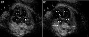
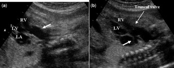
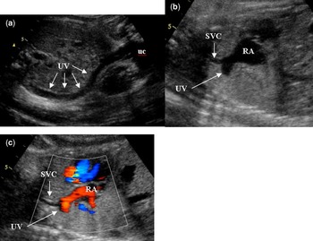
At first glance, foetal echocardiography revealed prominent cardiomegaly, a large peri-membranous ventricular septal defect, and atrial septal aneurysm on the modified four-chamber position (Fig 1). Detailed multi-planar scanning showed that there was a single arterial trunk, which leaved the heart directly above the ventricular septal defect. Just above the truncal valve the main pulmonary artery arose from the posterior aspect of the truncus and then divided into right and left pulmonary artery branches (Fig 2). The truncus continued as an aorta from the main pulmonary artery bifurcation, without interruption. The truncal valve was bicuspid and stenotic. We also detected a large, long aberrant vessel coursing posterior to the liver, crossing the diaphragm, and connecting to the superior caval vein just above the superior caval vein–right atrial junction. We tracked the vessel to the umbilical cord (Fig 3). The vessel was considered to be an aberrant umbilical vein. A ductus venosus could not be shown. The course and connection of the inferior caval vein, portal, and the hepatic veins were all normal. The foetus was followed weekly for foetal decompensation and signs of hydrops till the 37th week of gestation, and then elective caesarean section was performed. The male infant who was 2600 grams at birth. No abnormality was detected on post-natal abdominal ultrasonography related to the inferior caval vein, portal, and hepatic venous systems. His cardiac pathology was also confirmed by post-natal echocardiography. The truncal valve was mildly stenotic – there was only 30 millimetres of mercury systolic gradient at the truncal valve level – without any regurgitation. The atrial septum was aneurysmatic, allowing a little left to right shunt. He was operated upon at 2 months of age. Unfortunately, the patient died in the early post-operative period due to intractably low cardiac output syndrome.
(a) Echocardiography showed that all cardiac chambers were enlarged and atrial septal aneurysm protruding the left atrium (arrow), (b) perimembranous ventricular septal defect was shown by short arrow. There was an enlarged vessel connecting to right atrium (long arrow).

(a) There was a single arterial trunk leaved the heart directly above the ventricular septal defect (arrow). (b) Just above the truncal valve main pulmonary artery arised from the posterior aspect of the truncus and then divided into right and left pulmonary artery branches (thick arrow).

(a) The long aberrant vessel (umbilical vein-UV) starting from the umbilical cord (uc) coursing posterior to the abdomen, (b–c) crossing to the diaphragm and connecting to the superior vena cava (SVC) (arrows).

Discussion
An absent ductus venosus is a rarely defined pathology that results in anomalous umbilical venous return. Although it has been considered as a rare abnormality, little is known about its incidence, since visualisation of the ductus venosus is not a routine part of foetal ultrasonographic screening in most centres.Reference Hajdu, Marton and Kozsirek2–Reference Achiron, Hegesh, Yagel, Lipitz, Cohen and Rotstein5
The foetus reported herein has two unique features that have not been described in the foetuses with absent ductus venosus.
• Superior caval vein connection of umbilical vein, although theoretically possible, has not been defined previously.
• Persistent truncus arteriosus has also not been reported as an associated cardiac anomaly.
The drainage site of the umbilical vein is the main determinant of the outcome if any other cardiac or extra-cardiac anomaly does not exist. Three different types of abnormal venous connection sites have been described for the umbilical vein: umbilical vein connecting to the inferior caval vein or its branches; umbilical vein crossing the diaphragm and connecting directly to the heart generally via the right atrium, or exceptionally the left atrium or coronary sinus; and umbilical vein connecting to the portal venous system without giving rise to the ductus venosus.Reference Berg, Kamil, Geipel, Kohl, Knöpfle and Hansmann3, Reference Contratti, Banzi, Ghi, Perolo, Pilu and Visentin4
Prominent dilation of the right cardiac chambers and progressive cardiac decompensation have been described in cases in which umbilical venous drainage bypasses the liver, in the first two drainage sites, due to increased cardiac preload.Reference Berg, Kamil, Geipel, Kohl, Knöpfle and Hansmann3, Reference Sau, Sharland and Simpson6, Reference Jaeggi, Fouron and Hornberger7 In the foetus reported herein, not only the right, but all the chambers of the heart were found to be dilated. We thought that it was the consequence of associated cardiac malformation, that is, persistent truncus arteriosus, which decompressed the right cardiac chambers by way of a large ventricular septal defect.
Another important problem that potentially affects the long-term outcome of foetuses with absent ductus venosus and extra-hepatic umbilical venous drainage is the agenesis of portal venous system which occurs in nearly half of the patients. Some of them have serious post-natal complications including congestive cardiac failure, pulmonary hypertension, pulmonary oedema, focal nodular hyperplasia of the liver, and hepatic tumours.Reference Venkat-Raman, Murphy, Ghaus, Teoh, Higham and Carvalho8, Reference Peker, Ucar, Kuloglu, Ceyhan, Tutar and Fitoz9 Since the portal system of our case was normally developed we do not anticipate long-term hepatic complications.
Miscellaneous cardiac malformations, isolated or as part of the complex malformation syndrome, have been identified in a frequency ranging from 22% to 48% of the foetuses with absent ductus venosus.Reference Berg, Kamil, Geipel, Kohl, Knöpfle and Hansmann3 The reported cardiac malformations to date were isolated ventricular septal defect, tricuspid atresia, double outlet right ventricle, pulmonary atresia, transposition of great arteries, coarctation of the aorta, and persistent left superior caval vein.Reference Berg, Kamil, Geipel, Kohl, Knöpfle and Hansmann3 Among the aforementioned cardiac malformations we notice that the conotruncal malformations such as double outlet right ventricle, pulmonary atresia with ventricular septal defect, and transposition of great arteries are frequent. Our patient’s cardiac malformation of persistent truncus arteriosus has also been classified as a conotruncal malformation of the heart. We think that the associated cardiac malformations may be the result of changing blood flow distribution in these patients, or the same developmental insult causes both the ductus venosus agenesis and cardiac malformation.
In summary, different characteristics of the foetus, which are reported here, should be added to the clinical spectrum of the absent ductus venosus. We believe that if ductus venosus examination is a part of routine foetal ultrasonographic examination, the detection rate of the absent ductus venosus, and knowledge about it and its associated malformations would increase.