Implications
This study extends the available knowledge on the use of mixed bacterial and yeast probiotic preparations in the livestock industry. The study is focused on identifying the optimal dose of the mixed bacterial and yeast probiotic preparation used, in order to improve the welfare of birds in a poultry production setting, which may in turn reduce animal losses and thus increase cost effectiveness. Knowledge on the exact composition of the probiotic preparation and the areas of the gut in which the effects of the probiotic preparation are localised may contribute to the reduction of costs in a poultry breeding/production setting.
Introduction
From 1 January 2006, the European Union banned the use of antibiotics as growth promoters for farm animals (livestock) as a consequence of the growing concern about the development of antimicrobial resistance and the transference of antibiotic resistance genes from animal to human microbiota. However, the removal of these compounds from livestock diets has put tremendous pressure on livestock and poultry producers to find an effective alternative that would perform the same function, especially in the breeding of young animals (Agboola et al., Reference Agboola, Aroniyo, Suberu and Adeyemi2014; Hanczakowska et al., Reference Hanczakowska, Świątkiewicz, Natonek-Wiśniewska and Okoń2017). Thus there is a need to investigate the use of alternative products and strategies which would promote the growth of production animals, as well as carry out a prophylactic/therapeutic function, simultaneously. The abovementioned goals may be achieved through the use of specific feed additives, dietary raw materials or probiotics, which have been shown to favourably affect animal performance and welfare (Soccol et al., Reference Soccol, Porto de Souza Vandenberghe, Rigon Spier, Bianchi Pedroni Medeiros, Tiemi Yamaguishi, De Dea Lindner, Pandey and Thomaz-Soccol2013; Agboola et al., Reference Agboola, Aroniyo, Suberu and Adeyemi2014). The favourable effects of various feed additives, dietary raw materials and probiotics are thought to be attributed to their ability to regulate gut microbiota balance, which in turn plays a critical role in maintaining host health (Sekirov et al., Reference Sekirov, Russell and Antunes2010). Probiotics, which are viable microorganisms with no pathogenic properties consisting of either yeast or bacteria, are commonly added to food to improve the microbial balance in the gastrointestinal tract of the host. Probiotics also play a role in the intermediate control of host metabolism, as well as the provision of certain nutrients and regulatory substances, and hence, are associated with the beneficial effects for human and animal health (Marteau et al., Reference Marteau, De Vrese and Cellier2001; Soccol et al., Reference Soccol, Porto de Souza Vandenberghe, Rigon Spier, Bianchi Pedroni Medeiros, Tiemi Yamaguishi, De Dea Lindner, Pandey and Thomaz-Soccol2013). Probiotics favourably alter the balance of intestinal microflora by inhibiting the growth of harmful bacteria, producing metabolic substrates (e.g. vitamins and short-chain fatty acids) and stimulating the immune system in a non-inflammatory manner (Rahimi et al., Reference Rahimi, Grimes, Fletcher, Oviedo and Sheldon2009; Agboola et al., Reference Agboola, Aroniyo, Suberu and Adeyemi2014; Hanczakowska et al., Reference Hanczakowska, Świątkiewicz, Natonek-Wiśniewska and Okoń2017). Dietary probiotics make use of several modes of action within the gastrointestinal tract of animals including competition with pathogenic bacteria for intestinal adhesion sites and nutrients, production of antimicrobial substances including bacteriocins and microbial enzymes, modification of environmental conditions in the intestine by lowering pH through the increased production of organic acids, enhancement of the intestinal immune function and the enhancement of epithelial barrier integrity (Rahimi et al., Reference Rahimi, Grimes, Fletcher, Oviedo and Sheldon2009; Rawski et al., Reference Rawski, Kieronczyk, Dlugosz, Swiatkiewicz and Józefiak2016). All the abovementioned probiotic-induced changes within the gastrointestinal tract may be reflected in its histological structure. Probiotics affect the histology and ultrastructure of the gastrointestinal tract, as well as the synthesis and secretion of mucus (Deplancke and Gaskins, Reference Deplancke and Gaskins2001; Awad et al., Reference Awad, Böhm, Razzazi-Fazeli, Ghareeb and Zentek2006 and Reference Awad, Ghareeb, Abdel-Raheem and Böhm2009; Rahimi et al., Reference Rahimi, Grimes, Fletcher, Oviedo and Sheldon2009). Available data on changes in the intestinal morphology of turkeys also support the concept that poultry gut health and function, and ultimately bird performance, can be improved by probiotics (Rahimi et al., Reference Rahimi, Grimes, Fletcher, Oviedo and Sheldon2009; Agboola et al., Reference Agboola, Aroniyo, Suberu and Adeyemi2014). Gastrointestinal tract microstructure development has been linked to faecal microbiota numbers. There is a large diversity of bacterial (Lactobacillus sp. Pediococcus sp., Streptococcus sp., Bacillus sp., Bifidobacterium and Enterococcus faecium) and yeast (Saccharomyces sp.) strains, which have been studied at different doses, and they have been found to have a stimulating or modulating effect on structural features and the maturation of the gastrointestinal tract (de los Santos et al., Reference de los Santos, Donoghue, Farnell, Huff, Huff and Donoghue2007; Awad et al., Reference Awad, Ghareeb, Abdel-Raheem and Böhm2009; Kabir, Reference Kabir2009; Aliakbarpour et al., Reference Aliakbarpour, Chamani, Rahimi, Sadeghi and Qujeq2012; Soccol et al., Reference Soccol, Porto de Souza Vandenberghe, Rigon Spier, Bianchi Pedroni Medeiros, Tiemi Yamaguishi, De Dea Lindner, Pandey and Thomaz-Soccol2013; Chamani, Reference Chamani2016; Rawski et al., Reference Rawski, Kieronczyk, Dlugosz, Swiatkiewicz and Józefiak2016). Therefore, the current study was designed to test the hypothesis whether there is a relationship between the improvement of the intestinal structure and an increase in the dose of probiotic preparation. Thus, the aim of the present study was to examine the effects of elevated doses of a probiotic preparation on the structure of the small intestine of female turkeys and to establish an optimal dose, if possible.
Materials and methods
Animal breeding and experimental design
The experiment was performed on 80 three-day-old, healthy, female turkeys (Big-6 breed), randomly allocated to four weight-matched (59.70 ± 0.83 g) groups, according to probiotic treatment dose. Each group consisted of 20 birds divided into four replicates with five birds each. The animals were kept under standard rearing conditions (litter maintenance system) with optimal air temperature level depending on the age of the birds. The turkeys had free access to fresh water and appropriate feed was supplied ad libitum in accordance with the stage of production cycle. The birds were fed with a diet corresponding to the four periods of rearing (Supplementary Material S1, Table S1), as previously described (Tomaszewska et al., Reference Tomaszewska, Kwiecien, Dobrowolski, Klebaniuk, Muszynski, Olcha, Blicharski and Grela2016), for a total of 16 weeks. Diets were mixed and pelleted under temperature conditions not exceeding 45°C (since high temperatures may have killed the probiotic cells). At the age of 16 weeks, all birds were slaughtered by cutting the carotid arteries (Tomaszewska et al., Reference Tomaszewska, Kwiecien, Dobrowolski, Klebaniuk, Muszynski, Olcha, Blicharski and Grela2016). Mean weekly body weight gain, mean daily feed intake and average feed conversion ratio were calculated.
Probiotic culture and supplementation
The birds were administered with the probiotic culture (JHJ, Sp. z o.o. Gizałki, Poland), including four strains of bacterial isolates: Lactococcus lactis IBB500, Carnobacterium divergens S1, Lactobacillus casei LOCK 0915 and Lactobacillus plantarum LOCK 0862, and one yeast isolate: Saccharomyces cerevisiae LOCK 0141, all of poultry gastrointestinal origin (EFSA 2015). The microorganisms were non-genetically modified strains. The L. lactis IBB500 strain is deposited in the Polish Collection of Microorganisms (PCM, Wroclaw, Poland), the C. divergens S1 strain is deposited in the Collection of Industrial Microorganisms (IAFB, Warsaw, Poland) and the other three strains are deposited in the Centre of Industrial Microorganisms Collection (LOCK, Lodz, Poland).
Experimental groups differed according to the concentration of probiotic spores (if any) added to the diet at every stage of fattening: Control – 0, Group 1 – 107 cfu•g−1, Group 2 – 108 cfu•g−1, Group 3 – 109•cfu g−1, (cfu – a colony-forming unit) in an amount of 500 g•1000 kg−1 throughout the experimental period.
Histology preparation and histomorphometric analysis
At the end of the experiment all birds were weighed and sacrificed, and samples of small intestine segments (duodenum and jejunum) were obtained and processed for histology as previously described (Dobrowolski et al., Reference Dobrowolski, Huet, Karlsson, Eriksson, Tomaszewska, Gawron and Pierzynowski2012). Briefly, 25-mm long segments of duodenum (1.5 cm after stomach) and jejunum (from the middle portion of small intestine) were collected into a 35°C saline solution and gently cut open longitudinally, along the mesentery line. Samples were then placed flat (not stretched) into a standard histological cassette in such a way that the mucosa had no contact with any of the cassette walls, and fixed in 4% buffered formaldehyde (pH 7.0) for 24 h. After dehydration in graded ethanol solutions, samples were trimmed into three pieces (10 × 5 mm), allowing us to cut intestinal cross sections on the longer border of the samples. Following clearance in xylene, samples were then embedded in paraffin using a tissue processor (STP 120, Thermo Scientific, Waltham, MA, USA). Twenty cross sections (with 10 mm interval between each five-slice section), 4 mm thick, were then cut with a microtome (Microm HM 360, Microm, Walldorf, Germany) from every sample of the small intestine (Dobrowolski et al., Reference Dobrowolski, Huet, Karlsson, Eriksson, Tomaszewska, Gawron and Pierzynowski2012). Goldner’s trichrome staining was used to differentiate the small intestine wall layers (Suvarna et al., Reference Suvarna, Layton and Bancroft John2013; Dobrowolski et al., Reference Dobrowolski, Tomaszewska, Kurlak and Pierzynowski2016). Microscopic (two-dimensional) images of bright field (magnification ×50, ×200 and ×400) were collected using a confocal microscope (AXIOVERT 200 M, Carl Zeiss, Jena, Germany) equipped with a colour digital camera (AxioCam HRc, Carl Zeiss, Jena, Germany) and a halogen lamp (Dobrowolski et al., Reference Dobrowolski, Tomaszewska, Kurlak and Pierzynowski2016). Microscopic pictures of particular segments of small intestine were subjected to further histomorphometric analysis. The structure of the small intestinal wall was examined under microscopic observation and with the use of graphic analysis software (ImageJ 1.51, National Institutes of Health, Bethesda, MD, USA; available at: http://rsb.info.nih.gov/ij/index.html). The following parameters were analysed: the thickness of the inner and outer muscle layer, mucosa and submucosa thickness, crypt depth (defined as the depth of the invagination between adjacent villi, from the bottom of the crypt to the base of the villus) and width (measured in the middle of the crypt depth), villi height (from the tip of the villus to the villus–crypt junction) and width (measured in the middle of the villus height), total crypt number (opened and closed crypts), number of open (showing mitoses and an open internal space, with access to the intestinal lumen) and closed crypts (showing no mitoses and a closed internal space), number of villi per millimetre of mucosa, enterocyte height (measured as the distance from brush border membrane to the basolateral membrane) and number of enterocytes per 100 mm of villus epithelium (Dobrowolski et al., Reference Dobrowolski, Huet, Karlsson, Eriksson, Tomaszewska, Gawron and Pierzynowski2012). Only vertically oriented villi and crypts were measured. Small intestinal absorptive surface was also determined according to Kisielinski et al. (Reference Kisielinski, Willis, Prescher, Klosterhalfen and Schumpelick2002) (Supplementary Material S1). Villi height to crypt depth ratio was also calculated. Meissner as well as Auerbach plexus features including mean sectional area of nerve ganglia, ganglia perimeter, shape factor (where 0 refers to the rod-shaped objects and 1 refers to objects that are round or spherical in shape), minimal radius and diameter, mean diameter, sphericity and convexity were also analysed (Dobrowolski et al., Reference Dobrowolski, Huet, Karlsson, Eriksson, Tomaszewska, Gawron and Pierzynowski2012; Tomaszewska et al., Reference Tomaszewska, Dobrowolski and Puzio2012).
Statistical analysis
All results are expressed as mean (SD). Differences between means were tested with the use of one-way ANOVA and a post hoc Tukey’s honest significant difference test as a correction for multiple comparisons. Normal distribution of data was examined using the W Shapiro–Wilk test and equality of variance was tested by the Brown–Forsythe test. When data were not normally distributed and/or unequal variance of data, the Kruskal–Wallis test was used to analyse the difference between means and post hoc tests for mean range for every pair of groups. The two-sided significance level (P-value) of less than 0.05 was considered statistically significant. All statistical analyses were carried out using STATISTICA (data analysis software system), version 12. StatSoft, Inc. (2014). STATISTICA Power Analysis tool was used to calculate the sample size and the statistical power as a function of the error and the type and size of the effect for the one-way ANOVA. The effect size was calculated and interpreted according to Cohen’s f, as described by Kotrlik (Kotrlik et al., Reference Kotrlik, Williams and Jabor2011) (Supplementary Material S1, Tables S2 and S3). The general effect of probiotic dose on the small intestine parameters was calculated as a count of improvements (compared to the control group), compared using a χ 2 test.
Results
Body weight, body weight gain, feed intake and feed conversion ratio
No significant differences in body weight, body weight gain, feed intake and feed conversion ratios were observed between treatment groups (Table 1).
The effect of different doses a of dietary probiotic preparation on the body weight and rearing results of female turkeys

The results are presented as mean (SD). Control – animals not receiving probiotic preparation, Group 1 – 107 cfu•g−1, Group 2 – 108 cfu•g−1 and Group 3 – 109•cfu g−1 (cfu – a colony-forming unit).
Values with different superscripts within a single row are significantly different from one another (P < 0.05).
Histomorphometric analyses
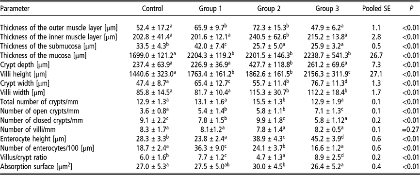
Duodenum. Several intestinal indices were significantly increased in the duodenum of birds receiving the lowest dose of the probiotic preparation (107 cfu•g−1) (Group 1), compared to the control group, namely: outer muscle thickness (26%), submucosa (25%) and mucosa thickness (30%), villi height (22%), crypt width (38%), number of open crypts (50%), total number of enterocytes (94%) and villus to crypt ratio (28%) (P < 0.01 in all cases). The number of closed crypts and enterocyte height were significantly decreased in Group 1 compared to the control group by 14% and 16% (P = 0.01 and P < 0.01), respectively (Table 2). Birds in Group 2 (receiving a higher dose of the probiotic preparation – 108 cfu•g−1) displayed significantly increased outer (38%, P < 0.01) and inner muscle thickness (19%, P = 0.03), mucosa thickness (30%, P < 0.01), crypt depth (80%, P < 0.01), villi height (29%, P < 0.01), crypt (18%, P = 0.02) and villi width (34%, P < 0.01), total crypt number (20%, P < 0.01), number of open crypts (61%, P < 0.01), enterocyte height (37%, P < 0,01), total enterocyte number (29%, P < 0,01), as well as small intestine absorptive surface (11%, P = 0.04), compared to the control group. On the contrary, submucosa thickness (23%, P < 0.01) and villus to crypt ratio (22%, P < 0.01) were significantly decreased compared to that observed in the control group, with no marked change in the number of closed crypts (Table 2).
The effect of different doses of a dietary probiotic preparation on the histomorphometry of the duodenum of female turkeys

The results are presented as mean (SD). Control – animals not receiving the probiotic preparation, Group 1 – 107 cfu•g−1, Group 2 – 108 cfu•g−1 and Group 3 – 109•cfu g−1 (cfu – a colony-forming unit).
Values with different superscripts within a single row are significantly different from one another (P < 0.05).
Interestingly, less duodenal parameters were affected by the highest dose of the probiotic preparation (109 cfu•g−1), since only mucosa thickness (32%), villi height (50%), crypt (62%) and villi width (31%), number of open crypts (97%), enterocyte height (60%) and villus to crypt ratio (48%) were significantly elevated in birds from Group 3 compared to the control group (P < 0.01 in all cases). Villi height, crypt width, number of open crypts, enterocyte height and the villus to crypt ratio in birds from Group 3 were the greatest of all the birds studied. Submucosa thickness and the number of closed crypts were significantly decreased in birds from Group 3 compared to the control group by 23% and 36% (P < 0.01), respectively (Table 2).
Birds from Group 2 had significantly thicker inner muscle layers and significantly deeper crypts compared to birds in Groups 1 (19% thicker and 88% deeper; P < 0.01) and 3 (12% thicker and 64% deeper; P = 0.02 – inner muscle layer; P < 0.01 – crypts).However, submucosa thickness was significantly decreased (by 63% (P < 0.01) in Groups 2 and 3 compared to Group 1.
Villi height was significantly increased in birds from Group 3 compared to those from Group 1 (by 22%, P < 0.01) and Group 2 (by 16%, P = 0.03). Birds from Group 1 had 17% (P = 0.04) wider crypts than those in Group 2, but 15% (P = 0.04) narrower crypts than those in Group 3. Villi width was significantly increased (P < 0.01) in birds from Groups 2 (by 41%) and 3 (by 37%) v. that observed in those from Group 1. The total number of crypts was highest (both P < 0.01) in birds from Group 2 compared to those from Groups 1 (by 18%) and 3 (by 20%). Female turkeys receiving the highest dose of the probiotic preparation (109 cfu•g−1) had more open crypts than those from Groups 1 (by 31%, P < 0.01) and 2 (by 22%, P = 0.02). Birds from Group 3 also had significantly less closed crypts (by 26%, P < 0.01) compared to those from Group 1.
The greatest enterocyte height was observed in birds from Group 3, with enterocytes that were 16% (P = 0.04) higher than those observed in birds from Group 2 and 90% (P < 0.01) higher than those observed in birds from Group 1. However, enterocyte number was greatest in birds from Group 1, with over 50% more enterocytes than that observed in birds from Group 2 (P < 0.01), and twice as much as that observed in birds from Group 3 (P < 0.01).
Jejunum
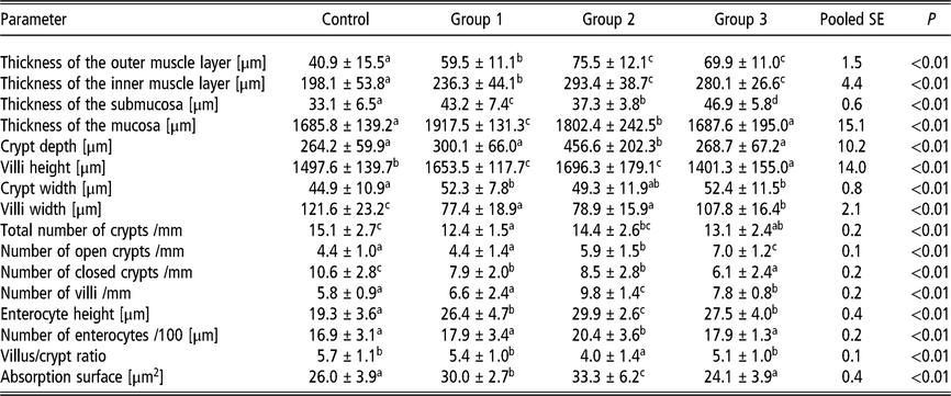
Over half of the histological parameters that were analysed in the jejunum were significantly elevated in birds receiving the lowest dose of the probiotic preparation (107 cfu•g−1) compared to the control group, namely: outer (45%) and inner muscle thickness (19%), submucosa (31%) and mucosa (14%) thickness, as well as villi height (10%), crypt width (16%), enterocyte height (37%) and small intestine absorptive surface (15%) (P < 0.01 in all cases). Villi width, total crypt number and the number of closed crypts were significantly decreased in birds from Group 1 compared to the control group, by 36%, 18% and 25% (P < 0.01), respectively (Table 3). Birds from Group 2, which received a higher dose of the probiotic preparation (108 cfu•g−1), displayed significantly greater outer (85%, P < 0.01) and inner muscle thickness (48%, P < 0.01), submucosa (13%, P = 0.03) and mucosa (7%, P = 0.04) thickness, crypt depth (73%, P < 0.01), villi height (13%, P < 0.01), number of open crypts (34%, P < 0.01), total villi number (69%, P < 0.01), total number of enterocytes (21%, P < 0.01), enterocyte height (55%, P < 0.01) and small intestine absorptive surface (28%, P < 0.01) compared to the control group. Villi width, the number of closed crypts, as well as the villus to crypt ratio were significantly decreased in birds from Group 2 (by 35%, 23% and 30%, respectively (all P < 0.01)) compared to the control group (Table 3).
The effect of different doses of a dietary probiotic preparation on the histomorphometry of the jejunum of female turkeys

The results are presented as mean (SD). Control – animals not receiving the probiotic preparation, Group 1 – 107 cfu•g−1, Group 2 – 108 cfu•g−1 and Group 3 – 109•cfu g−1 (cfu – a colony-forming unit).
Values with different superscripts within a single row are significantly different from one another (P < 0.05).
Less jejunal parameters were affected by the highest dose of the probiotic preparation (109 cfu•g−1). Only seven parameters, including outer (71%) and inner (41%) muscle layer thickness, submucosa thickness (42%), number of open crypts (59%), villi number (34%) and enterocyte height (42%) (P < 0.01 in all cases), as well as the width of the crypts (17%, P = 0.02) were all significantly elevated compared to that observed in the birds from the control group. Villi height and width, total number of crypts and the number of closed crypts were significantly decreased in birds from Group 3 compared to the control group, by 6% (P = 0.04), 11% (P = 0.02), 13% (P = 0.02) and 42% (P < 0.01), respectively (Table 3).
The thickness of the outer and inner muscle layers was significantly higher in birds from Groups 2 (by 27% and 24%, respectively, P < 0.01) and 3 (by 17% and 19%, respectively, P < 0.01 – outer muscle layer; P = 0.04 – inner muscle layer), compared to that observed in birds from Group 1. Birds from Group 3, which received the highest does of the probiotic preparation, displayed significantly thicker submucosa layers compared to those in Groups 1 (by 9%, P = 0.04) and 2 (by 26%, P < 0.01). The submucosa (16%, P < 0.01) and mucosa layers (6%, P = 0.03) were significantly thicker in birds from Group 1 compared to those from Group 2.
Birds from both Groups 1 and 2 had significantly longer villi than those in Group 3, by an average of 20% (both P < 0.01). Conversely, the villi width was significantly lower in both these groups compared to those from Group 3, by an average of 38% (P < 0.01). Crypt depth was significantly greater in birds from Group 2 compared to that observed in birds from Groups 1 (52%) and 3 (70%) (P < 0.01 in both cases). Total crypt number was significantly higher in birds from Group 2 (16%, P < 0.01) than those in Group 1. Furthermore, the number of open crypts was higher in birds from Group 2 (34%, P < 0.01) than those in Group 1; however, still lower (by 19%, P = 0.03) than that observed in Group 3. Additionally, birds from Group 3 had the lowest number of closed crypts compared to all other probiotic treatment groups, by an average of 35% (P < 0.01). Birds from Group 2 had significantly increased total villi and enterocyte numbers, as well as enterocyte height compared to birds from Groups 1 and 3, by an average of 37% (P < 0.01), 14% (P = 0.03) and 11% (P = 0.04), respectively. The absorptive surface of the jejunum was significantly higher in Groups 1 and 2 compared to the birds from Group 3, by an average of 23% (P < 0.01). Villus to crypt ratio was lowest in birds from Group 2.
Nervous innervation
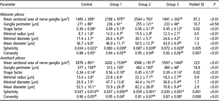
Duodenum. Duodenal Meissner and Auerbach plexus ganglia of female turkeys receiving the lowest dose of the probiotic preparation (107 cfu•g−1) had a significantly increased shape factor (by 63% and 65%, respectively, P < 0.01), minimal radius (by 53% and 78%, respectively, P < 0.01), minimal diameter (by 53% and 77%, respectively, P < 0.01), as well as mean diameter (by 25%, P = 0.02) and 41% (P < 0.01), respectively), compared to the control group (Table 4). Mean sectional area of nerve ganglia and sphericity were also significantly raised, by 135% (P < 0.01) and 47% (P = 0.02), respectively, but only in the Meissner plexus. No significant changes in mean sectional area of nerve ganglia, its perimeter as well as the sphericity and convexity indices were observed in the Auerbach plexus (Table 4). The intermediate dose of the probiotic preparation (108 cfu•g−1) exerted a similar effect on the nervous innervation of the duodenum as that observed in the control group (with no probiotic preparation administration). However, significantly higher mean sectional area of nerve ganglia was observed in birds from Group 2, in both the Meissner and Auerbach plexuses, compared to the control group (by 71% and 59%, respectively, both P < 0.01). Birds from Group 2 also displayed a significant increase (by 8%, P < 0.01) in convexity in the Meissner plexus compared to the control group. No signs of hypertrophy or hyperplasia of neurons were observed.
The effect of different doses of a dietary probiotic preparation on the morphology of duodenal Meissner and Auerbach plexuses in the small intestine of female turkeys

The results are presented as mean (SD). Control – animals not receiving probiotic preparation, Group 1 – 107 cfu•g−1, Group 2 – 108 cfu•g−1 and Group 3 – 109•cfu g−1 (cfu – a colony-forming unit).
Values with different superscripts within a single row are significantly different from one another (P < 0.05).
Fewer changes in nervous innervation of the small intestine were observed in female turkeys receiving the highest dose of the probiotic preparation (109 cfu•g−1). Significantly increased shape factor (50%), minimal radius (41%) and minimal diameter (41%) were observed in the Meissner plexus (P = 0.02 in all cases) compared to the control group (Table 4). Mean sectional area of nerve ganglia in the Auerbach plexus was almost twice as large as that observed in birds from the control group. However, no swollen ganglia were observed in any of the groups assessed.
Jejunum
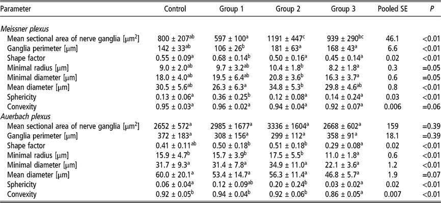
Significantly fewer changes with regard to nervous innervation were observed in jejunum compared to the duodenum (Table 5). The lowest does of the probiotic preparation (107 cfu•g−1) caused a significant increase in shape factor (by 24%, P = 0.04) and sphericity (by 177%, P < 0.01) in the Meissner plexus of birds from Group 1 compared to the control group. The intermediate dose of the probiotic preparation (108 cfu•g−1) caused a 49% (P < 0.01) increase in the mean sectional area of nerve ganglia in the Meissner plexus, compared to that observed in the control group. The highest dose of the probiotic preparation (109 cfu•g−1), surprisingly, decreased the minimal radius (by 31%, P = 0.01) and convexity (by 7%, P = 0.02) in the Auerbach plexus compared to that observed in the control group and the remaining treatment groups (Table 5).
The effect of different doses of a dietary probiotic preparation on the morphology of jejunal Meissner and Auerbach plexuses in the small intestine of female turkeys

The results are presented as mean (SD). Control – animals not receiving probiotic preparation, Group 1 – 107 cfu•g−1, Group 2 – 108 cfu•g−1 and Group 3 – 109•cfu g−1 (cfu – a colony-forming unit).
Values with different superscripts within a single row are significantly different from one another (P < 0.05).
The general effect of probiotic dose
The greatest stimulating effect was observed in animals receiving the moderate dose (108 cfu•g−1), where over 58% of parameters were significantly improved compared to the control, which corresponded to over 44% of all ameliorated parameters in the study. Moreover, duodenum (56% of ameliorated parameters) turned to be significantly (P < 0.01) more susceptible for probiotics than jejunum (31% of ameliorated parameters). Surprisingly, the weakest stimulating effect was observed in the highest used dose of probiotics (109 cfu•g−1), where only 30% of all parameters were ameliorated, especially compared to the moderate one (P < 0.01).
Discussion
Animals develop in association with complex commensal microbiota communities, which are essential for precise host organ function. Gastrointestinal microbiota, in particular, play a key role in overall host development, immunity, nutrient conversion, maintenance of host health and they serve as a barrier against exogenous pathogens (Sekirov et al., Reference Sekirov, Russell and Antunes2010; Montalban-Arques et al., Reference Montalban-Arques, De Schryver, Bossier, Gorkiewicz, Mulero, Gatlin and Galindo-Villegas2015). Alternative products and strategies that aid in the maintenance of animal gut health are constantly being investigated in order to prevent or reduce the prevalence of pathogens in livestock. This is specifically important nowadays, where the development of antibiotic resistance is on the increase (Yang et al., Reference Yang, Iji and Choct2009; Soccol et al., Reference Soccol, Porto de Souza Vandenberghe, Rigon Spier, Bianchi Pedroni Medeiros, Tiemi Yamaguishi, De Dea Lindner, Pandey and Thomaz-Soccol2013; Agboola et al., Reference Agboola, Aroniyo, Suberu and Adeyemi2014). The use of probiotics as an effective alternative approach could improve gut microbial balance, and therefore, the natural defence of livestock against pathogenic bacteria (Soccol et al., Reference Soccol, Porto de Souza Vandenberghe, Rigon Spier, Bianchi Pedroni Medeiros, Tiemi Yamaguishi, De Dea Lindner, Pandey and Thomaz-Soccol2013). The appropriate probiotic dose for use in livestock is also becoming of importance (Yang et al., Reference Yang, Iji and Choct2009).
Several aspects of host–microbiota interactions that promote functional and structural maturation of the gastrointestinal tract have been examined recently, including peristaltic motility, intestinal absorptive surface, blood supply, nutrient acquisition, microbial balance and gut integrity. The health-promoting effects of probiotics are dependent on the type and number of the different strains of bacteria or yeasts used in the probiotic preparation, especially in mixed preparations (Saarela et al., Reference Saarela, Lähteenmäki, Crittenden, Salminen and Mattila-Sandholm2002; Yang et al., Reference Yang, Iji and Choct2009; Soccol et al., Reference Soccol, Porto de Souza Vandenberghe, Rigon Spier, Bianchi Pedroni Medeiros, Tiemi Yamaguishi, De Dea Lindner, Pandey and Thomaz-Soccol2013; Agboola et al., Reference Agboola, Aroniyo, Suberu and Adeyemi2014; Rawski et al., Reference Rawski, Kieronczyk, Dlugosz, Swiatkiewicz and Józefiak2016). It must be stressed that all probiotic strains are different and their identification, characteristics and host relevance are important factors to consider when extrapolating results observed in different host species, which has to be done with great caution and makes the translation of data more complex (Saarela et al., Reference Saarela, Lähteenmäki, Crittenden, Salminen and Mattila-Sandholm2002; Chamani, Reference Chamani2016). Nevertheless, assessment of gastrointestinal tract morphology and histology is a broadly utilised technique in the investigation of the health-promoting effects of probiotic preparations, since intestinal morphology may be facilitated by the gut microflora and the digestive function of the small intestine is closely related to its mucosal architecture and villi structure (Liu et al., Reference Liu, She, Wang, Bao, Zhang, Luo, Hu, Ding, Wang and Peng2008; Rahimi et al., Reference Rahimi, Grimes, Fletcher, Oviedo and Sheldon2009; Sekirov et al., Reference Sekirov, Russell and Antunes2010; Agboola et al., Reference Agboola, Aroniyo, Suberu and Adeyemi2014; Rawski et al., Reference Rawski, Kieronczyk, Dlugosz, Swiatkiewicz and Józefiak2016). However, studies on the structural changes and development of the gastrointestinal tract in response to different doses of mixed bacteria and yeast preparations are scarce, especially in a turkey model.
Data from the detailed histomorphometric analyses performed in the present study confirm the effects of probiotics observed in previous studies. The significantly increased villi height, crypt depth, mucosa thickness and crypt and villi width observed in the present study, following administration of the probiotic preparation, are in agreement with results from previous studies (Pelicano et al., Reference Pelicano, Souza, Souza, Figueiredo, Boiago, Carvalho and Bordon2005; Awad et al., Reference Awad, Ghareeb, Abdel-Raheem and Böhm2009; Rahimi et al., Reference Rahimi, Grimes, Fletcher, Oviedo and Sheldon2009; Agboola et al., Reference Agboola, Aroniyo, Suberu and Adeyemi2014; Chamani, Reference Chamani2016; Rawski et al., Reference Rawski, Kieronczyk, Dlugosz, Swiatkiewicz and Józefiak2016). Improvement in epithelium features, villus to crypt ratio and small intestine absorptive surface were also in agreement with previous studies (de los Santos et al., Reference de los Santos, Donoghue, Farnell, Huff, Huff and Donoghue2007). However, the current study is unique in that it is the first study which compares, in detail, the effects of different doses of a mixed bacteria and yeast probiotic preparation on the development and structure of the small intestine of female turkey poults. Moreover, the results presented here also show, for the first time, an improvement in the nervous innervation of the small intestine following administration of the mixed probiotic preparation. In contrast to previous studies, more duodenal parameters were significantly improved in birds receiving the intermediate dose of the probiotic preparation compared to that observed in the jejunum, which was less susceptible to the favourable effects of the probiotic preparation (de los Santos et al., Reference de los Santos, Donoghue, Farnell, Huff, Huff and Donoghue2007).
The probiotic preparation used in the present study did not affect the body weight gain, feed intake or feed utilisation, which was also observed in previous studies (Chamani, Reference Chamani2016; Hanczakowska et al., Reference Hanczakowska, Świątkiewicz, Natonek-Wiśniewska and Okoń2017). However, Markovic et al. (Reference Markovic, Sefer, Krsti and Petrujki2009) observed an increase in body weight gain after administration of yeast additives to broilers (Markovic et al., Reference Markovic, Sefer, Krsti and Petrujki2009). Alternatively, in a study by Agboola et al. (Reference Agboola, Aroniyo, Suberu and Adeyemi2014), supplementation with probiotics increased the body weight of turkey poults; however, there was no influence on the performance of the turkeys, thus feed intake, feed conversion ratio and protein intake were not significantly changed (Agboola et al., Reference Agboola, Aroniyo, Suberu and Adeyemi2014). Intestinal bacteria have been suggested to regulate body weight through their effects on the host’s metabolic, neuroendocrine and immune functions, with some studies reporting a reduction in total body and abdominal fat (Florou-Paneri et al., Reference Florou-Paneri, Christaki, Bonos and Marcelino2013); however, these effects may largely depend on the variety of bacterial specific strains, their proportions and the doses used in these preparations. These issues still remain to be investigated. In the present study, the specific probiotic preparation was chosen based on the colonisation of a particular section of the intestine, since in the turkey duodenum, Lactobacillus sp. prevails and the rest of the bacterial strains that were used are largely present in further parts of the small intestine. The yeasts were used as a result of their nutritional content, their resistance to the acidic stomach environment and their immunomodulatory activities, which complement those of the naturally occurring microflora.
In conclusion, all doses of the probiotic preparation used in the present study exerted a beneficial effect on the histological structure of the small intestine; however, the observed effects were dose and region dependent. The structural development of particular parts of the gastrointestinal tract proved to be greatly accelerated by the 108 cfu•g−1 dose of the mixed probiotic preparation. Investigations into why the highest dose (109 cfu•g−1) of the probiotic preparation had the worst effect, as well as the mechanism of action of the probiotic preparation, will be carried out in future studies. Presented detailed data on the effects of different doses of probiotics on the histomorphometry of the small intestine, especially in turkey poults, may enrich poultry breeding practices.
Declaration of interest
None.
Ethics statement
The present study and the experimental procedures carried out were approved by the Local Ethics Committee on Animal Experimentation of the University of Life Sciences in Lublin, Poland.
Software and data repository resources
The data presented are not deposited in any official repository.
Supplementary material
To view supplementary material for this article, please visit https://doi.org/10.1017/S1751731119001149.