Introduction
Concha bullosa is the pneumatisation of the middle turbinate. It is one of the most common anatomic variations of the lateral nasal wall after agger nasi and deviated septum variations.Reference Dasar and Gokce1 Fadda et al.Reference Fadda, Rosso, Aversa, Petrelli, Ondolo and Succo2 documented a prevalence of 49.3 per cent for concha bullosa, Maru and GuptaReference Maru and Gupta3 reported a prevalence of 42.6 per cent and Bolger et al.Reference Bolger, Butzin and Parsons4 showed a prevalence of 53.6 per cent.
Extensive concha bullosa causes a crowded nose and obstruction of the middle meatus, resulting in obstruction of ventilation and the mucociliary drainage of the anterior paranasal sinuses group, including the maxillary, anterior ethmoid and frontal sinuses. Concha bullosa can also result in headaches or facial pain in the peri-orbital region due to its contact with the septum or the lateral nasal wall.Reference Kumral, Yıldırım, Cakır, Ataç, Berkiten and Saltürk5
There is no consensus on which method should be adopted or which side of the concha bullosa should be removed to promote a patient's nasal and olfactory functions. The available methods include crushing the middle turbinate, medial versus lateral laminectomy and/or transverse resection.Reference Apuhan, Yıldırım, Şimşek, Yılmaz and Yılmaz6
Middle turbinate resection may expose the patient to various complications, including increased post-operative synechia formation. Complete middle turbinate resection also increases the risk of olfactory affection and atrophic rhinitis, making it challenging to identify anatomical landmarks in revision cases.Reference Nurse and Duncavage7,Reference Soler, Hwang, Mace and Smith8
Although the classic lateral lamellectomy technique is less traumatic and more conservative than full-thickness resection,Reference Khalil, Aboel-naga, Adel Hussein and GadAllah9 it still has a risk of synechia formation, especially if carried out with functional endoscopic sinus surgery (FESS). While Canon et al. reported no synechia formation during isolated concha bullosa procedures, Dogru et al.Reference Dogru, Tuz, Uygur and Çetin10 reported synechia formation of up to 27 per cent when combined with osteo-meatal complex intervention.Reference Ahmed, Hanci and Üstün11 To prevent full-thickness middle turbinate resection complications, the submucosal resection technique, including removing prominent bone while leaving the overlying mucosa intact, was reported.Reference French and Goyal12
This study aimed to assess the difference between the lateral lamellectomy and submucosal conchoplasty techniques for managing concha bullosa regarding operating time, intra-operative applicability and turbinate stability. We also compared the post-operative endoscopic outcome of both techniques.
Patients and Methods
A prospective study of 56 patients undergoing primary FESS for chronic rhinosinusitis without nasal polyposis (CRSsNP) failed the maximal medical therapy.Reference Dubin, Liu, Lin and Senior13 Participants were gathered from the outpatient clinic at our tertiary care facility between October 2018 and April 2020.
The study included patients aged 18 years and older who were diagnosed with chronic sinusitis based on the criteria in the 2012 European Position Paper on Rhino-sinusitis and Nasal PolypsReference Fokkens, Lund, Mullol, Bachert, Alobid and Baroody14 and presented by bilateral concha bullosa (112 operated conchae). We excluded smokers and individuals with polyps, primary ciliary dyskinesia and nasal masses as well as revision cases. Sample size calculation was performed using the Kelsey formula.Reference Kelsey, Whittemore, Evans and Thompson15 A minimum of 110 operated conchae is required to detect a difference (α = 0.05, 80 per cent power) in the rate of post-operative synechia formation according to the study conducted by Semih Karaketir et al.Reference Ahmed, Hanci and Üstün11
All participants in the study provided informed written consent and the study was performed following the principles established in the Declaration of Helsinki with the hospital's ethics committee (approval code MKSU50-9-22).
The authors documented the participants’ characteristics. The nose and paranasal sinuses were scanned with high resolution computed tomography (HRCT) before surgery and evaluated using the Lund–Mackay score.Reference Hopkins, Browne, Slack, Lund and Brown16
Surgical procedure
According to the radiological score, the same surgeon (A.E.) performed primary bilateral FESS on all patients, including middle turbinate conchoplasty, middle meatal antrostomy, ethmoidectomy and frontal sinusotomy with or without sphenoidotomy.
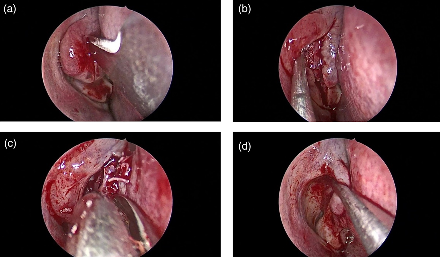
The concha bullosa was operated on at the beginning of the surgery to ease access to the middle meatus using one of two validated approaches: submucosal conchoplasty and lateral turbinectomy. The participants were divided into two equal groups of 28 patients with 56 operated conchae each. One group had submucosal conchoplasty and the other group had lateral lamellectomy. The method of distribution was determined using a computerised block randomisation system (Excel sheet randomisation). In the submucosal conchoplasty technique, the concha was locally infiltrated with adrenaline with a concentration of 1:200 000. The mucosa was incised by a blade 15 starting from the axilla posteriorly up to the attachment with the basal lamella. The mucosa covering the lateral lamella was dissected using a freer dissector. The lateral lamella was removed using a through-cut forceps then the mucosa was repositioned. The bony skeleton of the medial lamella was preserved (Figure 1).
Intra-operative endoscopic views of the right nasal cavity using a 0-angled 4-mm nasal endoscope illustrating the submucosal conchoplasty technique: (a) incision of the mucosa, (b) dissection of the mucosa, (c) removal of the lateral lamella and (d) repositioning of the mucosa.

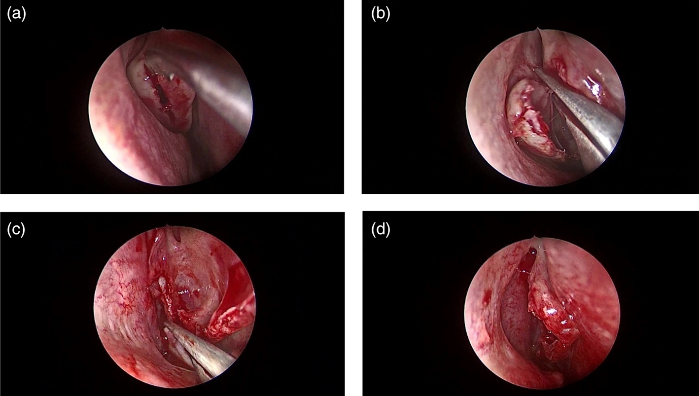
In the lateral lamellectomy technique, the concha was locally infiltrated with adrenaline with a concentration of 1:200 000. A sickle knife was used to open the concha then scissors were used to remove the lateral lamella with its covering mucosa in one block (Figure 2).
Intra-operative endoscopic views of the right nasal cavity using a 0-angled 4-mm nasal endoscope illustrating the lateral turbinectomy technique: (a) opening of the concha using a sickle knife, (b) excising the lateral lamella using scissors, (c) removal of the posterior attachment of the concha and (d) panoramic view.

Multiple intra-operative measurements were made, including the time taken to carry out the technique (calculated using the monitor's stopwatch) and the amount of bleeding (calculated using the Fromme Ordinal Scale).Reference Fromme, MacKenzie, Gould, Lund and Offord17
No middle meatus packing was applied. Systemic antibiotics and steroids (0.55 m mg/kg prednisolone) were given for 2 weeks after the surgery. The patients were instructed to use local nasal irrigations (22 m ml of budesonide 0.55 m mg/ml mixed with 2500 m ml of normal saline) twice daily for a month post-operatively. Patients were followed up at the end of each month throughout the six-month follow-up period.
The study assessed the clinical symptoms before and after the surgery through the 22-item Sino-Nasal Outcome Test (SNOT 22).Reference Hopkins, Gillett, Slack, Lund and Browne18 The patient's olfaction was tested using water as a control liquid (colourless and odourless) to exclude malingering and phenyl ethyl alcohol of 90 per cent concentration (colourless and odorous). The solutions were placed in separate bottles numbered 1 and 2. Both the examiner and the patients were unaware of the nature of the liquids within the bottles. A visual analogue score was used for olfaction assessment ranging from grade 1 to 10. The patients were instructed to assign a score of 1 if they could not smell the odour at all and a score of 10 if they could smell the odour clearly. This examination test was done pre-operatively and at the end of the sixth month post-operatively. The patient was blinded regarding the type of concha intervention in both nasal cavities.Reference Khafagy, Ghonim, Elgendy and Elzayat19
The study used two established endoscopic scores. Patients were evaluated using the Lund–Kennedy endoscopic score before the surgery as a baseline evaluation and at the first, third, and sixth months after the surgery. The Lund–Kennedy endoscopic score grades five parameters (polyps, discharge, oedema, scarring and crusting) on an ordinal scale from 0 to 2 for each side. For polyps, 0 = none, 1 = confined to middle meatus and 2 = beyond middle meatus. For discharge, 0 = none, 1 = clear and thin, and 2 = thick and purulent. For oedema, scarring and crusting, 0 = absent, 1 = mild and 2 = severe.Reference Lund and Kennedy20 To compare the endoscopic features of the frontal recess in the two techniques, peri-operative sinus endoscopy was conducted during the operation and then one, three and six months later. In peri-operative sinus endoscopy, the frontal recess or sinus was scored as patent or healthy (0), oedema or narrowed (1) or severely inflamed or infected or obstructed (2).Reference Wright and Agrawal21 Furthermore, the study evaluated the state of the middle turbinate six months post-operatively and categorised it as healthy (0), showing partial lateralisation (1) or showing severe lateralisation (2) using the peri-operative sinus endoscopy score to evaluate and compare the two methods used in the study.
The study was single-blinded so the patients were unaware of which technique was used. However, the investigator could not be blinded because of the nature of the technique, therefore two authors (A.E. and S.E.) conducted the post-operative endoscopic assessment in two separate settings.
The SPSS 23 program was used to analyse the results of this study. After applying the normal distribution tests, the quantitative data were analysed using the t-test of significance or the Wilcoxon signed ranks test. The Lund–Kennedy endoscopic scores were compared using paired and unpaired t-tests. The study compared olfactory function before and after surgery using the Wilcoxon signed ranks test. The qualitative information was analysed percentages using the Fisher test. The Fisher test was used to compare the degree of intra-operative bleeding and post-operative synechia of both groups.
Results
The study included 56 patients (25 males, 31 females) aged 34.68 ± 12.48 years old between October 2019 and April 2021. No major peri-operative complications were documented for either technique.
By the end of the sixth month after the surgical procedures, the overall SNOT 22 score had significantly improved (67.48 ± 12.589 pre-operatively vs 29.66 ± 15.687 post-operatively, p = 0.001*).
Both groups of patients showed significant improvement in the olfaction score post-operatively (p = 0.001*). There was no significant difference between the groups in the olfaction score net change (post-operative to pre-operative, 4.14 ± 1.268 vs 3.57 ± 1.372, p = 0.111 for the submucosal conchoplasty group and the lateral lamellectomy group, respectively).
The operative time (minutes) required was significantly higher in the submucosal conchoplasty group (11.86 ± 2.075 vs 6.29 ± 1.703, p = 0.001*).
The two groups showed no significant difference regarding the amount of intra-operative bleeding in both techniques according to the Fromme Ordinal Scale (Fisher exact test, 6.111; p = 0.086).
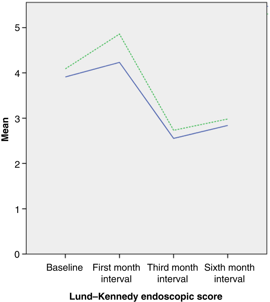
Considering the Lund–Kennedy endoscopic score, the differences observed between the two groups throughout the follow-up period were variable (Figure 3).
Lund–Kennedy endoscopic score at different follow-up intervals. *Significant difference <0.05. Dashed green line = lateral turbinectomy; solid blue line = submucosal conchoplasty.

There was no detectable distinction between the study groups at the baseline assessment time (3.91 ± 0.793 vs 4.09 ± 0.695, p = 0.208, for the submucosal conchoplasty and lateral turbinectomy sides, respectively).
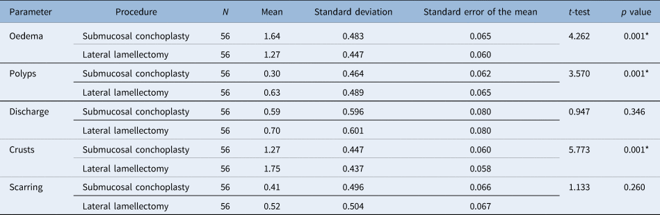
It was significantly higher in the lateral lamellectomy group at the first month interval (4.23 ± 1.191 vs 4.86 ± 1.034, p = 0.004*) (Table 1).
Detailed assessment of the post-operative Lund–Kennedy endoscopic score for the first month interval follow up

* Significant difference <0.05 according to the independent sample t-test
At the three- and six-month intervals, however, the groups did not differ significantly from each other (third month, 2.55 ± 0.952 vs 2.73 ± 0.981, p = 0.331; sixth month, 2.84 ± 1.398 vs 2.98 ± 1.368, p = 0.586) for the submucosal conchoplasty and lateral turbinectomy sides, respectively.
At the six-month interval, the Lund–Kennedy endoscopic scores had increased significantly from their baseline scores (p = 0.001*) for both groups (Table 2).
Level of significance for the Lund–Kennedy endoscopic score and peri-operative sinus endoscopy score changes between the two groups

* Significant difference <0.05 according to the independent sample t-test
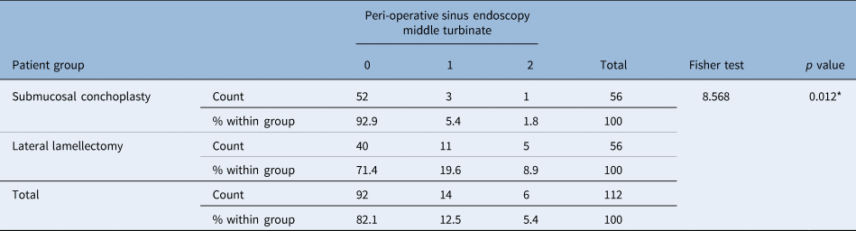
Regarding the middle turbinate peri-operative sinus endoscopy score in the sixth month, there was a statistically significant difference between the groups, with the lateral lamellectomy group showing more synechia formation (Fisher test, 8.568; degrees of freedom, 2; p = 0.012*) (Table 3).
Differences between the groups for post-operative synechia formation according to the peri-operative sinus endoscopy score at the six-month interval

* Significant difference <0.05 according to the Fisher test
Discussion
Concha bullosa is a common anatomical variant, but a large concha may contribute to many nasal problems, including nasal obstruction and sinusitis. Extensive concha bullosa narrows the middle meatus and hinders endoscopic accessibility during surgery. It is therefore essential to properly manage this anatomical variation at the beginning of the operation to have good access to the middle meatus. The turbinate mucosa plays a vital role in nasal function, such as humidification, temperature control, sensation of airflow and olfactory perception.Reference Nurse and Duncavage7
Although endoscopic lateral lamellectomy is the standard modality for its management, post-operative adhesions with subsequent failure of FESS may occur. Sigston et al. designed a modification to the partial lateral turbinectomy technique to decrease the post-operative exposed raw area by using the concha's lateral posterior pedicled mucosal flap after extracting its bony lamina (the submucosal conchoplasty technique).Reference Sigston, Iseli and Iseli22
Submucosal conchoplasty has the advantage of preserving the mucosa with its whole function. Although there was no significant difference between the two techniques regarding intra-operative bleeding, we observed that the concha's feeding blood supply of the concha bollusa was more liable to be injured during the excision of the lateral lamella of the middle turbinate at its posterior stump in the traditional technique. Fortunately, this bleeding can be easily controlled by bipolar cauterisation of the bleeding stump. This injury was less liable to occur with the submucosal turbinectomy technique because it included submucosal resection of the bony lamella with no mucosal tearing. The literature has reported an increase in blood loss during and after middle turbinate excision.Reference Nyquist, Anand, Brown, Singh, Tabaee and Schwartz23 It has also been reported that the transection of the arterial supply along the posterior portion of the middle turbinate could lead to bleeding during full-thickness middle turbinate resection.Reference Lee, Kim, Kim, Son, Kim and Cho24
We observed that the bony surface of the lateral lamella was rough with adherent mucosa, which made the mucosal dissection challenging and time-consuming. Although the time difference between the techniques was statistically significant, it was practically unimportant relative to the time needed for the whole surgery. Another disadvantage of the submucosal technique is that the repositioned mucosa of the lateral lamella may be repeatedly injured by the suction device during the operation or accidentally deflected during the application of the middle meatus pack.
The lateral turbinectomy group had higher crust and reactionary polyp formation rates than the submucosal conchoplasty group at the first month follow-up interval. Because of the increased surface area of the exposed bone, crusts were found primarily along the line of turbinate resection. This bone exposure induces more granulations and prolongs the time needed for complete healing. However, there was significant post-operative oedema on the side with submucosal conchoplasty compared to lateral turbinectomy on top of the preserved repositioned mucosal surface of the middle turbinate. Fortunately, these findings were temporary since they were eliminated once the recovery process was complete, with no unfavourable consequences.
The lateral lamellectomy group showed an increasing rate of synechia formation during the follow-up period and the peri-operative sinus endoscopy score also increased. The rate at which synechia formed between the middle turbinate and the lateral nasal wall was significantly higher in the lateral lamellectomy group because of the increased surface area exposure of the opposing raw tissues. The lateral lamellectomy group showed a post-operative synechia rate of 28.6 per cent, while the rate for the submucosal turbinectomy group was 7.1 per cent.
According to peri-operative sinus endoscopy at the final assessment, only one case of the submucosal turbinectomy group showed grade 2 synechia formation, restricted to one side only. This was due to the accidental deflection of the repositioned mucosa during merocel pack application at the end of the operation. This patient showed persistent symptoms and was a candidate for revision surgery. Regarding the lateral lamellectomy group, four patients showed complete middle meatus obliteration (one patient was bilaterally presented and three patients were unilateral). Only two of them showed persistent symptoms and needed revision surgery.
• Concha bullosa is the pneumatisation of the middle turbinate, one of the most common anatomic variations of the lateral nasal wall
• There is no consensus on which method is adopted to which side of the concha bullosa should be removed to promote nasal and olfactory functions
• A prospective, randomised study was conducted of 56 patients with bilateral concha bullosa who were undergoing primary functional endoscopic sinus surgery
• The patients were divided into two equal groups, with one group of patients being treated using the submucosal conchoplasty technique and the other group underwent the lateral lamellectomy
• Submucosal conchoplasty was significantly more time-consuming than lateral lamellectomy technique, which, according to the peri-operative sinus endoscopy score, showed a higher rate of synechia formation in the middle meatus
• Submucosal conchoplasty is a proper technique for managing concha bullosa with better post-operative endoscopic outcomes
One limitation of the study is that the net endoscopic scores combine the concha and ethmoid sinus interventions. Future studies should include the concha bollusa surgical procedure as a separate endoscopic intervention from other sinus surgical procedures.
Conclusion
Submucosal conchoplasty is a proper technique for managing concha bullosa with better post-operative endoscopic outcomes and no significant peri-operative co-morbidity.
Competing interests
None declared