Introduction
Wegener's granulomatosis is a primary systemic vasculitis characterised by granulomatous inflammation in the soft tissue as well as fibrinoid vasculitis in the small to medium sized vessels of the body. It was first described as a separate entity from periarteritis nodosa by Friderich Wegener in 1936 with the description of three cases he dissected.Reference Wegener and Wegener1, Reference Wegener2 It has recently been agreed by the rheumatology and vasculitis entities that the name of the condition should be changed to granulomatosis with polyangiitis (Wegener's), abbreviated to ‘GPA’.Reference Falk, Gross, Guillevin, Hoffman, Jayne and Jennette3 The new name will be used in the rest of the review.
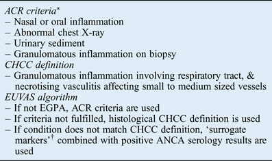
The condition affects approximately 10 people per million per year in the UK. It typically affects the ENT, renal and lower respiratory systems. It can occur at any age, with a peak incidence at 45 years of age. There is no simple diagnostic test, but clinical and pathological criteria exist to arrive at a diagnosis. These include the American College of Rheumatism classification,Reference Leavitt, Fauci, Bloch, Michel, Hunder and Arend4 the Chapel Hill Consensus Conference definitionsReference Jennette, Falk, Andrassy, Bacon, Churg and Gross5 and, most recently, an algorithm designed by the European Vasculitis Study Group.Reference Watts, Lane, Hanslik, Hauser, Hellmich and Koldingsnes6 This latter algorithm combines previous classification systems and adds serum anti-neutrophil cytoplasmic antibody (ANCA) levels to aid the classification of primary systemic vasculitides. The presence of ANCA provides support for the diagnosis of vasculitis although their absence does not exclude it. The target antigens associated with vasculitis are proteinase 3 (PR3-ANCA), commonly associated with granulomatosis with polyangiitis (Wegener's), and myeloperoxidase (MPO-ANCA), commonly associated with microscopic polyangiitis and to a lesser extent with Churg–Strauss syndrome.Reference Falk and Jennette7–Reference Sinico, Di Toma, Maggiore, Bottero, Radice and Tosoni9Table I describes all three sets of criteria for Wegener's granulomatosis.
AIDS to diagnosis of granulomatosis with polyangiitis

*Two criteria are required for diagnosis. †Markers suggestive of granulomatosis with polyangiitis in the absence of a biopsy (e.g. bronchial stenosis, nasal crusting, retro-orbital disease, nasal deformities). ACR = American College of Rheumatology; CHCC = Chapel Hill Consensus Conference; EUVAS = European Vasculitis Study Group; EGPA = Eosinophilic granulomatasis with polyangiitis; ANCA = anti-neutrophil cytoplasmic antibody
This review will concentrate on granulomatosis with polyangiitis (Wegener's), although there is a recognised overlap with other primary systemic vasculitides that are associated with ANCAs, namely microscopic polyangiitis and Churg–Strauss syndrome. Microscopic polyangiitis is predominantly a vasculitic disease that affects the kidneys, sparing the ENT system.Reference Guillevin, Durand-Gasselin, Cevallos, Gayraud, Lhote and Callard10 Churg–Strauss syndrome was also termed allergic angiitis as it is associated with eosinophilia in the tissues and serum, and often presents with asthma and nasal polyposis. Vasculitis affecting the lungs and kidneys, and the presence of mononeuritis multiplex, are serious and late complications of the disease.Reference Lanham, Elkon, Pusey and Hughes11
This article aimed to review the common presentation of ENT disease in granulomatosis with polyangiitis (Wegener's) and the management of the condition as illustrated by a representative case.
Case report
A 26-year-old lady presented to the ENT clinic in September 2007 with a 9-month history of flu-like symptoms, including generalised myalgia and arthralgia, purulent nasal discharge, and pain across the nasal bridge and the front teeth severe enough to keep the patient awake. The referral to the ENT clinic was triggered by an episode of ‘tonsillitis’ associated with vertigo. Prior to her referral, she had been treated with several courses of antibiotics, with limited effect.
When seen at the ENT clinic, the patient was found to have a 50 dB conductive hearing loss. Endoscopy and computed tomography (CT) of her sinuses showed nasal crusting, but no sinus involvement. She was initially treated conservatively with inhalations. After two months, a grommet was inserted into her right ear, with some relief of her symptoms. However, her symptoms returned within a month and in July 2007 she was re-referred to the ENT clinic. By this time, nine months had elapsed and her symptoms were similar to the initial ones, with the addition of mild nasal saddling and larger crusts (Figure 1c). She was previously fit and healthy apart from mild depression and polycystic ovary syndrome.
Endoscopic views showing: (a) left middle-ear granuloma during disease activity; (b) left retracted tympanic membrane following treatment; (c) ‘bombsite’ appearance of nose during disease activity, with bloody crusts and abnormal-looking mucosa; (d) nasal septal perforation and damage to lateral nasal wall; (e) irregular-looking subglottic stenosis in keeping with disease activity; and (f) fibrosed subglottic stenosis during remission.
The patient was seen at the ENT clinic and a biopsy and serology tests were organised. The biopsy showed acute on chronic inflammation with stratified squamous epithelium on the surface of general oedema. There were also neutrophils around blood vessels, but no fibrinoid scarring. Immunofluorescence testing for anti-neutrophil cytoplasmic antibodies (ANCA) was ‘weakly positive’, and enzyme-linked immunosorbent assay testing showed a PR3-ANCA level of 5 u/ml (normal range is 0–6 u/ml). C-reactive protein levels and erythrocyte sedimentation rate were mildly raised at 24 mg/l and 7 mm/hour respectively, and renal function was normal. The patient was then referred to the vasculitis unit at our tertiary centre; urine dipstick values were normal, and a chest X-ray closely followed by a chest CT showed normal mediastinum and clear lung fields.
Initial treatment consisted of methotrexate and high-dose oral corticosteroids. Unfortunately, the patient did not respond adequately and the disease progressed, causing significant subglottic, right upper lobe and bronchus intermedius stenosis (Figure 1e). The disease in the ears worsened, causing significant purulent discharge, hearing loss and transient facial nerve palsy. The nasal crusting and pain over her nose, controlled using morphine sulphate, did not improve. In January 2008, the treatment was changed to cyclophosphamide, and by April 2008, after six cycles, the only significant symptoms were dyspnoea and lethargy. Azathioprine has been used since that time for maintenance treatment.
On trying to reduce the glucocorticoid dose down from 5 mg, the patient developed otorrhoea and widespread arthralgia, and ANCA immunofluorescence results became positive (PR3-ANCA levels were 6 u/ml). All symptoms improved with a short course of 10 mg prednisolone.
Thirty-one months after the initial presentation, the patient remains in remission, but with significant damage to her hearing and middle ear (Figure 1b), a nasal septal perforation (Figure 1d) resulting in a saddle nose deformity (Figure 2a) and endobronchial fibrosed stenoses (Figure 1f). In an attempt to ease the hearing loss, myringotomies were performed and, because of adhesions, it was only possible to insert a grommet in the right ear with difficulty. This did not improve her hearing and so at present the patient wears hearing aids, having worn a body-worn aid for the first six months of her condition. The nasal crusting has settled almost completely and is managed by regular nasal douching. The patient underwent a limited correction of the saddle nose deformity using rib cartilage without re-activation of the disease and with good results (Figure 2b). Finally, the airway disease has been managed by repeated dilatations using bougies for the subglottis, and a combination of bougies and balloon dilatations for the lower airway. The last two interventions were combined with mitomycin C, and after three dilatations between May 2008 and September 2009, the patient's exercise tolerance has improved considerably. However, the treatment is ongoing and the patient had three staged dilatations to the right bronchus intermedius in 2011. Figure 3 demonstrates the change in flow–volume loops over time. The shuttle test also demonstrated an increase in the patient's walking distance from 460 m to 580 m, and the latest measurement for predicted forced expiratory volume in 1 second was 65 per cent.
Side views of the patient showing the saddle nose deformity (a), and following correction of the deformity (b). (Note: this young patient is also wearing hearing aids.) Published with patient's permission.
Flow–volume loops at first diagnosis of subglottic stenosis (in 2008) (a), and after treatment with immunosuppression and dilatations (in 2011) (b).
Discussion
Presentation
Granulomatosis with polyangiitis (Wegener's) can present with disease in any organ system and often with non-specific symptoms. A common feature, as illustrated by the case above, is a long prodromal course of flu-like symptoms (i.e. non-specific upper respiratory symptoms associated with lethargy and arthralgia).Reference Drinias and Florentzson12–Reference Takala, Kautiainen, Malmberg and Leirisalo-Repo17 Patients with disease limited to the head and neck tend to have their diagnosis made later than those with systemic disease, and often see an ENT surgeon at their first visit to hospital.Reference Srouji, Andrews, Edwards and Lund18–Reference Hoffman, Kerr, Leavitt, Hallahan, Lebovics and Travis20
Ear, nose and throat involvement in granulomatosis with polyangiitis (Wegener's) is associated with improved survival,Reference Bligny, Mahr, Toumelin, Mouthon and Guillevin21 but it is also associated with increased refractory disease (unresponsive to optimum treatment) and a relapsing course.Reference Walsh, Flossman, Berden, Westman, Manns and Jayne22, Reference Sproson, Jones, Al-Deiri and Lanyon23 Disease activity leads to fibrosis and/or necrosis, and in turn to deformities, such as saddle nose or impaired hearing. In a recent patient-reported outcome survey, the burden of ENT and constitutional symptoms was rated higher than severe manifestations such as dialysis and oxygen dependency.Reference Herlyn, Seo, Hellmich, Reimer and Merkel24
Disease in the ears
Otological symptoms are non-specific, with the exception of acute sensorineural hearing loss, which usually triggers a battery of tests including tests for autoimmune antibodies. In the acute phase, patients usually present with symptoms of acute otitis media or otitis media with effusion, and there may or may not be constitutional symptoms.Reference Illum and Thorling25–Reference Kempf28 In consequence, the most common type of hearing loss is mixed or conductive.Reference D'Cruz, Baguley, Asherson and Hughes13, Reference Sproson, Jones, Al-Deiri and Lanyon23, Reference Reinhold-Keller, Beuge, Latza, de Groot, Rudert and Nolle29, Reference Fauci, Haynes, Katz and Wolff30 In many of the reports it is also asymmetrical, but without a distinctive pattern.Reference Takagi, Nakamaru, Maguchi, Furuta and Fukuda26, Reference Kornblut, Wolff and Fauci27, Reference Ferri, Armato, Capuzzo, Cavaleri and Ianniello31
In the acute stages of granulomatosis with polyangiitis (Wegener's), conductive hearing loss is due to granulation tissue in the middle ear, or otitis media with effusion secondary to nasopharyngeal disease.Reference D'Cruz, Baguley, Asherson and Hughes13 If the patient is in remission, the differential diagnosis includes active or inactive chronic otitis media and the corresponding sequelae. Sensorineural hearing loss is typically bilateral with a sub-acute course, and is associated with tinnitus. Vertigo is not common in granulomatosis with polyangiitis (Wegener's) patients.Reference Pereira, Ayres do Amaral, Mansur Szajubok, Loduca Lima and Chahade32 The disease process is thought to be immunomediated, causing direct damage to the cochlea or as a result of vasculitis in the blood supply of the vestibulocochlear nerve.Reference Burlacoff and Wong33
It is challenging to distinguish active disease from infection using a patient's history alone as granulation in the middle ear and otorrhoea associated with tympanic perforations can be associated with vasculitis and infections. The main distinguishing feature is that vasculitis does not respond to conventional treatment and the pain may be persistent with few signs.Reference Ferri, Armato, Capuzzo, Cavaleri and Ianniello31, Reference Bucolo, Torre, Montemagno and Beatrice34–Reference Atula, Honkanen, Tarkkanen and Jero42 During follow up, other systemic features such as arthralgia should be considered, and the patient's own ability to distinguish previous symptoms that led to diagnosis or flare-ups.
One case series found a high incidence of sensorineural hearing loss, at 48 per cent, which is slightly higher than earlier reports.Reference Murty43 In that report, the incidence for any hearing loss was 56 per cent, but this varies widely in the literature, at 19–61 per cent.Reference Hoffman, Kerr, Leavitt, Hallahan, Lebovics and Travis20 In another study, 9 of 20 patients had sensorineural hearing loss, 8 had mixed hearing loss and 3 had conductive hearing loss. Seventeen of the 20 patients had bilateral hearing loss, but no comment was made about symmetry.Reference Bakthavachalam, Driver, Cox, Spiegel, Grundfast and Merkel44 The authors of that study recommend that all patients have an audiogram as part of their regular initial tests, particularly when assessing disease activity.Reference Bakthavachalam, Driver, Cox, Spiegel, Grundfast and Merkel44 Another case series reports findings in agreement with an increased proportion of sensorineural hearing loss (38 per cent for sensorineural hearing loss alone and 92 per cent if mixed hearing loss is included).Reference Kempf28 In 13 patients from the case reports reviewed, the distribution of pure sensorineural and conductive hearing loss was equal, while mixed hearing loss represented the higher proportion (10 out of 22, 45 per cent).Reference Ferri, Armato, Capuzzo, Cavaleri and Ianniello31, Reference Bucolo, Torre, Montemagno and Beatrice34–Reference Atula, Honkanen, Tarkkanen and Jero42 In addition, 70 per cent of the cases reported were female, while in the case series reported by Bakthavachalam et al., the sex distribution was equal.Reference Bakthavachalam, Driver, Cox, Spiegel, Grundfast and Merkel44
Disease in the nose
Presenting symptoms most often affect the nasal cavity and include non-specific symptoms, such as rhinorrhoea and nasal obstruction, although chronic rhinosinusitis symptoms can also occur.Reference Jennings, Jones, Dugar, Powell and Lowe45 Persistence of symptoms and signs despite treatment, and the presence of blood or crusts, should raise suspicion of vasculitis.Reference D'Cruz, Baguley, Asherson and Hughes13, Reference Kornblut, Wolff, deFries and Fauci46, Reference Stone47 Saddle nose has a prevalence of 12–28 per cent and usually presents later.Reference D'Cruz, Baguley, Asherson and Hughes13, Reference Hoffman, Kerr, Leavitt, Hallahan, Lebovics and Travis20, Reference Reinhold-Keller, Beuge, Latza, de Groot, Rudert and Nolle29, Reference Fauci, Haynes, Katz and Wolff30 Solid and/or blood-stained discharge are usually associated with active disease.Reference Pereira, Ayres do Amaral, Mansur Szajubok, Loduca Lima and Chahade32 Bone erosion through the palate or maxillary sinus is more consistent with conditions such as natural killer T-cell lymphoma (previously known as polymorphic reticulosis).Reference Fauci, Haynes, Katz and Wolff30
Examination of the nose consists of external appearance assessment for saddle nose deformities, alar collapse or epiphora. An endoscope is not only essential to make the diagnosis,Reference Yucel, Keles, Ozturk, Solmaz and Deger48 but also to assess disease activity and the extent of damage if present.Reference Sproson, Jones, Al-Deiri and Lanyon23 First, the mucosal lining is examined for evidence of rhinitis, friable mucosa, purple discolouration, granulomata and ulcers (often denoted by nasal crusting). At the same time, the presence and nature of discharge is noted. Then the anatomy is assessed, in particular checking whether the septum, turbinates and maxillary antrum wall are intact, or whether there are adhesions or stenoses.Reference McDonald, DeRemee, Kern and Harrison16, Reference Hoffman, Kerr, Leavitt, Hallahan, Lebovics and Travis20, Reference Sproson, Jones, Al-Deiri and Lanyon23, Reference Fauci, Haynes, Katz and Wolff30, Reference Pereira, Ayres do Amaral, Mansur Szajubok, Loduca Lima and Chahade32, Reference Kornblut, Wolff, deFries and Fauci49 Solid bone such as the hard palate or the anterior maxillary wall are rarely, if ever, eroded in granulomatosis with polyangiitis (Wegener's).Reference Fauci, Haynes, Katz and Wolff30
The differential diagnoses in the early phase of the disease include allergic or intrinsic rhinitis, or other vasculitides (e.g. Churg–Strauss syndrome or polyarteritis nodosa). As the disease progresses, granulomatous conditions feature more prominently in the differential diagnoses: tuberculosis, fungal rhinosinusitis, natural killer T-cell lymphoma and sarcoidosis are the main differential diagnoses.Reference Pereira, Ayres do Amaral, Mansur Szajubok, Loduca Lima and Chahade32
Disease in the throat
Isolated involvement of the pharynx and larynx is uncommon; painful mouth ulcers are the most common symptoms, followed by hoarseness.Reference D'Cruz, Baguley, Asherson and Hughes13, Reference Kornblut, Wolff, deFries and Fauci49 Airway obstruction secondary to subglottic stenosis tends to be a late complication. This occurs in younger patients, particularly children, and often occurs independently of disease activity.Reference D'Cruz, Baguley, Asherson and Hughes13, Reference Kornblut, Wolff, deFries and Fauci49–Reference Rottem, Fauci, Hallahan, Kerr, Lebovics and Leavitt51 Rarely, gingivitis presents with the classical appearance of strawberry gums (white-dotted, inflamed gums).Reference Cohen and Meltzer52 There are several isolated case reports of granulomatosis with polyangiitis (Wegener's) presenting with salivary gland involvement.Reference Berge, Niederhagen, von Lindern, Appel and Reich53–Reference Liu, Vlantis and Lee57 The differential diagnoses for salivary gland disease include Sjögren's syndrome and sialadenitis, although there are usually other ENT or endobronchial pathologies to aid the diagnosis of granulomatosis with polyangiitis (Wegener's).Reference Liu, Vlantis and Lee57
Stenosis is the most common pathology of the larynx in granulomatosis with polyangiitis (Wegener's) patients (7 per cent prevalenceReference Fauci, Haynes, Katz and Wolff30). The differential diagnoses of granulomata or stenoses involving the larynx are extensive and include trauma, vocal fold polyps or nodules, carcinoma, tuberculosis, amyloidosis, and lymphoma.Reference Pereira, Ayres do Amaral, Mansur Szajubok, Loduca Lima and Chahade32 With regard to chronic stenoses, conditions that present a bigger challenge to the clinician when assessing subglottic stenosis are sarcoidosis and relapsing polychondritis.Reference Hoare, Jayne, Rhys Evans, Croft and Howard58 However, as mentioned above, often the diagnosis of granulomatosis with polyangiitis (Wegener's) is made before subglottic or endobronchial disease manifests itself.
The presence of stenosis in the larynx and upper trachea is assessed using a flexible endoscope. This allows one to keep a sequential record of the images for discussion and comparison. Endobronchial and tracheal disease can be assessed and treated using sedation and flexible bronchoscopy. The management of airway stenosis is shared between ENT surgeons and respiratory physicians.
Other ENT-related disease sites
Retro-orbital disease, hypertrophic pachymeningitis and pituitary involvement are less common in granulomatosis with polyangiitis (Wegener's). Such disease is thought to occur as a result of contiguous invasion from sinonasal granulomatosis with polyangiitis (Wegener's). In an Italian longitudinal study, these three disease entities appeared before the systemic symptoms, but at the same time as the ENT symptoms of sinonasal disease.Reference Di Comite, Bonavida, Bozzolo, Bianchi, Ciboddo and Tresoldi59
Ocular involvement is present in up to 58 per cent of patients with granulomatosis with polyangiitis (Wegener's), and may represent the first sign of the condition.Reference Harper, Letko, Samson, Zafirakis, Sangwan and Nguyen60 Orbital disease tends to have a relapsing and/or refractory course. Conventional cytotoxic therapy is used to treat the condition as systemic disease must be controlled in order for retro-orbital disease to respond to treatment.Reference Robin, Schanzlin, Meisler, deLuise and Clough61
Hypertrophic pachymeningitis can be localised or present as diffuse thickening of the dura mater. The prevalence of pachymeningitis in granulomatosis with polyangiitis (Wegener's) is quoted as 1 per cent. Patients with the condition typically present with headaches and associated cranial nerve palsies.Reference Dorr, Elitok, Dieste, Vogel, Schneider and Wuerfel62 Pachymeningitis is usually associated with other granulomatosis with polyangiitis (Wegener's) signs affecting the airways, such as saddle nose or lung nodules.Reference Dorr, Elitok, Dieste, Vogel, Schneider and Wuerfel62–Reference Aries, Reinhold-Keller and Gross64 Although initially steroid-responsive, clinical manifestations frequently relapse, requiring the addition of immunosuppressive agents.Reference Aries, Reinhold-Keller and Gross64
Pituitary involvement affects approximately 1 per cent of patients with granulomatosis with polyangiitis (Wegener's) long-term. The most common manifestations of pituitary involvement are central diabetes insipidus and hypopituitarism,Reference Stone47, Reference Haynes and Fauci65 and 95 per cent have concomitant ENT symptoms.Reference Yong, Li, Amato, Mahadevan, Phillips and Coates66 The imaging modality of choice is a magnetic resonance imaging scan. The pituitary gland often does not return to normal function after disease control.Reference Haynes and Fauci65–Reference Lohr, Ryan, Toohill and Anderson67
Investigations
As mentioned above, patients presenting with non-specific features of common conditions, who do not respond to conventional treatment, should be investigated for vasculitis. As well as a detailed ENT assessment, examination of the hands for splinter haemorrhages or the skin for rashes may be appropriate. In addition, urinalysis should be carried out to exclude renal involvement. A chest X-ray is useful even in the absence of symptoms as nodules in the lungs are often silent.Reference Hoffman, Kerr, Leavitt, Hallahan, Lebovics and Travis20, Reference Fauci, Haynes, Katz and Wolff30, Reference McLaren, McRorie and Luqmani68, Reference Anderson, Coles, Crane, Douglas, Gibbs and Geddes69
Only these tests together with biopsies were used before the discovery of anti-neutrophil cytoplasmic antibody (ANCA), which has greatly facilitated the diagnosis of GPA.Reference van der Woude, Rasmussen, Lobatto, Wiik, Permin and van Es70 The typical finding on indirect immunofluorescence in granulomatosis with polyangiitis (Wegener's) is ANCA with a cytoplasmic distribution that binds to proteinase 3 (PR3-ANCA) on enzyme-linked immunosorbent assay testing. ANCA serology is positive in 73–94 per cent of cases.Reference Takala, Kautiainen, Malmberg and Leirisalo-Repo17, Reference Jennings, Jones, Dugar, Powell and Lowe45, Reference Stone47 Sensitivity and specificity is greatly enhanced when using both tests in conjunction.Reference Savige, Dimech, Fritzler, Goeken, Hagen and Jennette71 Other initial blood tests include full blood count to assess for eosinophilia or anaemia, and tests of non-specific inflammatory markers (erythrocyte sedimentation rate and C-reactive protein) and creatinine levels.Reference McLaren, McRorie and Luqmani68
Since the discovery of ANCA and improvements in the safety of renal biopsies, head and neck biopsies have been largely superseded. However, for the 6–26 per cent of patients with initially negative ANCA serology results and localised disease to the airway, they are still relevant. Current immunohistochemistry techniques have made it possible to exclude malignancies and infections with relative ease.Reference Andrassy and Rasmussen72, Reference Raynaud, Garrel, Rigau, Poizat, Vic and Cartier73 Nevertheless, the diagnosis of granulomatosis with polyangiitis (Wegener's) or other chronic inflammatory conditions remains difficult to establish with certainty. The reported biopsy results were compatible with a diagnosis of granulomatosis with polyangiitis (Wegener's) in 25–57 per cent of cases.Reference Jennings, Jones, Dugar, Powell and Lowe45, Reference Anderson, Coles, Crane, Douglas, Gibbs and Geddes69, Reference Raynaud, Garrel, Rigau, Poizat, Vic and Cartier73–Reference Rasmussen75 However, if only true positives are considered, the range of positive biopsy results is 16–18 per cent.Reference Raynaud, Garrel, Rigau, Poizat, Vic and Cartier73, Reference Devaney, Travis, Hoffman, Leavitt, Lebovics and Fauci74 Disease classification is largely based on a system proposed by Rasmussen et al. Specifically, the presence of small vessel vasculitis, giant cells (Langhans' or foreign body types) and epithelioid granulomas is diagnostic of granulomatosis with polyangiitis (Wegener's); the presence of two features is ‘compatible, but not diagnostic of granulomatosis with polyangiitis (Wegener's)’; and the presence of only one feature is considered ‘non-specific inflammation’.Reference Rasmussen, Petersen, Jensen and Andersen76 In terms of technique, most reports agree that larger biopsies are better (at least 5 mm). In addition, biopsies from actual lesions, particularly the paranasal sinuses, have a better diagnostic yield. In order to best achieve this, the patient should be under general anaesthesia.Reference McDonald, DeRemee, Kern and Harrison16, Reference Anderson, Coles, Crane, Douglas, Gibbs and Geddes69, Reference Raynaud, Garrel, Rigau, Poizat, Vic and Cartier73, Reference Del Buono and Flint77, Reference Diamantopoulos and Jones78 If suspicion persists despite negative tests, these should be repeated after a period of close observation of the patient.Reference Anderson, Coles, Crane, Douglas, Gibbs and Geddes69
Imaging of the head and neck is not considered helpful to make a diagnosis of granulomatosis with polyangiitis (Wegener's), but may be useful for excluding other conditions such as malignancy. Initial appearances in CT of the paranasal sinuses are similar to those of chronic rhinosinusitis, with opacification of the maxillary sinuses and bone thickening. Later, nasal septum and lateral nasal wall erosion and bone demineralisation occur. The main differential diagnoses of these appearances are cocaine abuse and trauma (iatrogenic or accidental). Sclerosing osteitis and osteoneogenesis are typical of later appearances in granulomatosis with polyangiitis (Wegener's).Reference Benoudiba, Marsot, Hadj, Cabanne, Bobin and Lasjaunias79–Reference Lloyd, Lund, Beale and Howard83 Sclerosing osteitis becomes apparent initially by an irregular line of corticated bone parallel to the lateral wall of the maxillary sinus; as the disease progresses, the maxillary sinus may become obliterated with abnormal bone formation.Reference Lohrmann, Uhl, Warnatz, Kotter, Ghanem and Langer82, Reference Lloyd, Lund, Beale and Howard83 Magnetic resonance imaging is only performed if there is suspicion of soft tissue mass, such as retro-orbital masses. The appearances are hyperintense on T2 sequencing during disease activity, but hypointense when disease is inactive.Reference Allen and Harvey80, Reference Silvera, Vignaux and Legmann81 Lloyd et al. demonstrated the fat-filled double line of cortical bone in the maxillary sinus using a T1 sequence with fat suppression (the hyperintense line between the cortical bones disappears).Reference Lloyd, Lund, Beale and Howard83
• A high index of suspicion is required for early diagnosis of granulomatosis with polyangiitis (Wegener's)
• Early diagnosis is essential to prevent scarring
• Patients with refractory ENT ‘infections’ and systemic symptoms should undergo urine dipstick, chest X-ray and serum anti-neutrophil cytoplasmic antibody (ANCA) tests
• If serum ANCA findings are negative but suspicion is high, repeated testing is required
• Only biopsies of abnormal-looking mucosa may be helpful
Mastoid air cells can also show sclerosing osteitis, bony thickening in similar proportions to the paranasal sinuses, and, to a lesser degree, mucosal thickening and opacification.Reference Allen and Harvey80, Reference Lohrmann, Uhl, Warnatz, Kotter, Ghanem and Langer82 For assessment of the airways, flexible endoscopies are very useful, but imaging in the form of CT with or without reconstruction can be helpful in assessing the extent and distribution of stenoses, and help decide the management plan.Reference Quint, Whyte, Kazerooni, Martinez, Cascade and Lynch84–Reference Screaton, Sivasothy, Flower and Lockwood86
Conclusion
Granulomatosis with polyangiitis (Wegener's) is a primary systemic vasculitis that commonly affects the kidneys, the respiratory system and the ENT system. Approximately two-thirds of affected patients present to ENT doctors, and over 90 per cent have ENT involvement long-term. The most common site affected is the nose. Patients often present with nasal obstruction, crusting and blood-stained discharge, followed by involvement of the ears and the throat, most commonly presenting as conductive hearing loss and subglottic stenosis respectively. Ear, nose and throat disease is characterised by a relapsing or a refractory course, and affects quality of life adversely.
This review demonstrates that there are several case series, case reports and some reviews discussing the presenting signs and long-term damage to the ENT system. In addition, recent research using patient-reported outcomes emphasises the negative impact that ENT disease has on patients' quality of life. This has prompted more interest in the inclusion of ENT disease as an outcome measure. This review supports the need for greater involvement of the ENT doctor throughout the course of the disease as a member of a multi-disciplinary team.