Introduction
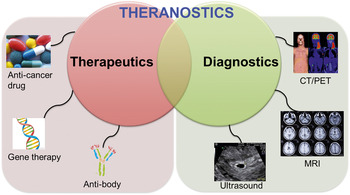
The term “theranostics,” a portmanteau word of therapeutics and diagnostics, is a proposed methodology for increasing treatment efficacy and safety and for monitoring the response to treatment at the same time. The integration of therapeutic and diagnostic functions within a single dose allows the development of effective curing protocols by simultaneously monitoring the results of a new medication test (Figure 1). In the last few years, an increasing number of articles on theranostics have reported imaging agents that can predict or monitor therapy. This image-guided therapy allows for the tailoring of optimized treatment for individual patients and would be of great interest to the field of personalized medicine.
Schematic representation of the term “theranostics,” a portmanteau word of therapeutics and diagnostics. The combined process of therapeutics (anti-cancer drug, gene, antibody, etc.) and diagnostics (CT, computed tomography; PET, positron emission tomography; MRI, magnetic resonance imaging; ultrasound, etc.) methodology proposes to increase treatment efficacy and safety, while monitoring the response to treatment at the same time.

Theranostics relies on emerging advances in nanotechnology, wherein the detection of nanomaterials could occur before, during, or after the treatment regimen.Reference Svenson1 Molecular imaging permits non-invasive monitoring of the distribution of theranostic nanocarriers. It can also provide very rapid feedback on specific targeting and delivery of therapeutic agents using real-time monitoring. Nanoparticles have specific advantages for theranostics that are not found for small molecules. They possibly have a longer circulation half-life, and they are useful for applications such as cardiac blood pool imaging, tumor contrast, diagnosis of inflammation, and sustained systemic drug delivery.Reference Fernandez-Fernandez, Manchanda and McGoron2
Gene therapy refers to an approach by which abnormal genes within an individual’s cells are supplemented or altered with normal ones to modulate the disease manifestation. Generally, two types of therapeutic strategies have been used in gene therapy: gain-of-function and loss-of-function.Reference Toscano, Romero, Munoz, Cobo, Benabdellah and Martin3 For gain-of-function, complementary DNA (cDNA) or messenger RNA (mRNA) needs to be delivered to increase expression of a target protein, which is mostly done by viral vectors (viruses for gene delivery, such as retrovirus, adenovirus, lentivirus, etc.).Reference van Beijnum, Eijgelaar and Griffioen4 In contrast, for loss-of-function, the function or synthesis of a target protein can be blocked when an antisense oligonucleotide (AS-ODN), a small interfering RNA (siRNA) or an aptamer, is delivered by non-viral vectors (for more information on AS-ODN, siRNA, and aptamer, see References 5 and 6).
Since the first conceptualization of gene therapy in 1972, a number of clinical trials have been conducted using various techniques.Reference Jyotil, Singh, Yashpal, Dwivedi and Shanker7 Clinical approval of Gendicine by the Chinese State Food and Drug Administration was the world’s first commercial gene therapy. Gendicine is a recombinant adenovirus engineered to express wild-type-p53, a tumor suppressor protein, for the treatment of cancer with mutated p53.Reference Tseng, Mozumdar and Huang8 The landmark clinical trials included the delivery of RPE65 (retinal pigment epithelium-specific 65 kDa protein) cDNA using adeno-associated virus for the treatment of Leber’s congenital amaurosis, a rare inherited eye disease.Reference Prabhu and Patravale9,Reference Rana, Bajaj, Mout and Rotello10
In recent years, it has been of interest and potential clinical value to combine the concept of a nanoscale delivery platform with gene therapy. However, there are major hurdles in nanoparticle-mediated delivery of genes, including: (1) uptake by the reticuloendothelial system (RES), a part of the immune system composed of phagocytic cells, should be avoided when delivering nanocarriers into specific target tissue.Reference Juliano, Bauman, Kang and Ming11 (2) To locate therapeutic genes at the site-of-action, such as the cytoplasm or nucleus, nanoparticles need to cross the cell membranes through endocytosis, and then endocytosed genes must escape the endosome.Reference Tseng, Mozumdar and Huang8 (3) In particular, the enhanced permeability and retention effect is considered as a tumor-targeting strategy because nanoparticles can extravasate into the tumor interstitium through the leaky vasculature and accumulate in a solid tumor; however, the accumulation level of nanomaterials depends on nanoparticle size and vascular density of the tumor tissue. The ideal size of nanoparticles is considered in the range of 20–200 nm.Reference Nie12
Physicochemical properties of nanocarriers, including surface charge and size, are optimized to overcome the biological barriers, as described previously. Surface modification of poly(ethylene glycol) (PEG) reduces the opsonization, a process by which foreign materials are coated with nonspecific proteins, thereby minimizing entrapment in RES. Positive surface charge of nanoparticles helps endocytosis by interaction with anionic proteoglycans on the cell surface. Further, transfection efficacy of nanoparticles can be increased by association with active targeting molecules, such as antibodies, ligands, and aptamers.
Despite many advances, the field of gene therapy is still facing many obstacles, such as the short-lived nature of gene therapy, immune response, and toxicity with viral vectors. Hence, most research has focused on the development of alternative nanocarriers for the effective delivery of genetic materials into desired tissues or cells.Reference Aryal, Pilla and Gong13 Representative non-viral vectors for gene delivery include lipid-based nanomaterials and polymeric nanoparticles. In the last five years, many scientists have also worked on gold nanomaterials, magnetic nanoparticles, and carbon-based nanomaterials. In parallel, there are increasing efforts to evaluate the efficacy of gene therapy by monitoring delivered nanoplatforms and identifying therapeutic responses in living subjects. This article focuses on the design and development of such theranostic agents and their applications to gene therapy.
Gold-based nanotheranostics for gene therapy
In recent years, gold-based nanomaterials, including gold nanoparticles and gold nanorods, have gained increasing interest as multifunctional carriers for various diagnostic and therapeutic agents, owing to their biocompatibility and facile surface functionalization methods via gold-thiol chemistry.Reference Ghosh, Han, De, Kim and Rotello14 They are attractive candidates for gene delivery, because their surfaces can be easily grafted with thiolated nucleic acid strands for enhancing cellular delivery and gene silencing efficiency.
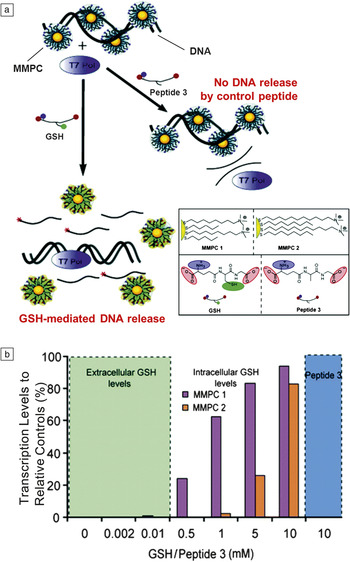
Surface-functionalized gold nanostructures with cationic molecules also bind to nucleic acids and play a role in gene delivery. Han et al. reported functionalized gold nanoparticles with cationic quaternary ammonium groups associated with DNA through electrostatic interactions, which released DNA via reaction with intracellular glutathione (GSH), a tripeptide that controls the redox environment in cells. After unpackaging DNA from the nanoparticles by GSH treatment, the DNA was efficiently transcribed by T7 polymerase (Figure 2).Reference Han, Martin and Rotello15,Reference Han, Chari, Verma, Hong, Martin and Rotello16 These non-covalent complexes of nanoparticles and DNA were also efficient in transfection of human embryonic kidney cells (293T).Reference Sandhu, McIntosh, Simard, Smith and Rotello17 Thomas and Klibanov covalently attached branched polyethylenimine (PEI, 2 kDa) to gold nanoparticles as vectors for the delivery of plasmid DNA, resulting in ternary complexes.Reference Thomas and Klibanov18 Wang et al. synthesized a novel DNA-binding vector by locating β-cyclodextrin at the external surface of oligo(ethylenediamino)-modified gold nanoparticles.Reference Wang, Chen, Li and Liu19 Accordingly, the surface functionalization of gold nanoparticles allows the successful incorporation of therapeutic genes. It may also offer opportunities for other functional moieties, such as imaging agents or targeting molecules.
(a) Schematic representation of DNA release from gold nanoparticle complexes after glutathione (GSH) treatment, and the structures of MMPC, GSH, and peptide 3 (control peptide). (b) Transcription level in the presence of GSH at various concentrations. GSH-mediated release of DNA enables efficient transcription of DNA by T7 polymerase. Reprinted with permission from Reference 14. © 2005 American Chemical Society. Note: T7 pol, T7 polymerase; MMPC, mixed monolayer-protected gold cluster.

Combining the imaging agent with gold nanoparticles enables the development of multifunctional nanostructures. Lu et al. showed that 64Cu-labeling of siRNA-conjugated gold nanoparticles allowed positron emission tomography (PET) imaging and gene therapy at the same time.Reference Lu, Zhang, Zhang, Flores, Huang, Gelovani and Li20 In the study, siRNA was released from the nanostructures when near infrared (NIR) wavelengths were irradiated, because strong surface plasmon resonance (SPR) absorption at NIR wavelengths, the unique properties of gold nanomaterials, resulted in the cleavage of the gold nanoparticles-biomolecules linkage. The NIR irradiation-induced release of nucleic acid from gold nanomaterials could be used for the selective delivery of siRNA as well as plasmid DNA.Reference Sokolov, Follen, Aaron, Pavlova, Malpica, Lotan and Richards-Kortum21−Reference Kawano, Yamagata, Takahashi, Niidome, Yamada, Katayama and Niidome23 This photothermal transfection genesis-RNA inhibited tumor growth only in the region irradiated by NIR, while circumventing undesired side effects.Reference Lu, Zhang, Zhang, Flores, Huang, Gelovani and Li20
Magnetic nanotheranostics for gene therapy
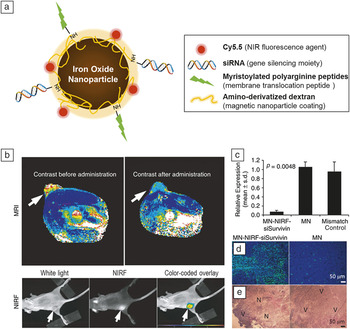
Magnetic nanoparticles (see the November 2013 issue of MRS Bulletin), which can be manipulated by magnetic field gradients, are advantageous for theranostic purposes because of their potential for biomedicine, magnetic resonance imaging (MRI), and magnetic particle imaging.Reference Gupta and Gupta24 As they can be used as carriers for in vivo gene delivery, magnetic nanoparticles simultaneously conjugated with siRNA molecules, a near-infrared dye, and a membrane translocation peptide were constructed as multifunctional probes (Figure 3).Reference Medarova, Pham, Farrar, Petkova and Moore25 Accumulation of nanoparticles in tumors was confirmed by both high-resolution MRI and fluorescence imaging, which were in good agreement with the efficiency of gene silencing. siRNA against survivin, a member of the inhibitor of the apoptosis protein family, inhibited the expression of the target gene, thereby increasing cell death in nanoparticle-delivered tumor tissue.
(a) Schematic illustration of dextran-coated magnetic nanoparticles linked with near-infrared Cy5.5, transmembrane translocation peptides, and small interfering RNA (siRNA). (b) Application of magnetic nanoparticles in a therapeutic tumor model. The accumulation of nanoparticles was detected by in vivo magnetic resonance imaging (MRI) and near-infrared fluorescent (NIRF) imaging (b). Arrows indicate tumor tissue. Suppression of gene expression was analyzed via RT-PCR (analysis of transcription level) (c), TUNEL assay (detection of apoptosis) (d), and H&E staining (histology analysis) (e). The expression of survivin was significantly inhibited, thereby increasing tumor apoptosis and necrosis. Reprinted with permission from Reference 23. © 2007 Nature Publishing Group. Note: MN, magnetic nanoparticle; siSurvivin, siRNA against survivin; N, tumor necrosis; V, viable tumor tissue; p, probability value.

Another magnetic-nanoparticle platform was also developed with siRNA, fluorescent dye, and arginine-glycine-aspartic acid (RGD)-peptide conjugated to the surface of MnFe2O4 nanoparticles.Reference Lee, Lee, Moon, Lee, Park and Cheon26 The formulation consisting of an oleic acid-coated magnetic nanocrystal core and cationic lipid shell was used to deliver siRNA targeting the epidermal growth factor receptor in a mouse tumor model.Reference Namiki, Namiki, Yoshida, Ishii, Tsubota, Koido, Nariai, Mitsunaga, Yanagisawa, Kashiwagi, Mabashi, Yumoto, Hoshina, Fujise and Tada27 Applying a magnetic field elevated the transfection efficacy, followed by target gene silencing and tumor suppression.Reference Namiki, Namiki, Yoshida, Ishii, Tsubota, Koido, Nariai, Mitsunaga, Yanagisawa, Kashiwagi, Mabashi, Yumoto, Hoshina, Fujise and Tada27
Polymeric nanotheranostics for gene delivery
Polymeric nanomaterials have been extensively used as nanotheranostics for imaging and drug delivery. Their physicochemical properties are dependent on the chain length of the polymer backbone and the morphology and mechanical properties of the nanomaterials. Their surface was chemically modified with targeting molecules.Reference Lee28 Nanomaterials were integrated with cationic polymers loaded with genetic materials through electrostatic interactions and could be further modified into theranostic nanomaterials.Reference Bryson, Fichter, Chu, Lee, Li, Madsen, McLendon and Reineke29,Reference Liu, Li, Hou, Xue, Yao, Ma, Feng, He, Lu, Wang and Zeng30
Plasmids were also integrated with lipids or polymers to form nanoparticles. Negatively charged nucleic acids were easily complexed with cationic polymers (e.g., PEI, biodegradable cationic polysaccharides, and cationic polypeptides), resulting in condensed nucleic acid complexes. The transfection efficiency of the resulting condensates was tuned by changing the effective size and charge of the nucleic acid.Reference Wolff and Rozema31 Positively charged complexes enhanced gene transfection efficiency. However, they also showed rapid capture by the RES and dose-dependent toxicity after systemic injection.Reference Zhang, Satterlee and Huang32
There is a consensus that nanoparticles in circulation bind with plasma proteins. Then, the nanoparticles can be recognized by the RES, thus resulting in rapid clearance of the nanoparticles by RES organs, including the liver, spleen, and bone marrow. One way to improve the pharmacokinetics is to modify the surface of the nanoparticles with PEG.Reference Li and Huang33 PEG forms a shielding layer on the surface of the nanocarriers and protects the carriers from degradation by steric hindrance.Reference Veronese and Pasut34 Furthermore, the increased hydrophilicity and hydrodynamic radius of nanomaterials after PEGylation enhance their solubility and reduce their clearance by the kidney, respectively. Polyion complexes of DNA with a block copolymer consisting of hydrophilic PEG and polycationic poly-L-lysine (PLL) form self-assembling nanoparticles with a core–shell structure through electrostatic interaction.Reference Harada-Shiba, Yamauchi, Harada, Takamisawa, Shimokado and Kataoka35 The polyion complex micelles are soluble in water and resistant to nuclease and show extended stability in the blood stream.
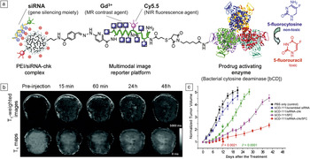
Incorporation of imaging agents into the nucleic acid/polymer complexes allowed the monitoring of gene therapy. For example, paramagnetic gadolinium (Gd3+) labeling enabled the visualization of delivery vehicles via MRI. Polymer beacons consisting of oligoethyleneamines and Gd3+ efficiently delivered plasmid DNA into cells in vitro, and the transfected cells exhibited higher T1-weighted MR image contrast.Reference Bryson, Fichter, Chu, Lee, Li, Madsen, McLendon and Reineke29 Li et al. developed PEI nanocomplexes containing both therapeutic agents (siRNA and prodrug enzyme) and an MRI reporter (Gd3+) (Figure 4).Reference Li, Penet, Wildes, Takagi, Chen, Winnard, Artemov and Bhujwalla36 The accumulation of nanocomplexes into tumor sites was detected by MRI, and the combination of siRNA and a prodrug enzyme resulted in the suppression of tumor growth.
(a) Schematic structure of polymeric nanocomplexes containing both therapeutic agents (small interfering RNA [siRNA] and prodrug enzyme) and an image reporter (MR contrast agent and NIR fluorescence dye). (b) In vivo T1-weighted MR images and T1 maps of a tumor before and after nanocomplex injection. The accumulation of nanocomplex in tumor tissue was visualized by MR imaging due to Gd3+. (c) Suppression of tumor growth after treatment with nanocomplexes. siRNA treatment or enzyme/prodrug strategy alone inhibited tumor growth; however, the combined strategy exhibited synergic therapeutic effects. Reprinted with permission from Reference 35. © 2010 American Chemical Society. Note: PEI, polyethylenimine; siRNA-chk, siRNA against choline kinase-α (chk); MR, magnetic resonance; NIR, near infrared; p, probability value; PBS, phosphate buffered saline; 5FC, 5-fluorcytosin; Cy5.5, NIR fluorescence agent.

In addition, NIR optical imaging of gene nanocarriers was conducted by conjugating NIR fluorescent dye to polymers. For example, FPR675 labeling of thiolated glycol chitosan/poly(siRNA) complexes allowed real-time monitoring of nanoparticle biodistribution in tumor-bearing mice.Reference Lee, Huh, Lee, Min, Lee, Koo, Chu, Lee, Jeon, Choi, Choi, Byun, Jeong, Park, Kim and Kwon37 The developed nanoparticles selectively accumulated in tumor tissue, and siRNA against vascular endothelial growth factor silenced the expression of target genes. PET was also used to track tumor localization of nanoparticles. 64Cu labeled polymer/siRNA nanocomplexes enabled non-invasive monitoring of pharmacokinetics of complexes using PET coupled with computed tomography (CT).Reference Bartlett, Su, Hildebrandt, Weber and Davis38,Reference Mudd, Trubetskoy, Blokhin, Weichert and Wolff39
Silica-based nanotheranostics for gene therapy
Silica-based nanomaterials have high surface area and can be readily suspended in an aqueous media. For effective theranostic application, silica-based nanomaterials were fabricated with anchoring groups or charge transferring functional groups using simple silane chemistry. Silica nanoparticles (SiNPs) modified with the cationic PLL were capable of binding to anionic AS-ODN via electrostatic interactions and enhancing the endocytotic cellular uptake of genetic materials.Reference Zhu, Xiang, Li, Shen, Lu, Zhou, Xiong, Zhang, Nie, Zhou, Tang and Li40 Fluorescently labeled SiNPs modified with sodium chloride also showed high in vitro transfection efficiency. They passed through the blood-brain, blood-prostate, and blood-testis barriers.Reference Chen, Xue, Zheng, Xia, Zhao, Liu, Long and Xia41 In order to optically track the delivery of DNA into cells, hydrophilic or hydrophobic dyes as well as various biomolecules were loaded into the core of the silica nanoparticles.Reference Jain, Roy, De and Maitra42 Using this approach, the adsorption of calf-thymus DNA onto the surface of nanoparticle was examined with fluorescence resonance energy transfer (FRET) occurring between the dye (energy donor) intercalated DNA and a second dye (energy acceptor) encapsulated in the nanoparticles.Reference Roy, Ohulchanskyy, Bharali, Pudavar, Mistretta, Kaur and Prasad43
Silica nanotubes are being increasingly studied because of their unique structure with an inner void that can load drug molecules and an outer surface that can present differential functional motifs, including imaging probes.Reference Martin and Kohli44 Mesoporous silica nanoparticles (MSNs), exhibiting uniform pore sizes of 2–2.5 nm, also exhibit superb potential for applications in gene delivery and imaging. The large pore surface area in MSNs facilitates loading of drug molecules and various functional moieties that improve cellular uptake through endocytosis. Meng et al. confirmed that PEI-modified MSNs loaded with both siRNA, silencing the expression of P-glycoprotein, and doxorubicin, an anticancer drug, were effective in overcoming multidrug resistance in cancer cells and the tumor-bearing animal model.Reference Meng, Liong, Xia, Li, Ji, Zink and Nel45,Reference Meng, Mai, Zhang, Xue, Xia, Lin, Wang, Zhao, Ji, Zink and Nel46 Furthermore, labeling of MSNs with NIR dye enabled monitoring of the location of therapeutic agent-loaded nanomaterials in vivo. Reference Meng, Mai, Zhang, Xue, Xia, Lin, Wang, Zhao, Ji, Zink and Nel46
Carbon nanotheranostics for gene therapy
The unusual properties of carbon nanoscale materials have attracted increasing attention for medical and biological applications. Based on the interatomic carbon–carbon bonds, carbon nanomaterials can be primarily classified into graphene, diamond, graphite, carbon nanotubes, and fullerenes. Graphene, a one-atom-thick layer of graphite, interacts with various biomolecules for applications in biosensors, drug delivery, and gene therapy.Reference Feng, Zhang and Liu47–Reference Tang, Wang and Loh49 Graphene oxide (GO) possesses chemically reactive oxygen groups such as carboxyl groups. Therefore, many hydrophilic macromolecules have been used for covalent functionalization of GO.Reference Park and Ruoff50 For instance, GO functionalized with PEI, chitosan, or 1-pyrenemethylamine was applied to load and deliver plasmid DNA or siRNA.Reference Bao, Pan, Ping, Sahoo, Wu, Li, Li and Gan51
Owing to their unique optical properties, graphene and its derivatives can be used not only for photothermal ablation of tumors, but also for applications in biomedical imaging.Reference Yang, Hu, Ma, Ye, Cheng, Shi, Li, Li and Liu52,Reference Sun, Liu, Welsher, Robinson, Goodwin, Zaric and Dai53 However, since visible fluorescence of GO has limited application in in vivo animal imaging due to autofluorescence interference from biological tissues, external fluorescent dyes should be coupled to the GO for imaging.Reference Yang, Zhang, Zhang, Sun, Lee and Liu54,Reference Peng, Hu, Zhou, Fan and Huang55
The biocompatibility of GO is a critical issue for its biomedical applications. GO at low concentrations (approx. 0–40 µg/ml) did not show obvious toxicity, but a high dose of GO caused oxidative stress in A549 cells or induced apoptosis of human fibroblasts.Reference Chang, Yang, Liu, Dong, Wang, Cao, Liu and Wang56,Reference Wang, Ruan, Song, Zhang, Wo, Guo and Cui57 This dose-dependent toxicity can be attenuated by functionalization with PEG or dextran.Reference Zhang, Yang, Feng and Liu58,Reference Wojtoniszak, Chen, Kalenczuk, Wajda, Łapczuk, Kurzewski, Drozdzik, Chu and Borowiak-Palen59 An in vivo distribution study of PEGylated nano-GO exhibited accumulation in RES after intravenous administration. A dose of 20 mg/kg did not induce any toxicity, as indicated by blood biochemistry, hematological analysis, and histological examinations.Reference Yang, Wan, Zhang, Zhang, Lee and Liu60 Although several studies reported biocompatibility of GO, a systematic investigation of the long-term health risks is required before its clinical application.
Exosomes in nanotheranostic gene delivery
Exosomes are cell-derived nanoparticles that are present in most biological fluidsReference Keller, Sanderson, Stoeck and Altevogt61 and may have functions in intercellular signaling, waste management, and coagulation.Reference van der Pol, Boing, Harrison, Sturk and Nieuwland62 Furthermore, previous studies reported that exosomes transfer mRNA or microRNA (miRNA) to other cells, termed “exosomal shuttle RNA.”Reference Valadi, Ekstrom, Bossios, Sjostrand, Lee and Lotvall63 The transferred mRNA or miRNA modify recipient cell protein production and gene expression, respectively. Thus, there is growing interest in the potential of exosomes as a gene delivery carrier. Kosaka et al. showed that intercellular transfer of tumor-suppressive miRNA by exosomes was effective in silencing genes in recipient cells, leading to inhibition of cancer cell growth.Reference Kosaka, Iguchi, Yoshioka, Takeshita, Matsuki and Ochiya64 Recently, Alhasan et al. demonstrated that exosomes encaged with gold nanoparticles and antisense DNA could assess the intracellular miRNA levels with fluorescent quenching by gold nanoparticles. Gold nanoparticles, which were hybridized with antisense DNA and fluorophore-labeled DNA, quenched the near fluorescence through energy transfer; the recognition of antisense DNA by target miRNA allowed the release of fluorophore-labeled DNA, thereby the recovery of fluorescence.Reference Alhasan, Patel, Choi and Mirkin65 Accordingly, exosomes will be a potential candidate for nanotheranostics when integrated with imaging contrast agents and therapeutic genes.
Summary
In this review, we have highlighted recent advances in theranostic nanoplatforms for gene therapy, including gold nanomaterials, magnetic nanoparticles, polyemeric complexes, silica nanoparticles, graphene derivatives, and exosomes. Nanotheranostic platforms exhibit great potential as therapeutic or diagnostic agents or as multifunctional agents. Recently, the use of gene therapy along with nanotechnology has allowed for the design and development of new theranostic tools. The detection modality, simultaneously with gene delivery, shows promise for monitoring the results of a new medication test before, throughout, or after the treatment regimen. However, it is important to investigate the long-term characteristics of nanotheranostic materials with respect to system complexity and stability. In addition, the toxicity of nanocarriers (e.g., cyto-, geno-, and immunotoxicity) should be evaluated, and cost-effectiveness and available testing systems need to be established. Nevertheless, the application of nanotheranostics to gene therapy is plausible and likely will be widely used in the field of personalized and predictive medicine.
Acknowledgments
This study was funded by the Intramural Research Program (Theragnosis, Global RNAi Carrier Initiative, and KIST Young Fellow) of KIST and the Global Innovative Research Center (GiRC) Program through the National Research Foundation of Korea (NRF).