Report
Long QT syndromes are the most common group of ion channelopathies with an estimated prevalence of 1 in 2500 live births.Reference Crotti, Celano, Dagradi and Schwartz 1 Affected individuals are at increased risk of sudden death.
We report a twin girl, of Chinese ethnicity, born at 26+5 weeks birth weight 796 g. Perinatal period was relatively uncomplicated necessitating <24 hours invasive ventilation. Early echocardiography demonstrated a moderate patent ductus arteriosus with left-heart dilatation. The mother was diagnosed with long QT syndrome in China in 2005, the family immigrated recently to Australia. The patient’s mother was on a beta blocker but was always asymptomatic and did not offer this information to the attending neonatal team.
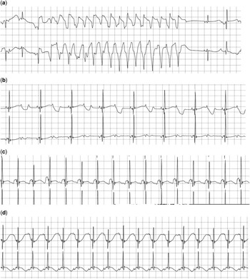
On day 35, the baby had several episodes of a wide complex tachycardia at 230 bpm, with associated haemodynamic compromise (Fig 1a) consistent with polymorphic ventricular tachycardia/torsades. The episodes were mostly self-limiting. There were also intermittent episodes of second-degree atrioventricular block owing to an excessively prolonged QT interval (Fig 1b). There was no central venous catheter in situ. The patient had been administered caffeine therapy, which was then discontinued. Serum electrolytes were normal, but potassium and magnesium supplementation were prescribed to maintain “high normal” serum values, although this did not alter the frequency of ventricular tachycardia.
( a – d ) Holter monitor recordings.

The resting electrocardiogram revealed normal sinus rhythm with abnormal repolarisation (Fig 1c). The QT interval was prolonged, up to 640 ms, and the P wave frequently coincided with the end of the T wave. The patient was emergently transferred to the congenital cardiac centre and commenced on propranolol 0.2 mg/kg tds orally, which was increased incrementally. However, the short but frequent episodes of polymorphic ventricular tachycardia continued. With an increasing dose of propanolol, the episodes of ventricular tachycardia somehow reduced in frequency, but 2:1 atrioventricular block became more frequent and caused haemodynamic compromise.
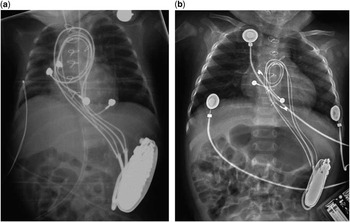
After 3 days of admission to the cardiac unit (corrected age=32+1 weeks, weight=1140 g), a DDD epicardial pacemaker was inserted and the patent ductus arteriosus was ligated. The pacemaker leads were placed via sternotomy at the left ventricular apex and right atrial wall. The pacemaker box was implanted in the left retroperitoneal space via a left flank oblique incision extending between the lower costal margin and the left lower quadrant of the abdomen (Fig 2a).
( a and b ) X-rays at time of pacemaker insertion and 8 months later.

Despite the large size of the pacemaker, the patient made an uncomplicated postoperative recovery.
The patient is now 20 months old (16 months corrected) and weighs 8.5 kg. Current medication is atenolol 10 mg bd. Repeated Holter monitoring demonstrates predominantly atrial pacing and ventricular sensing (Fig 1d), with occasional atrial and ventricular pacing. A recent chest X-ray demonstrates a satisfactory pacing box position and that the pacing leads have partially unravelled to accommodate increased patient length (Fig 2b). There have been no instances of ventricular tachycardia, atrioventricular block, or high-rate episodes since the concomitant pacemaker insertion and effective beta-blockade in the pacemaker memory or holter recordings. Genetic testing has confirmed a heterozygous mutation in KCNH2 gene, which is consistent with the family genotyping and long QT syndrome type 2.
Discussion
The management of long QT syndrome with 2:1 atrioventricular block is challenging. Medical therapy includes beta-blockade and possibly mexiletine.Reference Aziz, Tanel and Zelster 2 In the setting of 2:1 atrioventricular block, dual-chamber pacing is recognised as an effective adjunct in the management of long QT syndrome.Reference Crotti, Celano, Dagradi and Schwartz 1 , Reference Gillis, Russo and Ellenbogen 3 We initially thought that the extremely small size of this baby precluded the implantation of a dual-chamber pacemaker. However, A-V pacing seemed essential in our case because a small dose of propranolol appeared to exacerbate 2:1 atrioventricular block, and beta-blockade was necessary for effective suppression of ventricular arrhythmia. In addition, dual-chamber pacing permits A-V synchrony, guarantees a physiological heart rate with the facility for physiological tachycardia, prolongs the life of the generator box, and reduces the risk of R on T pacing and consequent torsades de pointes.Reference Aziz, Tanel and Zelster 2 DDD pacing also minimises the potential for pauses, which can be proarrhythmic. Temporary epicardial pacing was a possibility,Reference Filippi, Vangi, Murzi, Moschetti and Colella 4 but we have been disappointed by this strategy because of the lack of longer term reliability of temporary wires.
To the best of our knowledge, the smallest reported recipient of a dual-chamber pacemaker is 2 kg.Reference Kelle, Backer and Tsao 5
A single-chamber pacemaker implantation has been reported in a 1000 g baby.Reference Welch, Hannan and DeCampli 6 The creation of a retroperitoneal pocket via a flank incision was a key aspect to the success of the implantation of a proportionally huge box in this premature baby.
In conclusion, this case demonstrates that implantation of a dual-chamber pacemaker is possible, even in very premature babies. This may be valuable, not only in the management of specific cases of long QT syndrome but also in the management of high-grade atrioventricular block in premature and low birth weight children.