Introduction
Helminth parasite infections have been highly prevalent throughout history and are associated with immunomodulatory mechanisms that modify almost every facet of the host immune system. Trichinella spiralis (T. spiralis) both regulates and evades the immune system to maintain chronic infections in the host. Increasing evidence suggesting that cells of the innate and adaptive immune system could be regulated by helminths (Maizels and McSorley, Reference Maizels and McSorley2016). However, the precise mechanisms underlying the cellular responses regulated by T. spiralis has remained undetermined.
Functional proteins of parasite play an important role in the invasion and evasion of the host immune response. Immune modulation associated with cellular responses to T. spiralis molecules has also been widely reported (Bai et al., Reference Bai, Wu, Wang, Guan, Gao, Yu, Yu, Tang, Liu, Song, Wang, Radu, Boireau, Wang and Liu2012; Sun et al., Reference Sun, Zhao, Wang, Yang, Zhao, Zhan and Zhu2015; Chen et al., Reference Chen, Tang, Liang, Yang, Wu, Hu, Li, Zeng and Ma2016; Ding et al., Reference Ding, Bai, Wang, Wang, Shi, Rosenthal, Boireau, Wu, Liu and Liu2016; Della Bella et al., Reference Della Bella, Benagiano, De Gennaro, Gomez-Morales, Ludovisi, D'Elios, Luchi, Pozio, D'Elios and Bruschi2017; Xu et al., Reference Xu, Liu, Tang, Wang, Shi, Boireau, Liu and Bai2017). Glutathione-S-transferase (GST), which belongs to a family of detoxification enzymes, is classically considered to be part of the cell defence against harmful compounds (Mannervik et al., Reference Mannervik, Alin, Guthenberg, Jensson, Tahir, Warholm and Jornvall1985). The GST gene is expressed during all developmental stages of T. spiralis and the T. spiralis-GST gene is up-regulated in intestinal infective larvae compared to muscle larvae (Ren et al., Reference Ren, Cui, Yang, Liu and Wang2013). The recombinant GST of T. spiralis (rTs-GST) has been reported to have enzymatic activity (Cui et al., Reference Cui, Li, Jiang, Liu, Yang, Liu, Liu, Zhang and Wang2015), and appears to be a central protein for the development and survivals of the parasite in the host. However, it is not fully known whether Ts-GST exerts immune modulation on immune cells, and the underlying mechanisms have not been reported.
Dendritic cells (DCs) are the most potent antigen-presenting cells (APCs) and have the ability to regulate naïve T cells responses (Zhu et al., Reference Zhu, Yamane and Paul2010). Molecules of helminths can regulate DC phenotypes via modulating the expression of several types of pattern recognition receptors such as toll-like receptors (TLR). In response, a diverse range of DC phenotypes are induced, which is characterized by increased expression of surface co-stimulatory molecules and secretion of cytokines (Ilic et al., Reference Ilic, Gruden-Movsesijan, Cvetkovic, Tomic, Vucevic, Aranzamendi, Colic, Pinelli and Sofronic-Milosavljevic2018). However, immature or mature DCs induce distinct differentiation, activation and proliferation of T cells (Kapsenberg, Reference Kapsenberg2003). DCs treated with molecules of Heligmosomoides polygyrus showed significantly less MHC-II expression and were less effective for activating CD4+ T cells (Sun et al., Reference Sun, Liu, Li, Chen, Liu, Liu and Su2013). Stimulation of DCs with excretory-secretory products (ESP) and components (53 kDa protein) of ESP resulted in partially matured DCs which induced the Th2 response (Cvetkovic et al., Reference Cvetkovic, Sofronic-Milosavljevic, Ilic, Gnjatovic, Nagano and Gruden-Movsesijan2016). A recent study reported that T. spiralis ESP activated DCs to semi-mature status and subsequently induced expansion of regulatory T cells (Tregs) via both TLR 2 and 4 (Ilic et al., Reference Ilic, Gruden-Movsesijan, Cvetkovic, Tomic, Vucevic, Aranzamendi, Colic, Pinelli and Sofronic-Milosavljevic2018). GST of Fasciola hepatica induced partial activation of DCs (Dowling et al., Reference Dowling, Hamilton, Donnelly, La Course, Brophy, Dalton and O'Neill2010). These results indicate that different molecules induced distinct DC phenotypes associated with specific T cell responses.
This study first investigated the phenotypes of DCs exposed to rTs-GST. Subsequently, the regulation of rTs-GST-treated DCs on T cell responses was explored in vitro. Semi-mature DCs inhibited the activation and proliferation of OVA-specific CD4+ T cells derived from OT-II mice, indicating that DCs are a key target for the immunomodulation of Ts-GST. In addition, rTs-GST-treated DCs induced Th2-type responses and expanded the population of Tregs. These findings contribute to the explanation of the immunomodulatory effect of rTs-GST on cellular responses, which may be associated with chronic infections and the survival of T. spiralis in the host.
Materials and methods
Animals and parasites
C57BL/6 mice wild type (WT, female, 6–8 weeks old) were purchased from the Shanghai SLAC Company. OT-II ovalbumin (OVA)-specific T-cell receptor (TCR) transgenic mice (on C57BL/6 background) were purchased from the Nanjing University Model Animal Research Centre (Nanjing, China). Female Wistar rats were purchased from the Experimental Animal Centre of College of Basic Medical Sciences, Jilin University (Changchun, China). All animal experiments were performed according to regulations of the Administration of Affairs Concerning Experimental Animals in China. The protocol was approved by the Institutional Animal Care and Use Committee of Jilin University (20170318).
T. spiralis (ISS534) was prepared as previously described (Tang et al., Reference Tang, Liu, Wang, Yu, Shi, Boireau, Cozma, Wu and Liu2015). Briefly, Wistar rats were orally infected with 3000 infective larvae and T. spiralis muscle larvae were recovered at 35 days post-infection (dpi) via artificial digestion with pepsin-HCl (1% pepsin and 1% HCl at 37 °C for 2 h). All parasites were washed three times in saline solution.
Preparation and identification of rTs-GST
The Ts-GST gene (GenBank accession no. XM 003371707.1) was amplified by PCR using specific primers with Nde I and Xho I restriction enzyme sites (5′-CGCCATATGGCTCCTCTATACAAGC-3′; 5′-CCGCTCGAGTTAATTTAATCGAATTTTCTTG-3′). The purified PCR product was cloned into the pCold I vector (Takara, Dalian, China). The recombinant plasmid carrying the Ts-GST gene was transformed into Escherichia coli BL21 (DE3) (Novagen) and expressed under 0.1 mm IPTG induction. The rTs-GST was purified using Ni-NTA-Sefinose resin (Sangon Biotech, China), according to the manufacturer's instruction. The concentration of the purified rTs-GST was determined with Microplate BCA Protein Assay Kit-Reducing Agent Compatible (Thermo Fisher Scientific, Waltham, USA). Purity of the recombinant protein was identified by 12% SDS-PAGE analysis and the gel was stained with 0.25% Coomassie brilliant blue R-250 (AMRESCO). The contaminated endotoxin was removed by ToxOut High Capacity Endotoxin Removal Kit (Biovision, USA). The endotoxin was 0.1812 EU mg−1 in the purified rTs-GST, approximately equivalent to 20 pg mg−1 endotoxin in rTs-GST, which is lower than the minimal amount that could stimulate TLR2/4 based on the standard LPS O55: B5 (Invivogen, USA) (Schwarz et al., Reference Schwarz, Schmittner, Duschl and Horejs-Hoeck2014).
Generation of bone marrow-dendritic cells and CD4+ T cells
Bone marrow-dendritic cells (BMDCs) were generated from bone marrow cells of mice as previously described (Lutz et al., Reference Lutz, Kukutsch, Ogilvie, Rossner, Koch, Romani and Schuler1999). Briefly, bone marrow cells were obtained from C57BL/6 mice and cultured for at 37 °C, 5% CO2 in RPMI 1640 medium containing the growth factors recombinant GM-CSF (20 ng mL−1) and IL-4 (20 ng mL−1; Sigma–Aldrich) and 10% FBS. Immature DCs were harvested on day 7 for further experiments.
CD4+ T cells of OT-II mice were purified by magnetic sorting, using anti-CD4 magnetic beads (Miltenyi Biotec, Auburn, CA, USA) from spleen cells. The purified CD4+ T cells had >90% purity.
In vitro BMDCs stimulation assay
To analyse the effects of rTs-GST on DC maturation, immature DCs were isolated. DCs from mice were stimulated with sterile phosphate-buffered saline (PBS), rTs-GST (10 µg mL−1) and LPS (100 ng mL−1, Sigma–Aldrich) alone or in combination, in vitro for 48 h. Cell culture supernatants were collected and stored at −80 °C. The cytokine in the supernatants was measured by enzyme-linked immunosorbent assay (ELISA) kits (Huamei, Wuhan, China) according to the manufacturer's instructions. Stimulated DCs were stained with FITC-conjugated monoclonal antibody (mAb) to CD11c and PE-conjugated mAbs to MHC-II, CD40, CD80 or CD86 respectively (Biolegend, USA). Appropriately labelled isotype-matched antibodies were used as controls. Cells were analysed using a BD FACS Calibur Flow Cytometer and FlowJo software (Tree star Inc, Ashland, OR). Results are shown as mean ± s.d. (n = 3) of three individual different experiments.
Co-culture of BMDC with CD4+ T cells
CD11c+ DCs were typically of >90% purity as determined by flow cytometry. The purified CD4+ T cells had >90% purity. To determine the roles of rTs-GST-pulsed DC in CD4+ T-cell proliferation, the DCs (2 × 10⁵ per well) were firstly treated with PBS or rTs-GST (10 µg mL−1) for 8 h (using PBS as the control group). After washing three times with sterile PBS, DCs were stimulated either with or without OVA (1 mg mL−1; Calbiochem) for 2 h. The DCs were then treated with 50 µg mL−1 mitomycin (Sigma–Aldrich) for 30 min and washed with medium. CD4+ T cells (2 × 10⁶ per well) stained with 5-and 6-carboxyfluorescein diacetate succinimidyl ester (CFSE) (eBioscience, San Diego, CA, USA) were co-cultured with DC for 72 h. Then, cell samples were analysed using a BD FACS Calibur Flow Cytometer and FlowJo software (Tree star Inc). Results are shown as means ± standard deviation (s.d.) (n = 3) of three individual different experiments.
In addition, the Treg populations induced by rTs-GST-treated DCs were measured. CD4+ T cells were co-cultured with DC treated with PBS, OVA (1 mg mL−1) alone or OVA + rTs-GST for 72 h, respectively. Co-cultured cells were collected and firstly pre-incubated with Fc Blocker (Anti-Mouse CD16/CD32, BD Biosciences, USA) for 15 min to decrease non-specific binding of labelled antibodies. Cells were then stained with APC-labeled anti-CD25, fixed and permeabilized using the Foxp3 Transcription Factor staining buffer kit (eBioscience) according to the manufacturer's instructions. Cells were then blocked in 5% rat serum prior to intracellular staining with Percp Cy5.5-labelled anti-Foxp3. Appropriately labelled isotype-matched antibodies were used as controls. Samples were analysed using BD FACS Calibur Flow Cytometer and FlowJo software (Tree star Inc, Ashland, OR). Results are shown as means ± standard deviation (s.d.) (n = 3) of three individual different experiments.
To determine cytokine production, CD4+ T cells were co-cultured with DC treated with PBS, OVA (1 mg mL−1) alone or OVA + rTs-GST for 48 h respectively, and the DC and CD4+ T-cell co-culture was stimulated by Concanavalin-A (Con-A) (2.5 µg mL−1) (Sigma–Aldrich) for the final 18 h of co-culture. The supernatants in DCs and CD4+ T-cells co-culture were harvested for cytokine analysis by ELISA kits (Huamei, Wuhan, China). Results are shown as means ± standard deviation (s.d.) (n = 3) of three individual different experiments.
Statistical analysis
All results were expressed as mean ± s.d. Statistical analysis was performed using the GraphPad Prism 5 for Windows. One-way and two-way analysis of variance (ANOVA) were used to compare statistical differences at different conditions. P values are expressed as *P < 0.05, **P < 0.01 and ***P < 0.001.
Results
Expression and identification of rTs-GST
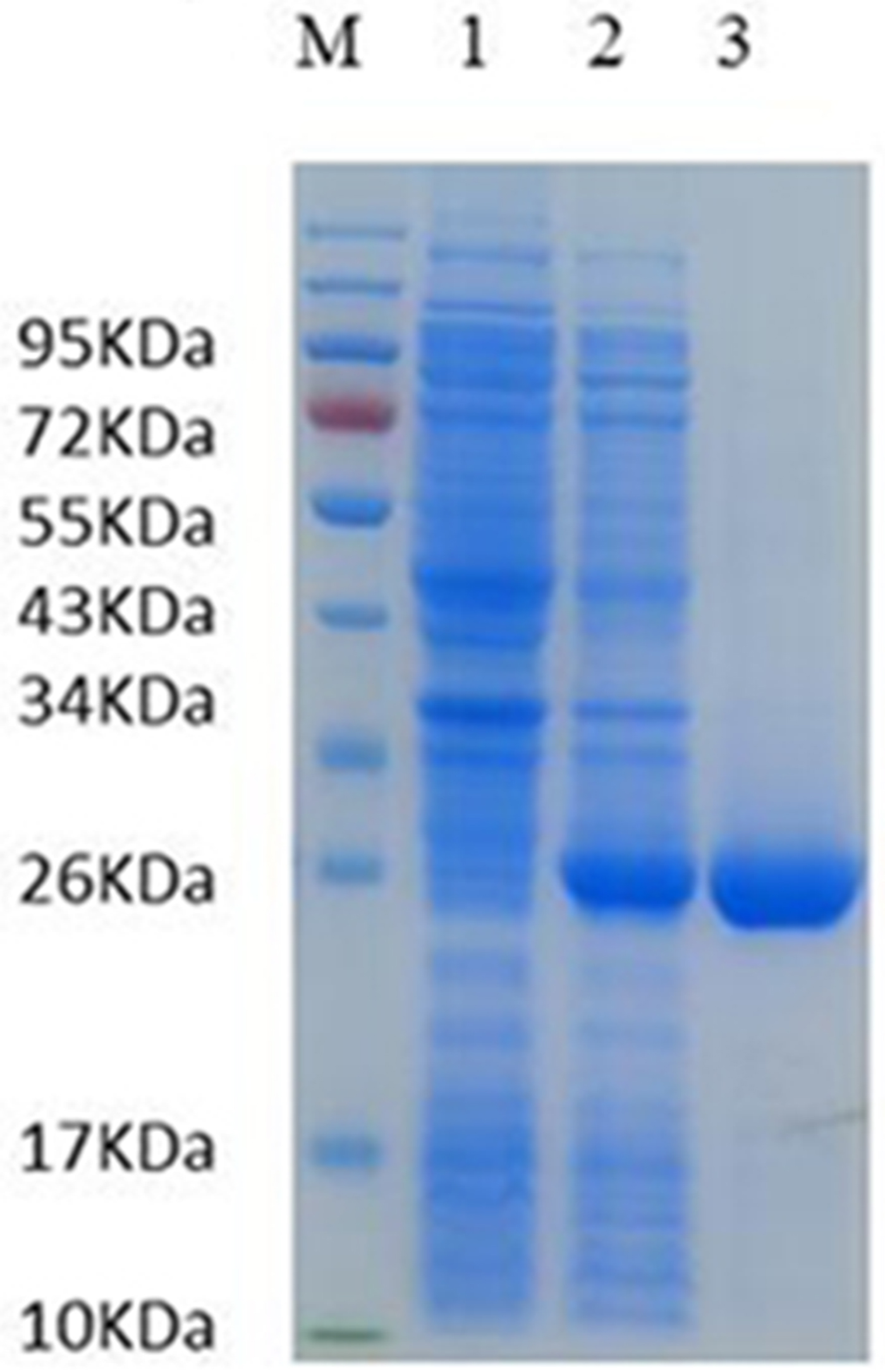
The Ts-GST gene was cloned into the pCold I vector and rTs-GST was successfully expressed in Escherichia coli BL21 (DE3) under IPTG induction. After purification, soluble rTs-GST was obtained, with an approximate molecular weight of 26 kDa (Fig. 1).
Identification of rTs-GST. rTs-GST was purified using Ni-NTA-Sefinose resin, according to the manufacturer's instruction. Purity of the recombinant protein was identified by 12% SDS-PAGE analysis and the gel was stained with 0.25% Coomassie brilliant blue R-250. SDS-PAGE analysis of rTs-GST. M: protein molecular weight marker; 1: lysis of recombinant bacteria; 2: lysis of induced recombinant bacteria harbouring pCold I vector Ts-GST after ultrasonication and 3: purified rTs-GST.

rTs-GST induced semi-matured DCs and impair TLR ligand-induced DC maturation in vitro
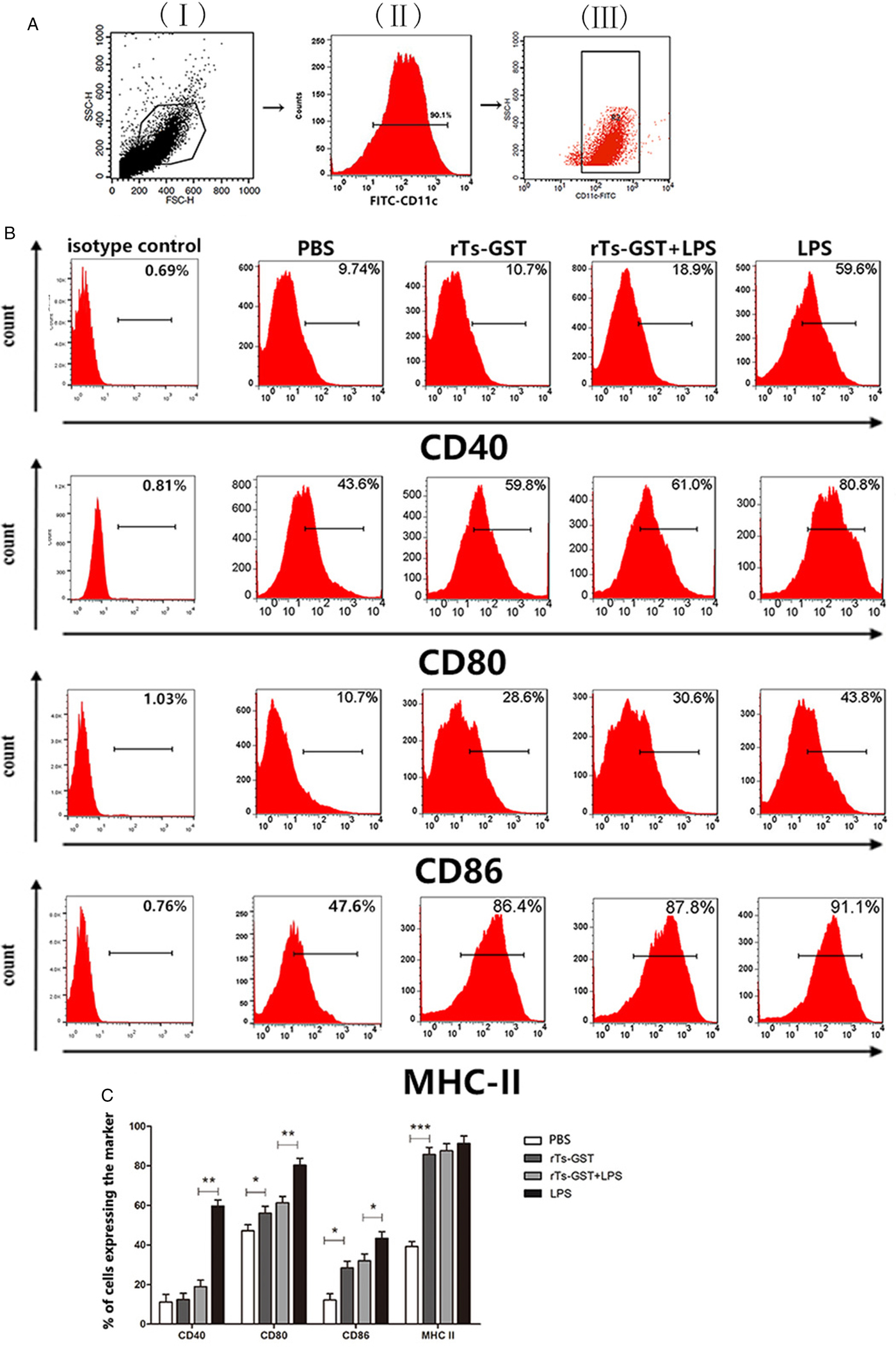
To investigate the effect of rTs-GST on the maturation of DCs, surface co-stimulatory molecules on CD11c+ DCs (>90% CD11c+, Fig. 2A) were analysed. Expression of surface molecules CD40, CD80, CD86 and MHC-II on LPS stimulated CD11c+ DCs were significantly upregulated compared to PBS or rTs-GST alone. In contrast, rTs-GST inhibited upregulation of CD40, CD80 and CD86 on LPS stimulated DCs (Fig. 2B and C). However, rTs-GST was not able to down-regulate the expression of MHC-II. Moreover, the expression of CD40 on CD11c+ DCs was similar to PBS and expressions of CD80, CD86 and MHC-II significantly increased by rTs-GST compared to the PBS group.
Expression of surface co-stimulatory molecules on DCs induced by LPS, rTs-GST or PBS. DCs were stimulated with sterile phosphate-buffered saline (PBS), rTs-GST (10 µg mL−1), LPS (100 ng mL−1) alone or rTs-GST+LPS, in vitro for 48 h. (A) Expression of CD11c+ DCs was measured by FACS. Representative dot plots for the gating strategy: (I) gating on viable cells, (II) expression of CD11c+ and (III) gating on CD11c+ cells. (B) Expression of CD40, CD80, CD86 and MHC-II measured by FACS. (C) Data represent means ± standard deviations (n = 3) of the results of three individual experiments. *P < 0.05, **P < 0.01 and ***P < 0.001 vs control groups.

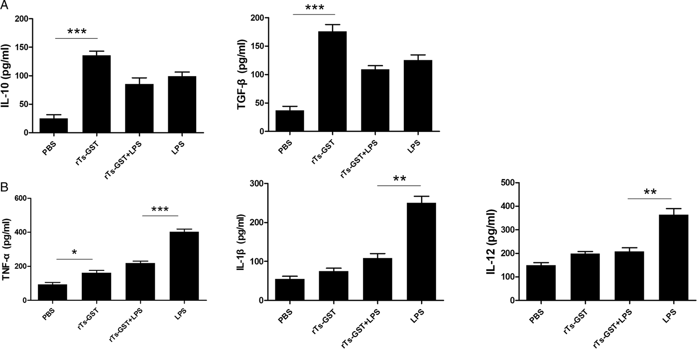
Then, the production of inflammatory cytokines of DCs was examined. Stimulation of LPS resulted in increased cytokine production including pro-inflammatory cytokines and anti-inflammatory cytokines. Stimulation of rTs-GST alone increased the levels of IL-10 and TGF-β, which are regulatory cytokines. The levels of IL-10 and TGF-β in rTs-GST-treated LPS-matured DCs were not significantly different, compared to LPS-matured DCs (Fig. 3A). In addition, rTs-GST decreased the production of TNF-α, IL-1β and IL-12 induced by LPS (Fig. 3B). In summary, these results indicate that rTs-GST inhibited the LPS-induced proinflammatory response on DCs.
Cytokine production by DCs treated with LPS or rTs-GST. DCs were cultured for 48 h and supernatants were measured by ELISA. (A) Regulatory cytokine (IL-10 and TGF-β) production was measured. (B) The production of pro-inflammatory cytokines (TNF-α, IL-1β and IL-12) was measured. Results are presented as mean ± standard deviation (n = 3) from three individual experiments. *P < 0.05; **P < 0.01, ***P < 0.001 (one-way ANOVA with Tukey's post-test).

DCs treated with rTs-GST regulated the proliferation of OVA-specific CD4+ T cells
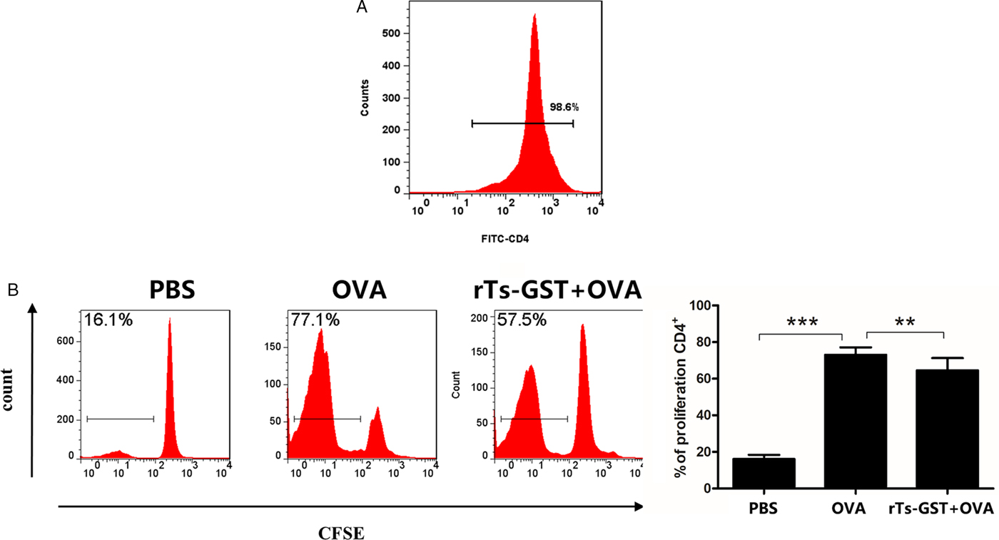
The immunoregulatory activity of CD11c+ DCs on T cells proliferation was determined. OVA-specific CD4+ T cells were labelled with CFSE and co-cultured with DCs that had been treated with or without rTs-GST in the presence of OVA. FACS results showed that proliferation of OVA-specific CD4+ T cells was significantly boosted by DCs treated with OVA alone compared to PBS (Fig. 4B). This effect was diminished when cells were treated with rTs-GST, suggesting that DCs treated with rTs-GST impaired OVA-induced T cell proliferation.
Proliferation of T cells by rTs-GST-pulsed CD11c+ DCs. (A) The purity of CD4+ T cells was analysis by FACS after magnetic sorting using anti-CD4 magnetic beads. (B) To assess the effect of rTs-GST-treated DCs for the proliferation of OVA-specific CD4+ T cells, CD4+ T cells were stained with CFSE prior to co-culture with DCs. Proliferation of CD4+ T Cells was determined by CFSE labelling and flow cytometry. Results are shown as means ± standard deviation (s.d.) (n = 3) of three individual different experiments. *P < 0.05, **P < 0.01, ***P < 0.001 (one-way ANOVA with Tukey's post-test).

DCs treated with rTs-GST expanded the Treg cells population
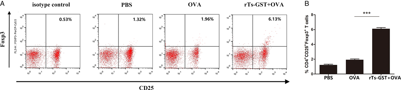
To determine the effect of DCs treated by rTs-GST on the generation of Tregs, rTs-GST-treated DCs were cultured with T cells isolated from spleens of OT-II (C57BL/6 background) for 48 h. The FACS results demonstrated that the population of CD4+CD25+Foxp3+ T cells significantly increased when OVA-specific CD4+ T cells were co-cultivated with rTs-GST-treated CD11c+ DCs in the presence of OVA, compared to PBS or the OVA alone treatment group (Fig. 5).
Population of Tregs induced by rTs-GST-treated CD11c+ DCs. Treg populations induced by rTs-GST-treated DCs. CD4+CD25+Foxp3+ T cells were determined by flow cytometry. (A) Representative analysis of Tregs are shown and (B) the summarized results are presented as means ± standard deviation (s.d.) (n = 3) of three individual different experiments. *P < 0.05, **P < 0.01, ***P < 0.001 (one-way ANOVA with Tukey's post-test).

DCs treated with rTs-GST regulated production of inflammatory cytokines of CD4+ T cells
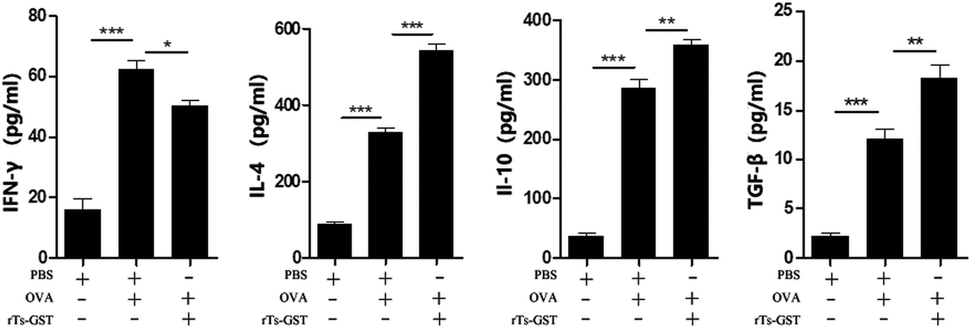
The culture supernatants from co-culture were collected, and the secretion levels of the cytokines associated with each type of T cell were determined by ELISA. Production of cytokines significantly increased by OVA compared to the group without OVA. Then, treatments with or without rTs-GST in the presence of OVA were compared. Compared to the PBS-treated DCs group, rTs-GST-treated DCs not only induced OVA-specific CD4+ T cells to secrete cytokine IL-4 (Th2) (P < 0.001), but also induced higher levels of IL-10 and TGF-β (IL-10, P < 0.01; TGF-β, P < 0.01), which is consistent with the increase of the Tregs population observed by FACS. Furthermore, the levels of IFN-γ (Th1) were significantly inhibited (P < 0.01) by rTs-GST-pulsed DCs, compared to the OVA group (Fig. 6).
Cytokine production of T cells induced by rTs-GST-treated CD11c+ DCs. DCs (2 × 10⁵ per well) were treated with PBS or rTs-GST (10 µg mL−1) for 8 h. DCs were washed three times with sterile PBS and resuspended with RPMI 1640 medium. Then DCs and CD4+ T cells (2 × 10⁶ per well) were co-cultured with or without OVA (1 mg mL−1) for 48 h. During the last 18 h, 2.5 µg mL−1 Concanavalin-A (Con-A) was added. The supernatants in DCs and CD4+ T-cells co-culture were harvested for cytokine analysis by ELISA. Results are shown as means ± standard deviation (s.d.) (n = 3) of three individual different experiments. *P < 0.05, **P < 0.01, ***P < 0.001 (one-way ANOVA with Tukey's post-test).

Discussion
GSTs form a family of detoxification enzymes and are major immune-modulatory molecules. GSTs from helminths, such as T. spiralis (Cui et al., Reference Cui, Li, Jiang, Liu, Yang, Liu, Liu, Zhang and Wang2015), Schistosoma (Grezel et al., Reference Grezel, Capron, Grzych, Fontaine, Lecocq and Capron1993), Fasciola (Morrison et al., Reference Morrison, Colin, Sexton, Bowen, Wicker, Friedel and Spithill1996), Setaria cervi (Gupta et al., Reference Gupta, Bhandari, Reddy, Harinath and Rathaur2005), Ancylostoma caninum (Zhan et al., Reference Zhan, Liu, Perally, Xue, Fujiwara, Brophy, Xiao, Liu, Feng, Williamson, Wang, Bueno, Mendez, Goud, Bethony, Hawdon, Loukas, Jones and Hotez2005) and Necator americanus (Zhan et al., Reference Zhan, Perally, Brophy, Xue, Goud, Liu, Deumic, de Oliveira, Bethony, Bottazzi, Jiang, Gillespie, Xiao, Gupta, Loukas, Ranjit, Lustigman, Oksov and Hotez2010) indicated immunoprotective potential. The GST is a T. spiralis functional protein, and the GST gene of T. spiralis is expressed during all developmental stages of T. spiralis. T. spiralis-GST gene is up-regulated in intestinal infective larvae compared to muscle larvae (Ren et al., Reference Ren, Cui, Yang, Liu and Wang2013). And the recombinant GST of T. spiralis (rTs-GST) has been reported to exert enzymatic activity (Cui et al., Reference Cui, Li, Jiang, Liu, Yang, Liu, Liu, Zhang and Wang2015). It has been reported that Ts-GST expressed in the stichosome which are secretory cells in Trichinella (Rojas et al., Reference Rojas, Rodriguez-Osorio and Gomez-Garcia1997; Li et al., Reference Li, Wang, Liu, Yang, Liu, Sun, Jiang, Zhang, Zhang and Cui2015). The presence of anti-Ts-GST antibodies in infected animals agrees with its localization on the secretory organ of the parasite. Furthermore, variable levels of GST are observed in the blood from infected animals at different stages and the first peak corresponds to the presence of adults during the intestinal phase, whereas larvae have already settled when the higher levels are reached (Rojas et al., Reference Rojas, Rodriguez-Osorio and Gomez-Garcia1997). And the presence of Ts-GST in sera from infected animals was confirmed after its isolation by GSH affinity chromatography. These results indicated that the Ts-GST directly interact with host immune system and could play an important role in regulating immune response. It has also been also previously reported that the GST of F. hepatica showed immunomodulatory activity for cell response (Dowling et al., Reference Dowling, Hamilton, Donnelly, La Course, Brophy, Dalton and O'Neill2010). However, whether Ts-GST is immune-modulatory for immune cells remains unknown.
DCs play an important role in the balance between tolerance and immunity. Protein expressed by nematode parasites can modulate differentiation and activation of DCs. Paramyosin, one of the proteins expressed by T. spiralis, has been shown to induce semi-maturation in DCs (Guo et al., Reference Guo, Sun, Gu, Wang, Huang and Zhu2016). The results of the present study show that rTs-GST significantly inhibited LPS-induction of DC maturation, as indicated by the attenuation of CD40, CD80 and CD86 markers by rTs-GST. Moreover, rTs-GST induced low CD40 expression and moderate CD80, CD86 and MHC-II expression, suggesting that rTs-GST regulated DCs to a semi-maturation phenotype. Between the rTs-GST and LPS group, no significant changes in the levels of MHC-II were observed, which is related to the recognition of the T cell antigen receptor, indicating that DCs treated with rTs-GST retained the capacity of antigen presentation to the T cell. Secretion of cytokines related analysis indicated that rTs-GST alone induced low levels of pro-inflammatory cytokines (TNF-α, IL-1β and IL-12) on DCs. rTs-GST was able to inhibit the increasing pro-inflammatory cytokine levels (TNF-α, IL-1β and IL-12) induced by LPS. The observed low production of IL-12 and upregulation of IL-10 secreted by rTs-GST-treated-DCs might be related to Treg expansion, as previously reported (Rutella et al., Reference Rutella, Bonanno, Procoli, Mariotti, de Ritis, Curti, Danese, Pessina, Pandolfi, Natoni, Di Febo, Scambia, Manfredini, Salati, Ferrari, Pierelli, Leone and Lemoli2006). Furthermore, IL-10 cytokine production in DCs has been reported to be related to Th2 differentiation (Williams et al., Reference Williams, Tjota, Clay, Vander Lugt, Bandukwala, Hrusch, Decker, Blaine, Fixsen, Singh, Sciammas and Sperling2013), which is also critical for the polarization of naïve T cells into Tregs (Villablanca et al., Reference Villablanca, Russo and Mora2008). In the present study, IL-10 and TGF-β, as regulatory cytokines, increased in DCs treated with rTs-GST alone, compared to the PBS group. IL-10 and TGF-β were able to maintain the tolerogenic function of DCs and the ability of IL-10-producing Tregs (Torres-Aguilar et al., Reference Torres-Aguilar, Aguilar-Ruiz, Gonzalez-Perez, Munguia, Bajana, Meraz-Rios and Sanchez-Torres2010). Thus, our results indicated that DCs treated with rTs-GST may be involved in both the Th2 immune response and Tregs population.
Furthermore, investigation of DC function showed that DCs treated with rTs-GST not only induced OVA-specific CD4+ T cells to secrete increased levels of cytokine IL-4 (Th2), IL-10 and TGF-β, but also significantly inhibited IFN-γ (Th1) compared to the OVA group, indicating that T cell immune responses switched toward Th2 and Treg by DCs treated by rTs-GST. Furthermore, FACS results demonstrated that rTs-GST-treated-DCs increased the percentage of Tregs, further confirming that Ts-GST has the ability to regulate Treg differentiation and to maintain immunologic tolerance. Several studies showed that ES of T. spiralis muscle larvae, or its components, possess the ability to induce semi-mature DCs, which can induce Tregs expansion in vitro (Guo et al., Reference Guo, Sun, Gu, Wang, Huang and Zhu2016; Ilic et al., Reference Ilic, Gruden-Movsesijan, Cvetkovic, Tomic, Vucevic, Aranzamendi, Colic, Pinelli and Sofronic-Milosavljevic2018). Tregs are a fundamental mechanism of immune regulation during helminth infections, and the induction of Foxp3+ Treg responses is of principal importance for the design of both prophylactic helminth treatments and therapies for autoimmunity (Shevach, Reference Shevach2018). Moreover, rTs-GST-treated DCs were less effective in stimulating CD4+ T cell proliferation. These results suggest that rTs-GST often inhibit the T cell response via DCs during infection to establish chronic infections. A similar result was reported for H. polygyrus cysteine protease inhibitor (CPI), which was shown to induce a weaker proliferation of OVA-specific CD4+ T cells (Sun et al., Reference Sun, Liu, Li, Chen, Liu, Liu and Su2013).
In conclusion, this study demonstrated that rTs-GST exert immunomodulation on the phenotypes of DCs and T cells differentiation. rTs-GST induced semi-maturation of DCs, by decreasing the expressions of co-stimulatory molecules on DCs surface and cytokines release. Moreover, the proliferation of CD4+ T cells was reduced by DCs treated with rTs-GST, resulting in modulated T cell response into Th2 and Tregs. These findings contribute to the understanding of chronic T. spiralis infections in the host through the immunomodulatory effect of Ts-GST on cellular responses. In addition, semi-matured DCs treated with parasitic antigens attenuate immune responses in autoimmune disorders (Blum et al., Reference Blum, Hang, Setiawan, Urban, Stoyanoff, Leung and Weinstock2012; Sofronic-Milosavljevic et al., Reference Sofronic-Milosavljevic, Radovic, Ilic, Majstorovic, Cvetkovic and Gruden-Movsesijan2013; Xiao et al., Reference Xiao, Shi, Qiu, Huang, Zeng, Zou, Zhan, Liang, Yang and Xu2016; Matisz et al., Reference Matisz, Faz-Lopez, Thomson, Al Rajabi, Lopes, Terrazas, Wang, Sharkey and McKay2017). Schistosome derived GST protected against inflammatory bowel diseases through a Th2-type response (Driss et al., Reference Driss, El Nady, Delbeke, Rousseaux, Dubuquoy, Sarazin, Gatault, Dendooven, Riveau, Colombel, Desreumaux, Dubuquoy and Capron2016). Whether rTs-GST is a potential therapeutic anti-inflammatory agent for the treatment of inflammatory bowel disease or other autoimmune diseases requires further study.
Financial support
This study was supported by The National Key Research and Development Program of China (2017YFC1601206, 2017YFD0501302); Jilin Provincial Science, Technology Development Project (20180520042JH) and the Fundamental Research Funds for the Central Universities.
Conflict of interests
The authors declare that they have no competing interests.
Ethical standards
All animal experiments were carried out in accordance with the European Communities Council Directive 86⁄609⁄EEC for animal welfare and were approved by the Institutional Animal Care and Use Committee of Jilin University.