Introduction
Cognitive impairment in general is a core feature of psychotic disorders, particularly in schizophrenia (Heinrichs & Zakzanis, Reference Heinrichs and Zakzanis1998; Ma et al. Reference Ma, Wang, Sham, Liu, Rabe-Hesketh, Sun, Hu, Meng, Chen, Chen, Deng, Chan, Murray, Collier and Li2007) but also in bipolar (Arts et al. Reference Arts, Jabben, Krabbendam and van Os2008; Bora et al. Reference Bora, Yucel and Pantelis2010) and major depressive disorder (Zakzanis et al. Reference Zakzanis, Leach and Kaplan1998; Bora et al. Reference Bora, Yucel and Pantelis2010). In an attenuated degree, it can also be found in unaffected relatives of patients with psychotic disorders (Tiihonen et al. Reference Tiihonen, Haukka, Henriksson, Cannon, Kieseppa, Laaksonen, Sinivuo and Lonnqvist2005; Ma et al. Reference Ma, Wang, Sham, Liu, Rabe-Hesketh, Sun, Hu, Meng, Chen, Chen, Deng, Chan, Murray, Collier and Li2007; Arts et al. Reference Arts, Jabben, Krabbendam and van Os2008). Among the different cognitive domains, attentional and executive dysfunctions have been considered as the most prominent impairments and the most promising familial vulnerability markers in schizophrenia (Nestor & O'Donnell, Reference Nestor, O'Donnell and Parasuraman1998; Michie et al. Reference Michie, Kent, Stienstra, Castine, Johnston, Dedman, Wichmann, Box, Rock, Rutherford and Jablensky2000; Zalla et al. Reference Zalla, Joyce, Szoke, Schurhoff, Pillon, Komano, Perez-Diaz, Bellivier, Alter, Dubois, Rouillon, Houde and Leboyer2004; Mulet et al. Reference Mulet, Valero, Gutierrez-Zotes, Montserrat, Cortes, Jariod, Martorell, Vilella and Labad2007) and psychotic disorders in general (Zalla et al. Reference Zalla, Joyce, Szoke, Schurhoff, Pillon, Komano, Perez-Diaz, Bellivier, Alter, Dubois, Rouillon, Houde and Leboyer2004; Mulet et al. Reference Mulet, Valero, Gutierrez-Zotes, Montserrat, Cortes, Jariod, Martorell, Vilella and Labad2007).
Attention is a heterogeneous concept and in recent theories it has been divided into three subdomains: alerting, orienting and executive control (Posner & Petersen, Reference Posner and Petersen1990; Fan et al. Reference Fan, Raz, Posner, Aminoff and Daroff2003c). Alerting is characterized by the cognitive control of wakefulness and arousal (intrinsic alertness) and the ability to increase response readiness for a short period of time subsequent to external cues (phasic alertness; Sturm & Willmes, Reference Sturm and Willmes2001). Orienting implies the overt or covert directing of spatial attention to unattended stimuli. Executive control of attention means the ability to respond to one aspect of a stimulus while ignoring another (more dominant) aspect.
In several studies, these attentional domains have been verified to be impaired in schizophrenia (Zahn et al. Reference Zahn, Rosenthal and Shakow1963; Posner et al. Reference Posner, Early, Reiman, Pardo and Dhawan1988; Wang et al. Reference Wang, Fan, Dong, Wang, Lee and Posner2005; Gooding et al. Reference Gooding, Braun and Studer2006; Gouzoulis-Mayfrank et al. Reference Gouzoulis-Mayfrank, Balke, Hajsamou, Ruhrmann, Schultze-Lutter, Daumann and Heekeren2007), bipolar disorder (Robinson et al. Reference Robinson, Thompson, Gallagher, Goswami, Young, Ferrier and Moore2006) and major depressive disorder (Veiel, Reference Veiel1997; Politis et al. Reference Politis, Lykouras, Mourtzouchou and Christodoulou2004; Pardo et al. Reference Pardo, Pardo, Humes and Posner2006; van der Meere et al. Reference van der Meere, Borger and van Os2007). Because impaired attention can also be found to a lesser degree in relatives of psychotic patients, attention deficits are thought to index genetic liability (Cornblatt & Keilp, Reference Cornblatt and Keilp1994; Chen & Faraone, Reference Chen and Faraone2000; Sitskoorn et al. Reference Sitskoorn, Aleman, Ebisch, Appels and Kahn2004; Snitz et al. Reference Snitz, Macdonald and Carter2006; Gallinat et al. Reference Gallinat, Bauer and Heinz2008). The mechanisms regarding how genetic variation leads to attention deficits on a neural network level remain unclear but might be elucidated by functional imaging studies.
Distinct neural networks have been shown to underlie each of the three attentional domains. Alerting has been associated with neural activity of thalamic, frontal and parietal areas (Pardo et al. Reference Pardo, Fox and Raichle1991; Sturm et al. Reference Sturm, de Simone, Krause, Specht, Hesselmann, Radermacher, Herzog, Tellmann, Muller-Gartner and Willmes1999; Sturm & Willmes, Reference Sturm and Willmes2001; Fan et al. Reference Fan, McCandliss, Fossella, Flombaum and Posner2005). Orienting has been shown to rely on the superior parietal lobe, temporal parietal junction, and frontal eye fields (Corbetta et al. Reference Corbetta, Miezin, Shulman and Petersen1993, Reference Corbetta, Kincade, Ollinger, McAvoy and Shulman2000; Kastner et al. Reference Kastner, Pinsk, De Weerd, Desimone and Ungerleider1999; Fan et al. Reference Fan, McCandliss, Fossella, Flombaum and Posner2005). Executive control of attention has been related to activation of the medial and lateral prefrontal cortex (Botvinick et al. Reference Botvinick, Nystrom, Fissell, Carter and Cohen1999; Carter et al. Reference Carter, Botvinick and Cohen1999; Macdonald et al. Reference Macdonald, Cohen, Stenger and Carter2000; Fan et al. Reference Fan, Flombaum, McCandliss, Thomas and Posner2003a, Reference Fan, McCandliss, Fossella, Flombaum and Posner2005). Networks of alerting and orienting are lateralized to the right hemisphere (Posner & Petersen, Reference Posner and Petersen1990; Corbetta et al. Reference Corbetta, Kincade, Ollinger, McAvoy and Shulman2000; Sturm & Willmes, Reference Sturm and Willmes2001) whereas prefrontal activity in executive control shows a left hemisphere bias (Bush et al. Reference Bush, Luu and Posner2000; Macdonald et al. Reference Macdonald, Cohen, Stenger and Carter2000).
Functional imaging studies with schizophrenia patients have found alterations in the above-mentioned networks during attentional tasks requiring alerting, orienting or executive control (Carter et al. Reference Carter, Mintun, Nichols and Cohen1997; Nestor et al. Reference Nestor, Kubicki, Gurrera, Niznikiewicz, Frumin, McCarley and Shenton2004, Reference Nestor, Kubicki, Spencer, Niznikiewicz, McCarley and Shenton2007). Dysfunctional attention networks were also found in patients with bipolar disorder (Gruber et al. Reference Gruber, Rogowska and Yurgelun-Todd2004; Strakowski et al. Reference Strakowski, Adler, Holland, Mills and Delbello2004) and major depression (Wagner et al. Reference Wagner, Sinsel, Sobanski, Kohler, Marinou, Mentzel, Sauer and Schlosser2006).
In relatives of schizophrenia patients, altered neural activity was detected in various cognitive domains (Macdonald et al. Reference Macdonald, Thermenos, Barch and Seidman2009), including the attention systems (Macdonald et al. Reference Macdonald, Becker and Carter2006; Becker et al. Reference Becker, Kerns, Macdonald and Carter2008; Filbey et al. Reference Filbey, Russell, Morris, Murray and McDonald2008). These alterations were similar to those in schizophrenia patients and therefore it was suggested that reduced blood oxygen level-dependent (BOLD) activity in attention networks may be an intermediate marker for schizophrenia (Filbey et al. Reference Filbey, Russell, Morris, Murray and McDonald2008).
Given the genetic liability, the contribution of single genetic variants to cognitive deficits and their neural correlates remains to be elucidated. In the past, several susceptibility genes for schizophrenia, bipolar disorder and major depressive disorder have been detected, including CACNA1C, ankyrin G (ANK3), dysbindin 1 (DTNBP1), neuregulin 1 (NRG1), disrupted-in-schizophrenia 1 (DISC1), regulator of G-protein signalling 4 (RGS4), G72, proline dehydrogenase (PRODH) and d-amino acid oxidase (DAAO) (Owen et al. Reference Owen, Williams and O'Donovan2004; Harrison & Weinberger, Reference Harrison and Weinberger2005; O'Tuathaigh et al. Reference O'Tuathaigh, Babovic, O'Meara, Clifford, Croke and Waddington2007; Sklar et al. Reference Sklar, Smoller, Fan, Ferreira, Perlis, Chambert, Nimgaonkar, McQueen, Faraone, Kirby, de Bakker, Ogdie, Thase, Sachs, Todd-Brown, Gabriel, Sougnez, Gates, Blumenstiel, Defelice, Ardlie, Franklin, Muir, McGhee, MacIntyre, McLean, VanBeck, McQuillin, Bass, Robinson, Lawrence, Anjorin, Curtis, Scolnick, Daly, Blackwood, Gurling and Purcell2008; Ferreira et al. Reference Ferreira, O'Donovan, Meng, Jones, Ruderfer, Jones, Fan, Kirov, Perlis, Green, Smoller, Grozeva, Stone, Nikolov, Chambert, Hamshere, Nimgaonkar, Moskvina, Thase, Caesar, Sachs, Franklin, Gordon-Smith, Ardlie, Gabriel, Fraser, Blumenstiel, Defelice, Breen, Gill, Morris, Elkin, Muir, McGhee, Williamson, MacIntyre, MacLean, St Clair, Robinson, Van Beck, Pereira, Kandaswamy, McQuillin, Collier, Bass, Young, Lawrence, Ferrier, Anjorin, Farmer, Curtis, Scolnick, McGuffin, Daly, Corvin, Holmans, Blackwood, Gurling, Owen, Purcell, Sklar and Craddock2008; Green et al. Reference Green, Grozeva, Jones, Jones, Kirov, Caesar, Gordon-Smith, Fraser, Forty, Russell, Hamshere, Moskvina, Nikolov, Farmer, McGuffin, Holmans, Owen, O'Donovan and Craddock2009).
The effect of single gene variants on neural networks in healthy subjects has recently been shown for several cognitive domains (Krug et al. Reference Krug, Markov, Eggermann, Krach, Zerres, Stocker, Shah, Schneider, Nothen, Treutlein, Rietschel and Kircher2008, Reference Krug, Markov, Sheldrick, Krach, Jansen, Zerres, Eggermann, Stocker, Shah and Kircher2009; Jansen et al. Reference Jansen, Krach, Krug, Markov, Eggermann, Zerres, Stocker, Shah, Nothen, Treutlein, Rietschel and Kircher2009; Kircher et al. Reference Kircher, Krug, Markov, Whitney, Krach, Zerres, Eggermann, Stocker, Shah, Treutlein, Nothen, Becker and Rietschel2009; Markov et al. Reference Markov, Krug, Krach, Whitney, Eggermann, Zerres, Stocker, Shah, Nothen, Treutlein, Rietschel and Kircher2009; Thimm et al. Reference Thimm, Krug, Markov, Krach, Jansen, Zerres, Eggermann, Stocker, Shah, Nothen, Rietschel and Kircher2010a). One study explicitly investigated gene effects on attentional subdomains in healthy subjects (Fan et al. Reference Fan, Fossella, Sommer, Wu and Posner2003b), and found that polymorphisms in dopamine receptor (DRD4) and monoamine oxidase A (MAOA) genes led to impaired executive control of attention. This was associated with reduced neural activity of the anterior cingulate cortex.
So far, relatively little is known about the effects of CACNA1C on cognition and its underlying networks. A mutation in this gene causes, in an autosomal recessive way, the Timothy syndrome, a multi-organ dysfunction including cognitive impairments (Splawski et al. Reference Splawski, Timothy, Sharpe, Decher, Kumar, Bloise, Napolitano, Schwartz, Joseph, Condouris, Tager-Flusberg, Priori, Sanguinetti and Keating2004, Reference Splawski, Timothy, Decher, Kumar, Sachse, Beggs, Sanguinetti and Keating2005). Genome-wide association studies have detected the single nucleotide polymorphism (SNP) rs1006737 of the CACNA1C gene as a risk factor for schizophrenia, bipolar disorder and major depressive disorder (Ferreira et al. Reference Ferreira, O'Donovan, Meng, Jones, Ruderfer, Jones, Fan, Kirov, Perlis, Green, Smoller, Grozeva, Stone, Nikolov, Chambert, Hamshere, Nimgaonkar, Moskvina, Thase, Caesar, Sachs, Franklin, Gordon-Smith, Ardlie, Gabriel, Fraser, Blumenstiel, Defelice, Breen, Gill, Morris, Elkin, Muir, McGhee, Williamson, MacIntyre, MacLean, St Clair, Robinson, Van Beck, Pereira, Kandaswamy, McQuillin, Collier, Bass, Young, Lawrence, Ferrier, Anjorin, Farmer, Curtis, Scolnick, McGuffin, Daly, Corvin, Holmans, Blackwood, Gurling, Owen, Purcell, Sklar and Craddock2008; Sklar et al. Reference Sklar, Smoller, Fan, Ferreira, Perlis, Chambert, Nimgaonkar, McQueen, Faraone, Kirby, de Bakker, Ogdie, Thase, Sachs, Todd-Brown, Gabriel, Sougnez, Gates, Blumenstiel, Defelice, Ardlie, Franklin, Muir, McGhee, MacIntyre, McLean, VanBeck, McQuillin, Bass, Robinson, Lawrence, Anjorin, Curtis, Scolnick, Daly, Blackwood, Gurling and Purcell2008; Green et al. Reference Green, Grozeva, Jones, Jones, Kirov, Caesar, Gordon-Smith, Fraser, Forty, Russell, Hamshere, Moskvina, Nikolov, Farmer, McGuffin, Holmans, Owen, O'Donovan and Craddock2009; Nyegaard et al. Reference Nyegaard, Demontis, Foldager, Hedemand, Flint, Sorensen, Andersen, Nordentoft, Werge, Pedersen, Hougaard, Mortensen, Mors and Borglum2010). In recent studies it has been shown that the rs1006737 variant influenced the neural networks underlying reward and emotional processing (Wessa et al. Reference Wessa, Linke, Witt, Nieratschker, Esslinger, Kirsch, Grimm, Hennerici, Gass, King and Rietschel2010) and semantic language production (Krug et al. Reference Krug, Nieratschker, Markov, Krach, Jansen, Zerres, Eggermann, Stocker, Shah, Treutlein, Muhleisen and Kircher2010).
CACNA1C knock-out mice show a defect in long-term potentiation. The effect of CACNA1C on cognitive functions seems to be mediated by the L-type voltage-gated calcium channel Cav1.2, which is expressed in the cardiovascular system and also in the entire nervous system (Striessnig et al. Reference Striessnig, Koschak, Sinnegger-Brauns, Hetzenauer, Nguyen, Busquet, Pelster and Singewald2006). In the brain, Cav1.2 affects synaptic plasticity, learning and memory in an N-methyl-d-aspartate (NMDA) receptor-independent manner by activating cyclic adenosine monophosphate (cAMP) response element binding (CREB)-dependent transcription (Woodside et al. Reference Woodside, Borroni, Hammonds and Teyler2004; Moosmang et al. Reference Moosmang, Haider, Klugbauer, Adelsberger, Langwieser, Muller, Stiess, Marais, Schulla, Lacinova, Goebbels, Nave, Storm, Hofmann and Kleppisch2005a, Reference Moosmang, Lenhardt, Haider, Hofmann and Wegenerb; White et al. Reference White, McKinney, John, Powers, Kamp and Murphy2008).
The present study aimed to investigate the effects of the SNP rs1006737, located in the CACNA1C gene, on the neural attention networks of alerting, orienting and executive control in healthy subjects. We used a modified version of the Attention Network Test (ANT; Fan et al. Reference Fan, McCandliss, Sommer, Raz and Posner2002) as the activation paradigm. In this test the three attention networks were assessed in a single reaction-time (RT) task. Risk allele carriers were expected to have reduced neural activity in brain areas known to be involved in alerting, orienting or executive control and to be impaired in patients suffering from psychosis. From the above-mentioned literature we deduced the following regions of interest (ROIs):
(1) Frontal and parietal areas (with a right hemisphere bias) associated with alerting (Posner & Petersen, Reference Posner and Petersen1990; Pardo et al. Reference Pardo, Fox and Raichle1991; Sturm et al. Reference Sturm, de Simone, Krause, Specht, Hesselmann, Radermacher, Herzog, Tellmann, Muller-Gartner and Willmes1999; Sturm & Willmes, Reference Sturm and Willmes2001; Fan et al. Reference Fan, McCandliss, Fossella, Flombaum and Posner2005).
(2) Superior parietal areas and the temporal parietal junction (with a right hemisphere bias) associated with orienting (Posner & Petersen, Reference Posner and Petersen1990; Corbetta et al. Reference Corbetta, Miezin, Shulman and Petersen1993; Kastner et al. Reference Kastner, Pinsk, De Weerd, Desimone and Ungerleider1999; Corbetta et al. Reference Corbetta, Kincade, Ollinger, McAvoy and Shulman2000; Fan et al. Reference Fan, McCandliss, Fossella, Flombaum and Posner2005).
(3) Anterior cingulate cortex and lateral frontal areas (with a left hemisphere bias) associated with executive control of attention (Botvinick et al. Reference Botvinick, Nystrom, Fissell, Carter and Cohen1999; Carter et al. Reference Carter, Botvinick and Cohen1999; Macdonald et al. Reference Macdonald, Cohen, Stenger and Carter2000; Fan et al. Reference Fan, Flombaum, McCandliss, Thomas and Posner2003a, Reference Fan, McCandliss, Fossella, Flombaum and Posner2005).
Method
Subjects
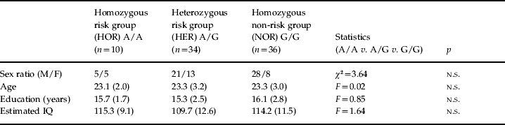
Five hundred and twenty-one healthy subjects (268 men, 253 women) were recruited from RWTH Aachen University to investigate associations between genotypes, personality traits and cognitive functions. From this large sample, a subsample of 80 subjects performed the functional magnetic resonance imaging (fMRI) task and were included in the present study (see Table 1). Inclusion criteria were right-handedness (assessed by the Edinburgh Laterality Scale; Oldfield, Reference Oldfield1971), age 18–55 years, and no psychiatric disorder according to ICD-10. After a description of the procedure, each participant gave written informed consent. The study protocol was approved by the local ethics committee according to the Declaration of Helsinki.
Sociodemographic variables of the sample (n=80)

M, Male; F, female; n.s., non-significant (p>0.05).
Values given as mean (standard deviation).
IQ was estimated using the Multiple-Choice Vocabulary Intelligence Test [Mehrfachwahl-Wortschatz-Intelligenztest (MWT-B); Lehrl, Reference Lehrl2005].
Genetic analysis
DNA was extracted from peripheral lymphocytes according to standard procedures. The SNP rs1006737 was genotyped on an Applied Biosystems 7900HT Fast Real-Time PCR System, using a TaqMan 5′ nuclease assay (TaqMan® SNP Genotyping Assay ID C_2584015_10; Applied Biosystems, USA). Genotyping accuracy was assessed by running 15% of the total sample in duplicate, resulting in a concordance of 100%. No deviation of genotype distribution from Hardy–Weinberg equilibrium (HWE) was detected using Haldane's exact test (Elston & Forthofer, Reference Elston and Forthofer1977).
fMRI task and stimuli
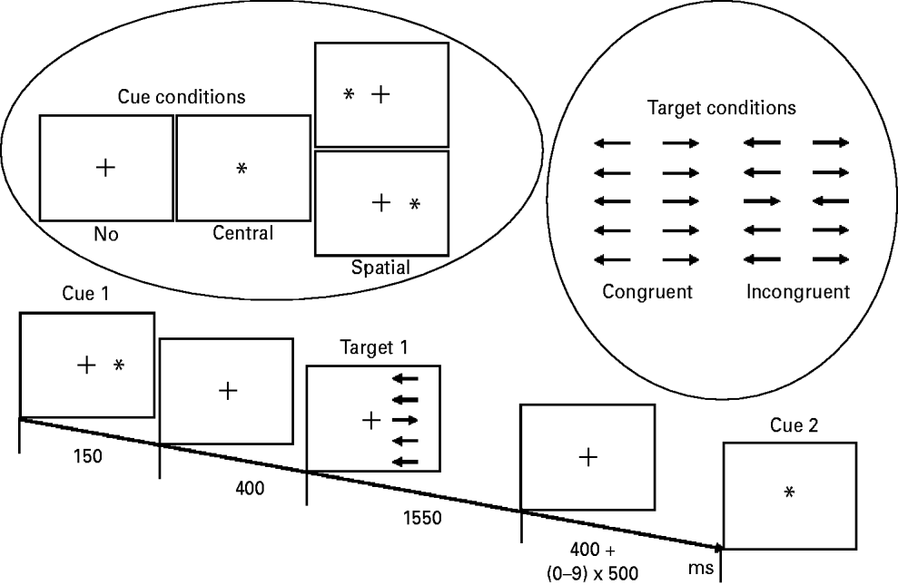
The fMRI task was a modified version of the original ANT (Fan et al. Reference Fan, McCandliss, Sommer, Raz and Posner2002). It was designed as an event-related paradigm using Presentation software (Neurobehavioral Systems Inc., USA). Three cue conditions (center cue, spatial cue, no cue), two target conditions (congruent, incongruent) and two visual field conditions (left, right) were used (see Fig. 1). An asterisk served as a cue, appearing either at the center of the screen (center cue), where it replaced a fixation cross, or lateralized next to the fixation cross, where it indicated the location of the following target stimulus (spatial cue). Targets consisted of blocks of five parallel horizontal lines, with arrowheads pointing leftward or rightward. These blocks were displaced approximately 4° from the center, either in the left or right visual field (visual field condition). The middle of the five arrows (at the horizontal midline) was the target stimulus that had to be attended to. The other four arrows (two above and two below the target stimulus) served as distractors. In each trial, all distractor arrows pointed either to the same direction (congruent condition) or to the opposite direction (incrongruent condition) as the target arrow. Stimulus material was presented in black against a white background.
Stimulus material and example for the course of one trial (right spatial cue/incongruent target).

Each trial started with a 150 ms presentation of either a cue or just the fixation cross in the ‘no cue’ condition. This was followed by an inter-stimulus interval (ISI) of 400 ms when the fixation cross was presented. Then the target block appeared (comprising target arrow and distractor arrows). Participants were required to indicate the direction of the target arrow by pressing a corresponding button with the left or right index finger. Targets were presented for 1550 ms and followed by a variable interval until the end of the trial when the fixation cross was presented. The duration of this interval was 400 ms plus a pseudo-randomized multiple (0–9 times) of 500 ms, resulting in a total ISI of 400–4900 ms. Twenty trials were presented for each of the 12 possible permutations of the three conditions (3×2×2) in a pseudo-randomized order, resulting in a total of 240 trials.
Data acquisition
fMRI took place on a 3-T Trio MR scanner (Siemens, Germany) at the Institute of Neuroscience and Biophysics – Medicine, Research Centre Jülich, using a T2*-weighted echo planar imaging (EPI) sequence (time repetition=2200 ms, echo time=30 ms, flip angle=90°). Slices covered the whole brain and were positioned transaxially parallel to the anterior–posterior commissural (AC–PC) line. A total of 391 functional images were acquired, each consisting of 36 slices (3 mm thickness, 20×20 cm field of view, 64×64 image matrix). The initial three images were excluded from further analysis so as to remove the influence of T1 stabilization effects.
fMRI data analysis
Analyses of fMRI data were calculated using SPM5 (www.fil.ion.ucl.ac.uk/spm). Functional images were realigned, normalized (to a voxel size of 2×2×2 mm), smoothed (6 mm isotropic Gaussian filter) and high-pass filtered (cut-off period 128 s). Twelve onset regressors (related to the onset of the targets) were defined resulting from the permutation of all conditions comprising three cues (no/center/spatial), two targets (congruent/incongruent) and two target positions (left/right). The hemodynamic response to each different event type was modeled using a canonical synthetic hemodynamic response function. The six head movement parameters were included as confounds. To identify the effects of (a) alerting, (b) orienting and (c) executive control, first-level linear contrasts were calculated for each subject, comparing (a) events of center cue versus no cue, (b) events of spatial cue versus center cue and (c) events of incongruent targets versus congruent targets. At the second level, the individual β-contrasts of the first-level analyses were used to calculate two-sample t tests to investigate for genotype effects (random effects analyses); for each attentional effect, group differences were examined between the homozygous non-risk group (NOR=G/G), the heterozygous risk group (HER=G/A) and the homozygous risk group (HOR=A/A).
Gene effects were expected to occur in brain areas known to be involved in alerting, orienting or executive control and to be impaired in patients with psychosis (Carter et al. Reference Carter, Mintun, Nichols and Cohen1997; Nestor et al. Reference Nestor, Kubicki, Gurrera, Niznikiewicz, Frumin, McCarley and Shenton2004, Reference Nestor, Kubicki, Spencer, Niznikiewicz, McCarley and Shenton2007).
According to the findings of previous studies, for each attentional domain, specific ROIs were defined by using masks from the WFU Pickatlas (Maldjian et al. Reference Maldjian, Laurienti, Kraft and Burdette2003): alerting=right hemisphere prefrontal and parietal areas (Posner & Petersen, Reference Posner and Petersen1990; Pardo et al. Reference Pardo, Fox and Raichle1991; Sturm et al. Reference Sturm, de Simone, Krause, Specht, Hesselmann, Radermacher, Herzog, Tellmann, Muller-Gartner and Willmes1999; Sturm & Willmes, Reference Sturm and Willmes2001; Fan et al. Reference Fan, McCandliss, Fossella, Flombaum and Posner2005); orienting=right hemisphere parietal areas (Posner & Petersen, Reference Posner and Petersen1990; Corbetta et al. Reference Corbetta, Miezin, Shulman and Petersen1993; Kastner et al. Reference Kastner, Pinsk, De Weerd, Desimone and Ungerleider1999; Corbetta et al. Reference Corbetta, Kincade, Ollinger, McAvoy and Shulman2000; Fan et al. Reference Fan, McCandliss, Fossella, Flombaum and Posner2005); executive control of attention=left hemisphere prefrontal areas (Botvinick et al. Reference Botvinick, Nystrom, Fissell, Carter and Cohen1999; Carter et al. Reference Carter, Botvinick and Cohen1999; Bush et al. Reference Bush, Luu and Posner2000; Macdonald et al. Reference Macdonald, Cohen, Stenger and Carter2000; Fan et al. Reference Fan, Flombaum, McCandliss, Thomas and Posner2003a, Reference Fan, McCandliss, Fossella, Flombaum and Posner2005). The prefrontal areas included the superior, middle and inferior frontal gyrus and also the anterior cingulate cortex. Analyses were performed with a predefined threshold of p<0.001 (uncorrected).
Behavioral data analysis
Attentional effects were calculated as differential (d) scores from RTs as follows: (1) alerting=(no cue) – (center cue); (2) orienting=(center cue) – (spatial cue); (3) executive control=(incongruent targets) – (congruent targets). Group differences for each attentional effect were proved by two-sample t tests.
Results
Subjects
Genetic analysis of SNP rs1006737 showed the following distribution: 10 subjects were homozygous for the risk allele (A/A), 34 subjects were heterozygous (A/G) and 36 subjects belonged to the non-risk group (G/G). Groups did not differ regarding sex ratio, age, education and estimated IQ (see Table 1).
Behavioral data
Accuracy was comparably high in each group: NOR 98.0±2.4%, HER 97.8±2.3% and HOR 98.8±1.2%. Error trials were discarded from further analyses.
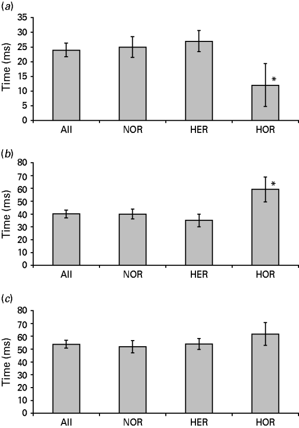
RT analyses of the sample in total showed significant effects for each of the three attentional domains: RTs were faster at center cue (617±92 ms) than no cue (641±94 ms) trials (alerting effect: d=24±21 ms, t=10.1, p<0.001) and slower at center cue than spatial cue (577±95 ms) trials (orienting effect: d=40±28 ms, t=12.9, p<0.001). The executive control of attention effect resulted from slower RTs (d=54±26 ms, t=18.8, p<0.001) at incongruent (638±93 ms) than congruent (584±93 ms) trials.
For each attentional effect, group differences were calculated between the NOR, HER and HOR groups (see Fig. 2 a–c). Alerting: the HOR group did profit less from the cue than the HER group (d=15 ms, t=1.9, p<0.05) and the NOR group (d=13 ms, t=1.7, p<0.05). Orienting: the HOR group was impaired in orienting compared to the HER (d=23 ms, t=2.2, p<0.05) and NOR (d=19 ms, t=2.1, p<0.05) groups. Executive control: the HOR group showed higher cost by the conflict condition than the HER (d=8 ms, t=0.9, p=0.18) and NOR (d=10 ms, t=1.0, p=0.16) groups but these effects did not reach significance.
Behavioral effects for (a) alerting, (b) orienting and (c) executive control of attention. * Significant differences compared to the other groups (p<0.05). NOR, Homozygous non-risk group; HER, heterozygous risk group; HOR, homozygous risk group.

A two-way ANOVA did not reveal any significant (p<0.05) main effect for sex (alerting: F=2.12; orienting: F=0.82; executive control: F=2.37) or sex/genotype interactions (alerting: F=2.66; orienting: F=0.42; executive control: F=0.93) for any of the three attentional domains.
fMRI data
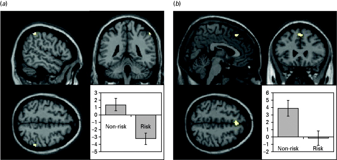
Group differences were investigated for alerting, orienting and executive control in the predefined ROIs. No group differences were found between the NOR, HER and HOR groups. To increase statistical power (because there were only 10 subjects in the HOR group), the HER and HOR groups were unified to a single risk group and compared to the NOR group. During orienting, the group of risk allele carriers showed significantly reduced neural activity in the right Brodmann area (BA) 40 of the inferior parietal lobule [Talairach coordinates (TC) 55, −38, 52, cluster size 23 voxels, T=3.88, p<0.001 uncorrected; see Fig. 3 a]. During executive control, risk allele carriers had reduced neural activity in the left BA 8 of the medial frontal gyrus (TC 0, 28, 47, cluster size 50 voxels, T=4.03, p<0.001 uncorrected; see Fig. 3 b). No group differences were found for alerting and for the reverse contrasts (risk>non-risk). No significant (p<0.001 uncorrected) main effects for sex or sex/genotype interactions were found for any of the three attentional domains.
Decreased neural activity during (a) orienting and (b) executive control of attention: risk group<non-risk group.

Discussion
In the current fMRI study we investigated the effects of the CACNA1C gene variant SNP rs1006737 on attention networks as defined by Posner & Petersen (Reference Posner and Petersen1990). As an activation paradigm we used an adapted version of the well-established ANT (Fan et al. Reference Fan, McCandliss, Fossella, Flombaum and Posner2005) to examine the three attentional subdomains of alerting, orienting and executive control.
At the behavioral level, homozygous risk allele carriers showed impaired performance in phasic alerting and orienting measured by RT differences. They profited less from a central cue in the alerting condition and showed prolonged reactions in the orienting condition compared to the heterozygous risk group and the homozygous non-risk group. These results are in line with general findings that cognitive deficits have a heritable component and can be found in patients with psychosis and also in their close relatives. We show that this holds true for a specific cognitive domain (attention) on a single genetic variant/SNP level. Although genome-wide association studies have implicated the SNP rs1006737 in the CACNA1C gene as a susceptibility factor for bipolar disorder, schizophrenia and major depression, our study suggests one way in which this effect might evolve at a physiological level. By means of functional imaging, reduced neural activity was found when homozygous and heterozygous risk allele carriers were subsumed to a single risk group and compared to the homozygous non-risk group. The risk group showed decreased neural activity in the right BA 40 of the inferior parietal lobule during orienting and in the left BA 8 of the medial frontal gyrus during executive control of attention. No genotype effect was found for the alerting condition. The levels of significance of the above-mentioned clusters did not survive statistical correction for multiple comparisons. However, these clusters are within the hypothesized areas according to the literature and might therefore be ascribed to genotype effects.
BA 40 belongs to the posterior attention system as described by Posner & Petersen (Reference Posner and Petersen1990). It has been shown that damage to the posterior parietal lobe affects the ability to disengage from an attentional focus to a target located at a position other than the expected one (Posner et al. Reference Posner, Walker, Friedrich and Rafal1984). Exactly this ability was impaired in our CACNA1C risk group, possibly based on the dysfunction of a small cluster in the right BA 40. It is well known that the right inferior parietal lobe is an important area for the development of hemispatial neglect, a syndrome that is characterized by a disturbed ability to orient attention to the left hemispace (Mort et al. Reference Mort, Malhotra, Mannan, Rorden, Pambakian, Kennard and Husain2003).
Medial frontal areas including BA 8 have often been reported to be involved in executive control functions. According to the response-conflict theory (Botvinick et al. Reference Botvinick, Braver, Barch, Carter and Cohen2001), this region plays a crucial role whenever a choice between incompatible actions is required, which results in a response conflict and an inhibition of one response in favor of another (Ullsperger & von Cramon, Reference Ullsperger and von Cramon2001; Rushworth et al. Reference Rushworth, Hadland, Paus and Sipila2002; Garavan et al. Reference Garavan, Ross, Kaufman and Stein2003; Nachev et al. Reference Nachev, Rees, Parton, Kennard and Husain2005, Reference Nachev, Wydell, O'Neill, Husain and Kennard2007).
The comparison with a recent genetic imaging study (Thimm et al. Reference Thimm, Krug, Kellermann, Markov, Krach, Jansen, Zerres, Eggermann, Stöcker, Shah, Nöthen, Rietschel and Kircher2010b) suggests that the effects on attention networks seem to be gene specific; that is, attention networks are not influenced in the same way by every gene that confers risk to psychiatric disorders. In that fMRI study we investigated the effect of the SNP rs1018381 of the DTNBP1 gene on the three attentional subdomains by using the same paradigm. No genotype effect was found for the alerting and orienting network. However, in the executive control condition, the risk group showed reduced neural activity in the left superior frontal gyrus (BA 9) associated with a significant behavioral deficit compared to the non-risk group. At the behavioral level, specifically increased executive control effects have also been reported previously for Met allele carriers of the catechol-O-methyltransferase (COMT) gene (Opgen-Rhein et al. Reference Opgen-Rhein, Neuhaus, Urbanek, Hahn, Sander and Dettling2008).
The detected neural activity of our present study is also specific for the particular cognitive domain (attention). A study by Krug et al. (Reference Krug, Nieratschker, Markov, Krach, Jansen, Zerres, Eggermann, Stocker, Shah, Treutlein, Muhleisen and Kircher2010) investigated the effects of CACNA1C on neural correlates of a verbal fluency task. In contrast to the attention task of the present experiment, that study found increased activation in risk allele carriers in the left inferior frontal gyrus and the left precuneus. This was interpreted as an inefficient recruitment of prefrontal networks or as a compensatory activation.
Our data suggest that the behavioral and physiological effects of the CACNA1C gene variant SNP rs1006737 differ with respect to the attentional domain and the neural networks involved in the tasks we used. It seems that the different subdomains we investigated might have different genetic underpinnings. Independence of heritability between alerting, orienting and executive control was demonstrated in a study by Fan et al. (Reference Fan, Wu, Fossella and Posner2001), who found heritability specifically for the executive attention network. However, because their study used another methodical approach (twin study) and did not control for the distribution of single genotypes, their findings are not necessarily contradictory to our study, which explicitly investigated the effects of a single genotype.
It is likely that genetic variants found to be associated with psychiatric disorders are not associated with the disorder per se, but with more subtle endophenotypes underlying these disorders. However, it is also likely that the effect of a given variant may not only influence one domain but also have pleiotropic effects, depending on the given individual genetic make-up. We therefore assume that the effect of this variant is one way in which it confers risk to bipolar disorder, schizophrenia and major depression, but it is possible that it may also act through impairment of other neuronal functions.
Limitations
In our study we found effects at the behavioral and functional imaging levels, but these effects were not completely congruent. In the alerting condition we did not find a neural correlate for the behavioral deficit that was detected in the risk allele carriers. This deficit concerned the automatic increase of response readiness triggered by a preceding visual cue. It might be assumed that the alerting network was not impaired in the sense of a hypofunction of neuronal clusters but rather in the sense of a general slowing down. If the network is intact but just working more slowly, the BOLD response does not necessarily differ. Another explanation would be that the performance difference in phasic alerting was relatively small and not sufficient to find a neural correlate. This finding is in line with the results of a study with schizophrenia patients that showed deficits in the executive and orienting network but not in the alerting network (Wang et al. Reference Wang, Fan, Dong, Wang, Lee and Posner2005).
By contrast, in the executive control condition we found decreased neural activity in the risk group without a significant correlate at the behavioral level. However, there were numerical differences between the three groups in the expected direction and it has been suggested that physiological indicators (such as changes in neural activity) may be more sensitive than behavioral measures (Fink et al. Reference Fink, Marshall, Weiss, Toni and Zilles2002; Wilkinson & Halligan, Reference Wilkinson and Halligan2004).
The orienting condition was the only one that revealed significant effects at both the behavioral and functional imaging levels. At the behavioral level, the HOR group showed significant impairment compared to the HER and the NOR groups. However, at the functional imaging level significant effects were only found when the HOR and HER groups were merged to form a single risk group and compared to the NOR group. This was done to increase the statistical power because the HOR group consisted of only 10 subjects. For these reasons, our findings can be regarded as exploratory and conclusions should be drawn with caution. Further investigations with a larger HOR group would be desirable and might lead to a better congruency between behavioral and functional imaging data.
In summary, we investigated the effect of CACNA1C rs1006737 on three subdomains of attention. Risk allele carriers showed performance deficits in alerting and orienting in addition to reduced neural activity during orienting (BA 40) and executive control (BA 8). Our results suggest that attention deficits in psychosis might be co-determined by CACNA1C rs1006737 allele status and might be based on functional deficits that are already emerging in healthy risk allele carriers and affecting well-known key regions of attention networks.
Acknowledgements
This work was supported by the Federal Ministry of Education and Research (BMBF; Brain Imaging Centre West, 01GO0204 and 01GW0751) and by the START Program of the Medical Faculty of RWTH Aachen University.
Declaration of Interest
None.