Introduction
Haemoptysis can be caused by diseases affecting either the tracheobronchial tree or the upper aerodigestive tract.Reference Tremble1 The latter is of principal concern to the otolaryngologist though the tracheobronchial tree is the main source of pathology. Haemoptysis patients may present to the ENT department through a direct referral or via a respiratory service following inconclusive investigation. The aetiological cause of haemoptysis is usually benign but may be sinister.Reference Tsoumakidou, Chrysofakis, Tsiligianni, Maltezakis, Siafakas and Tzanakis2, Reference Abal, Nair and Cherian3
Our article aims to provide an evidence-based clinical approach to managing patients presenting with haemoptysis. It also reviews the causes of haemoptysis (Table I).Reference Bidwell and Pachner4 The following is a suggested strategy for history-taking, and examining, investigating and managing patients with haemoptysis.
Haemoptysis aetiology

Materials and methods
The data in this article are based on a systematic literature search performed in August 2013 using PubMed. The keywords used included ‘haemoptysis’ in combination with ‘otolaryngology’, ‘ENT’, ‘head & neck’, ‘diagnosis’, ‘management’, ‘investigations’ and ‘treatment’. The abstracts of these articles were reviewed.
Results
A total of 967 articles were reviewed. Twenty-eight articles were relevant to the management of otolaryngological aspects of haemoptysis; 939 articles were excluded as they applied to respiratory-related aspects of haemoptysis.
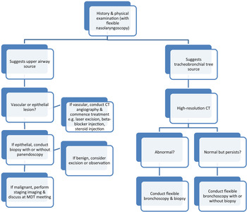
Figure 1 shows an algorithm for haemoptysis management based on the findings of the literature search.
Haemoptysis management algorithm. CT = computed tomography; MDT = multidisciplinary team

Discussion
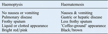
Haemoptysis is usually intermittent rather than continuous, though the frequency does not indicate a cause. Haemoptysis should be clearly differentiated from haematemesis, the aetiology of which arises from the oesophago-gastric tract (Table II).Reference Bidwell and Pachner4 Unless there is a clear trigger event for the haemoptysis, the duration will not otherwise give an indication of the cause either.Reference Wolfe and Simmons5 The amount of haemoptysis reported by the patient is an unreliable indicator of cause too; haemoptysis can be difficult to quantify as blood may be swallowed or aspirated. Additionally, even small amounts of blood can indicate serious disease.Reference Wolfe and Simmons5
Differentiating features of haemoptysis and haematemesis

Specific associations can give an indication of the cause. Onset of haemoptysis from an older age with associated airway symptoms (e.g. cough, wheeze and expiratory stridor) is more suggestive of tracheobronchial tree carcinoma, usually in patients with a history of smoking.Reference Bidwell and Pachner4, Reference Abbate, Lancella, Contini and Scotti6, Reference Naqiyah, Zulkarnaen, Rohaizak and Das7 Intermittent haemoptysis following epistaxis suggests severe post-nasal bleeding that requires intervention such as mucosa moisturisation, cautery or vessel ligation. Onset following tracheostomy may precede an impending haemorrhage from a tracheo-innominate artery fistula (a herald bleed).Reference Wang, Xu, Tang and Yue8 Development in the presence of a growing anterior neck mass with compressive symptoms (e.g. dyspnoea, stridor and dysphagia) suggests local invasion from a thyroid mass.Reference McCaffrey and Lipton9 Haemoptysis following airway surgery can be attributed to that intervention.Reference Armstrong, Chiosea, Carty, Hodak and Yip10, Reference Hassani and Bhananker11 Development in conjunction with fever or pleuritic chest pain suggests an infective process, including pneumonia, bronchiectasis or tuberculosis.
Association with cyclical menses in women describes idiopathic catamenial haemoptysis due to endometrial tissue deposition in the lungs. Association with orthopnoea and paroxysmal nocturnal dyspnoea suggests congestive cardiac failure.Reference Bidwell and Pachner4 Development following a diagnosis of breast, renal or colon cancer suggests pulmonary metastases with mucosal invasion and blood vessel erosion.Reference Bidwell and Pachner4 Pulmonary embolism will be associated with pleuritic chest pain and shortness of breath.
Bleeding from oral cavity lesions, gum disease and poor dentition can occur.Reference Kruse, Zwahlen, Bredell, Gengler, Dannemann and Grätz12 Particular concern should be paid to bleeding from non-healing tooth sockets following extraction, which can indicate an underlying malignancy, especially in the presence of pain over the area affected. Two separate patterns of haemorrhage from tonsils during episodes of tonsillitis have been described: diffuse parenchymal bleeding and more localised bleeding from small blood vessels on the tonsil surface.Reference Levy, Brodsky and Stanievich13 The commonest associated infective organism in cases of haemorrhagic tonsillitis is group B haemolytic streptococcus.Reference Levy, Brodsky and Stanievich13, Reference Calubiran, Zackson and Cunha14
History of foreign travel may be useful to assess for infective causes, including tuberculosis.Reference Abal, Nair and Cherian3, Reference Kuehnemund and Bootz15 Injuries to the neck and those affecting the larynx may result in subtle haemoptysis.Reference O'Neill, Shine and Timon16 Penetrating injuries can be accompanied by subcutaneous emphysema and hoarseness in cases of recurrent laryngeal nerve injury. Sharp foreign bodies that are ingested penetrate the mucosal linings causing localised pain and bleeding.Reference Bolzoni, Peretti, Piazza, Pedruzzi and Nicolai17 Symptoms more indicative of head and neck cancer (e.g. sore throat, voice hoarseness, otalgia, neck lump and dysphagia) should be established. These may suggest the presence of oropharyngeal and laryngeal lesions, as well as compression and erosion of surrounding nerves. Stridor develops on airway obstruction.
Ear examination can detect middle-ear effusion, most commonly unilateral, which may indicate the presence of eustachian tube dysfunction due to an occluding nasopharyngeal lesion.Reference Tabba, Anderson and Lamb18, Reference Chu, Wu, Tunkel and Ishman19 Anterior rhinoscopy can reveal blood clots or prominent blood vessels on either the nasal septum or lateral nasal wall. Septal perforations may be a bleeding source and can indicate a variety of conditions. Ulcerating lesions, granulomas and polyps may be visualised too, the latter suggesting a sinonasal malignancy if unilateral. Foreign bodies in the nose may be the cause of recent or long-standing haemoptysis, as can ectopic teeth within the maxillary antrum.Reference Saleem, Khalid, Hameed and Ghaffar20 Tongue, oral cavity and tonsil lesions may appear as ulcers or swellings. These usually represent primary pathology, though very rarely can be a source of distant metastases.Reference Tueche, Nguyen, Larsimont and Andry21 Oral cavity masses should be bimanually palpated to assess the location and depth of spread, though this may prompt further bleeding. Foreign bodies lodged in the tonsil or tongue may be visualised if suspected in the history. Poorly fitting dentures can be assessed too. Tonsils may appear enlarged with visible crypts or appear shrunken from fibrosis following recurrent episodes of tonsillitis.
Examination of the nasopharynx, tongue base, hypopharynx and larynx with a flexible nasolaryngoscope is essential. Lesions at these sites can be localised and biopsy samples taken if possible. Nasopharyngeal, tongue base, larynx and pharyngeal lesions can all be viewed, with the size and precise location documented.Reference Martines and Immordino22–Reference Yu, Qu, Kong, Pan, Wang and Lv26 A flexible nasolaryngoscope can be used following topical anaesthesia to view the trachea and main bronchi and to cannulate the upper oesophagus, allowing brief mucosal inspection during swallowing. Reference Abbate, Lancella, Contini and Scotti6, Reference Meyer, Becker and Quinones27, Reference Kuschill-Dziurda, Mastalerz, Grzanka and Niżankowska-Moglinicka28 Fibre-optic transnasal oesophagoscopy is increasingly employed by ENT surgeons. Lymphadenopathy may indicate advanced metastatic disease or be part of an infective process. A thyroid mass may also account for haemoptysis due to local invasion of the airway. Such thyroid tumours are rare, but are more likely to be undifferentiated histological types, with a poor prognosis and a greater propensity for regional spread and metastasis.Reference Naqiyah, Zulkarnaen, Rohaizak and Das7, Reference Armstrong, Chiosea, Carty, Hodak and Yip10
Ear, nose and throat clinicians can rely on their general medical knowledge and experience when managing haemoptysis. Reduced chest expansion may reveal a foreign body or pleural effusion as a consequence of pneumonia. The latter produces a stony dull note on percussion. Auscultation can detect reduced air entry suggesting bronchial obstruction, and crepitations suggesting infection (e.g. pneumonia, bronchiectasis and cystic fibrosis). Digital clubbing can reveal bronchial carcinoma and cyanotic heart disease. Peripheral and central cyanosis indicates disease location and severity. Raised jugular venous pressure can be caused by superior vena caval obstruction from bronchial carcinoma.
All patients with haemoptysis not clearly defined by an aerodigestive disorder should have a chest X-ray performed. Lung lesions can be detected, as can infective processes. However, chest X-rays are considerably less sensitive than high-resolution computed tomography (CT).Reference Gavelli and Giampalma29 At the very least, it is a useful screening tool. However, further investigation of patients with haemoptysis who have a normal chest X-ray is justified and necessary in smokers. This is because of the higher incidence of lung malignancy in such patients, and because small lung tumours not detected on chest X-ray can still account for a varying level and frequency of haemoptysis.Reference Wolfe and Simmons5, Reference Thirumaran, Sundar, Sutcliffe and Currie30 Facial X-rays may detect foreign bodies in the nose and paranasal sinuses accounting for haemoptysis.Reference Tabba, Anderson and Lamb18
High-resolution CT is the ‘gold standard’ radiological investigation for the detection of lung lesions. Sensitivity and specificity for lung tumour detection using high-resolution CT is 83 per cent and 78 per cent respectively.Reference Clin, Morlais, Guittet, Gislard, Marquignon and Paris31, Reference Pastorino32 Low-dose scanning has been proposed as a new screening investigation.Reference Golice33 Mediastinal involvement can be assessed. Computed tomography scanning allows tissue sampling for analysis (via a biopsy) and staging of any malignant disease. Metastatic head and neck malignancy can also be detected, as can lung metastases from other primary tumour sites. Foreign bodies within the tracheobronchial tree can be detected on CT scanning too. Upper aerodigestive tract malignancy should be properly staged using CT neck imaging to assess for deeper tissue involvement and regional lymphatic spread. In those patients in whom a pulmonary embolism is suspected, a CT pulmonary angiogram should be performed.
• Haemoptysis has a varied and potentially sinister aetiology due to pathology from both upper and lower aerodigestive tracts
• A history and thorough clinical examination can identify haemoptysis aetiology, without need for radiological imaging
• Once a cause has been identified, appropriate treatment should be offered to achieve symptom resolution
Neck ultrasound allows masses to be defined and suspicious features recorded. Though the investigation is operator-dependent, it does allow aspiration cytology for analysis and clinical decision-making. Flexible bronchoscopy permits visualisation of the upper part of the tracheobronchial tree, and cytological diagnosis through endobronchial washings, brushings or even direct biopsy. The procedure is well established, with a low morbidity, and can be performed under local anaesthesia. It is justified in all patients with haemoptysis, even those with a normal chest X-ray.Reference Heaton34 Rigid bronchoscopy permits haemorrhage control and foreign body removal, whilst ventilating the anaesthetised patient and providing a secure airway. A full upper airway endoscopy allows visualisation of the upper aerodigestive tract and the tracheobronchial tree to the level of the main bronchi. Disease location and extent can be assessed, which contributes to staging in the case of malignancy. Biopsy samples can also be taken. Particular attention should be paid to the pyriform fossa, posterior pharyngeal wall and post-cricoid region. A tympanogram can demonstrate a type B picture with normal ear canal volume in those patients with a post-nasal space lesion obstructing the eustachian tube.
Conclusion
Haemoptysis remains an uncommon presenting symptom that the ENT surgeon may encounter at various stages of investigation. The aetiology usually originates from the lungs or tracheobronchial tree, and so ENT specialists should be familiar with these. However, based on our review, the three main ENT causes that should be considered are foreign bodies, upper aerodigestive tract tumours and thyroid tumours. A thorax CT should be considered in all cases of unexplained haemoptysis.Reference Hill, Pasteur, Cornford, Welham and Bilton35, 36
A full history and examination can aid diagnosis, whilst endoscopic examination and imaging allow full assessment of disease. If a lower respiratory tract cause is suspected, CT imaging of the thorax and direct visualisation of the airway through bronchoscopy should be arranged.Reference Tsoumakidou, Chrysofakis, Tsiligianni, Maltezakis, Siafakas and Tzanakis2 If an upper respiratory tract cause is suspected, flexible nasolaryngoscopy should be urgently arranged to identify the site and possible aetiology. In a proportion of cases, the cause may never be identified. A chest X-ray may provide information that correlates with endoscopic and CT imaging findings. Idiopathic haemoptysis can be encountered, though no studies reporting the incidence of this have been published.