Children and adolescents who experience significant adversity are at greater risk for developing mental health problems (Edwards, Holden, Felitti, & Anda, Reference Edwards, Holden, Felitti and Anda2003; Evans, Li, & Whipple, Reference Evans, Li and Whipple2013; Kessler et al., Reference Kessler, McLaughlin, Green, Gruber, Sampson, Zaslavsky and Williams2010). Adversity characterized by repeated exposure to threat may alter the functioning of neurophysiological systems involved in the processing of threat (McCrory, De Brito, & Viding, Reference McCrory, De Brito and Viding2010; McLaughlin, Sheridan, & Lambert, Reference McLaughlin, Sheridan and Lambert2014; Sheridan & McLaughlin, Reference Sheridan and McLaughlin2014; VanTieghem & Tottenham, Reference VanTieghem and Tottenham2016), changing the way emotional information is processed (Cicchetti & Curtis, Reference Cicchetti and Curtis2005; Dodge, Pettit, Bates, & Valente, Reference Dodge, Pettit, Bates and Valente1995; Guyer et al., Reference Guyer, Kaufman, Hodgdon, Masten, Jazbec, Pine and Ernst2006; Masten et al., Reference Masten, Guyer, Hodgdon, McClure, Charney, Ernst and Monk2008; McLaughlin & Sheridan, Reference McLaughlin and Sheridan2016; Pollak, Reference Pollak2003), and increasing risk for internalizing problems (e.g., depression and anxiety; Cicchetti & Toth, Reference Cicchetti and Toth2005; Miller et al., Reference Miller, Sheridan, Hanson, McLaughlin, Bates, Lansford and Dodge2018; Mrug & Windle, Reference Mrug and Windle2010). Threats encountered in peer and community contexts may be particularly influential for adolescents, due to their greater autonomy and orientation toward peers compared to younger children (Brown & Larson, Reference Brown, Larson, Lerner and Steinberg2009; Updegraff, McHale, Whiteman, Thayer, & Crouter, Reference Updegraff, McHale, Whiteman, Thayer and Crouter2006). While previous research has demonstrated relations between specific neurobiological systems and adversity in adolescence, little work has examined how adversity impacts the coordination between neurobiological systems. The flexible, predictive regulation of autonomic nervous system (ANS) activity is central to emotion processing and regulation (Seth & Friston, Reference Seth and Friston2016; Smith, Thayer, Khalsa, & Lane, Reference Smith, Thayer, Khalsa and Lane2017). The brain's emotional circuitry, including brain regions involved in the regulation of ANS activity, undergoes considerable development during adolescence, potentially making it a sensitive period for environmental influences on emotion regulation and dysregulation (Nelson, Jarcho, & Guyer, Reference Nelson, Jarcho and Guyer2016). The goal of the present study was to examine how the coordination of brain and ANS activity during emotion processing varies as a function of Mexican-origin adolescents’ exposure to threats in the community and peer contexts, and relates to internalizing problems.
Threat Exposure and Latinx Adolescents’ Internalizing Problems
The peer and community context are particularly salient sources of threat for Latinx adolescents. Adolescents spend more unsupervised time in their communities than younger children and their behavior is more susceptible to its influence (Brown & Larson, Reference Brown, Larson, Lerner and Steinberg2009). Within the neighborhood context, adolescents increasingly spend time with peers that is not supervised or monitored by caregivers (Csikszentmihalyi, Larson, & Prescott, Reference Csikszentmihalyi, Larson and Prescott2014; Updegraff et al., Reference Updegraff, McHale, Whiteman, Thayer and Crouter2006). School and neighborhood crime therefore have a greater opportunity to influence adolescents’ socioemotional development and adjustment. Moreover, violent crime exposure is more strongly associated with internalizing problems among adolescents than among children (Fowler, Tompsett, Braciszewski, Jacques-Tiura, & Baltes, Reference Fowler, Tompsett, Braciszewski, Jacques-Tiura and Baltes2009). Latinx adolescents are exposed to more community crime than their European American counterparts (Slopen et al., Reference Slopen, Shonkoff, Albert, Yoshikawa, Jacobs, Stoltz and Williams2016), suggesting that although it is not culturally specific, community crime exposure may be a particularly prevalent threat for Mexican-origin adolescents.
Given adolescents’ increased time spent with peers, peer victimization is another potent threat in an adolescent's environment. Rates of peer victimization have been found to increase from late childhood into middle adolescence (Troop-Gordon, Reference Troop-Gordon2017). In addition, the association between peer victimization and internalizing problems is stronger for adolescents than for children (Cook, Williams, Guerra, Kim, & Sadek, Reference Cook, Williams, Guerra, Kim and Sadek2010) and may be particularly strong during the early adolescent years (Sheppard, Giletta, & Prinstein, Reference Sheppard, Giletta and Prinstein2016). Peer victimization also predicts increases in internalizing symptoms over time (Reijntjes, Kamphuis, Prinzie, & Telch, Reference Reijntjes, Kamphuis, Prinzie and Telch2010). It is therefore a highly threatening stressor for adolescents, increasing their risk for developing internalizing problems (Prinstein, Cheah, & Guyer, Reference Prinstein, Cheah and Guyer2005) through impacts on the developing brain (Casement et al., Reference Casement, Guyer, Hipwell, McAloon, Hoffmann, Keenan and Forbes2014).
Peer victimization also increases emotional distress among Mexican-origin adolescents (Espinoza, Gonzales, & Fuligni, Reference Espinoza, Gonzales and Fuligni2013). One salient form of peer victimization for Latinx adolescents is discrimination (Seaton, Neblett, Cole, & Prinstein, Reference Seaton, Neblett, Cole and Prinstein2013). Discrimination is the experience of unfair treatment from others because of one's identity or status. Discrimination is an important and stressful feature of life for socially disadvantaged groups (Kessler, Mickelson, & Williams, Reference Kessler, Mickelson and Williams1999). Experiencing discrimination from peers based on one's ethnicity can disrupt the process of developing one's ethnic identity during adolescence, delaying commitment to one's ethnic identity, with negative consequences for self-esteem and mental health (Umaña-Taylor & Updegraff, Reference Umaña-Taylor and Updegraff2007). Discrimination has been found to predict later internalizing problems (Delgado, Updegraff, Roosa, & Umaña-Taylor, Reference Delgado, Updegraff, Roosa and Umaña-Taylor2011; Smokowski & Bacallao, Reference Smokowski and Bacallao2007). Moreover, Latinx adolescents’ perceived discrimination has been found to predict physical and relational victimization by peers (Seaton et al., Reference Seaton, Neblett, Cole and Prinstein2013). Facing ethnic discrimination therefore composes an important portion of a constellation of threats with negative consequences for Mexican-origin adolescents’ emotional functioning.
School and neighborhood crime, peer victimization, and discrimination are common and frequently co-occurring threats that are especially prevalent and salient for Mexican-origin youth, and each has been shown to relate to subsequent internalizing problems. Identifying the mechanisms through which these threats influence adolescents’ emotion functioning may therefore be essential for understanding individual differences in risk for developing internalizing problems. One key mechanism to consider is emotion dysregulation due to adaptation of adolescents’ neurophysiological functioning to threats in the peer and neighborhood contexts.
Threat, Emotion Dysregulation, and Adolescent Brain Development
Because of the confluence of neurodevelopmental changes coupled with increasing autonomy and complexity of social relationships, the adolescent brain is theorized to be uniquely attuned to the social environment (Nelson et al., Reference Nelson, Jarcho and Guyer2016), and particularly sensitive to influences from that environment on its development and later functioning (Blakemore & Mills, Reference Blakemore and Mills2014; Schriber & Guyer, Reference Schriber and Guyer2016; Šimic, Reference Šimic2012). Adolescence is a period of increased emotional volatility and intensity, especially in social contexts (Arnett, Reference Arnett1999; Larson, Moneta, Richards, & Wilson, Reference Larson, Moneta, Richards and Wilson2002; Monahan, Guyer, Silk, Fitzwater, & Steinberg, Reference Monahan, Guyer, Silk, Fitzwater, Steinberg and Cicchetti2016). This increased emotionality is at least partially mediated by increased reactivity and plasticity in the amygdala, striatum, and medial prefrontal cortex (mPFC), among other regions (Guyer, Silk, & Nelson, Reference Guyer, Silk and Nelson2016). In particular, structural development of the mPFC has been found in animal models to confer greater sensitivity to both threat and safety cues (Pattwell et al., Reference Pattwell, Liston, Jing, Ninan, Yang, Witztum and Lee2016). In humans, the magnitude of amygdala–mPFC coupling increases across adolescence (Gabard-Durnam et al., Reference Gabard-Durnam, Flannery, Goff, Gee, Humphreys, Telzer and Tottenham2014; Heller, Cohen, Dreyfuss, & Casey, Reference Heller, Cohen, Dreyfuss and Casey2016). This increasing connectivity facilitates inhibition of amygdala reactivity by mPFC during emotion processing, as indicated by increasingly negative task-based connectivity (Gee, Humphreys, et al., Reference Gee, Humphreys, Flannery, Goff, Telzer, Shapiro and Tottenham2013; Silvers et al., Reference Silvers, Insel, Powers, Franz, Helion, Martin and Ochsner2017). Thus, because the mPFC circuitry involved in regulating affective responding undergoes significant development during adolescence, it may be particularly sensitive to the influence of threats experienced at that time.
In the presence of early life adversity, children's neurophysiology may become specialized for detecting threat (Blair & Raver, Reference Blair and Raver2012; Frankenhuis & de Weerth, Reference Frankenhuis and de Weerth2013). For example, children who experience child maltreatment, a severe form of threat in early life, detect anger in faces with greater sensitivity (Pollak, Cicchetti, Hornung, & Reed, Reference Pollak, Cicchetti, Hornung and Reed2000; Pollak & Kistler, Reference Pollak and Kistler2002; Pollak & Sinha, Reference Pollak and Sinha2002). The neural circuitry for processing threat is calibrated by adversity, such that the magnitude of the brain's reactivity to threatening stimuli and the regulation of those responses vary as a function of prior adversity (Evans et al., Reference Evans, Swain, King, Wang, Javanbakht, Ho and Liberzon2016; Maheu et al., Reference Maheu, Dozier, Guyer, Mandell, Peloso, Poeth and Ernst2010; Suzuki et al., Reference Suzuki, Luby, Botteron, Dietrich, McAvoy and Barch2014; Swartz, Williamson, & Hariri, Reference Swartz, Williamson and Hariri2015; VanTieghem & Tottenham, Reference VanTieghem and Tottenham2016). Exposure to threats in the sensitive period of adolescence may also contribute to the ongoing calibration of coordinated neurobiological responses. For example, stressful life events experienced in adolescence have been found to predict subsequent threat-related increases in amygdala reactivity to fearful faces (Swartz et al., Reference Swartz, Williamson and Hariri2015), and cumulative adversity in childhood and early adolescence have been found to relate to amygdala hyperreactivity to neutral faces in early adulthood (Evans et al., Reference Evans, Swain, King, Wang, Javanbakht, Ho and Liberzon2016). While this hypervigilance to threat may be protective against bodily harm and thereby promote immediate survival, it has trade-offs.
Emotion dysregulation is a pattern of emotional responses and regulation that interferes with an individual's goals (Beauchaine, Reference Beauchaine2015a; Cole, Hall, & Hajal, Reference Cole, Hall, Hajal, Beauchaine and Hinshaw2017). Variations in emotion functioning that can serve as adaptations to threatening contexts can disrupt an individual's ability to effectively regulate his or her emotional responses in nonthreatening contexts. Exposure to violence in childhood is associated with emotion dysregulation as indicated by behavioral observations and parent and self-reports (Heleniak, Jenness, Vander Stoep, McCauley, & McLaughlin, Reference Heleniak, Jenness, Vander Stoep, McCauley and McLaughlin2016; Kim & Cicchetti, Reference Kim and Cicchetti2010; Maughan & Cicchetti, Reference Maughan and Cicchetti2002; Shields & Cicchetti, Reference Shields and Cicchetti1997). In adolescence, emotion dysregulation is associated with exposure to threats in the peer and community context, and mediates their association with later internalizing problems (Heleniak, King, Monahan, & McLaughlin, Reference Heleniak, King, Monahan and McLaughlin2018; McLaughlin, Hatzenbuehler, & Hilt, Reference McLaughlin, Hatzenbuehler and Hilt2009). Adolescence may be a particularly sensitive period for the influence of extrafamilial threat on emotion dysregulation.
Recent conceptualizations of the neural mechanisms of emotion dysregulation suggest that threat-related increases in amygdala reactivity may reflect an exacerbation of trait emotional lability through disruptions of PFC regulation of subcortical activity (Beauchaine, Reference Beauchaine2015a; Beauchaine & Zisner, Reference Beauchaine and Zisner2017). Childhood adversity has been linked with less structural integrity in the uncinate fasciculus, the white matter tract connecting the mPFC with the amygdala, which then confers greater vulnerability to developing internalizing problems (Hanson, Knodt, Brigidi, & Hariri, Reference Hanson, Knodt, Brigidi and Hariri2015). Further, negative task-based functional connectivity between the amygdala and mPFC during emotion processing has been found to be reduced in adults with histories of childhood violence exposure (Jedd et al., Reference Jedd, Hunt, Cicchetti, Hunt, Cowell, Rogosch and Thomas2015), and amygdala–mPFC circuitry has been found in several studies to confer vulnerability to psychopathology in adolescence following early adversity (Gee, Gabard-Durnam, et al., Reference Gee, Gabard-Durnam, Flannery, Goff, Humphreys, Telzer and Tottenham2013; Herringa et al., Reference Herringa, Birn, Ruttle, Burghy, Stodola, Davidson and Essex2013, Reference Herringa, Burghy, Stodola, Fox, Davidson and Essex2016; Pagliaccio et al., Reference Pagliaccio, Luby, Bogdan, Agrawal, Gaffrey, Belden and Barch2015; VanTieghem & Tottenham, Reference VanTieghem and Tottenham2016). Disruption of the regulation of emotional reactivity via mPFC inhibition is therefore an important candidate mechanism contributing to emotion dysregulation. Such a disruption may signify emotion dysregulation that can manifest in the functioning of the ANS.
Responses to Threat and Safety by the ANS
The ANS functions to maximize survival by allocating bodily resources to meet environmental demands. In contexts that require immediate mobilization of resources, like the presence of a threat, the sympathetic nervous system (SNS) is activated. Conversely, in safe contexts, parasympathetic nervous system (PNS) activity facilitates conservation of energy and engagement in social and restorative activities, which promote future health and fitness (Porges, Reference Porges2009). However, the ANS is not a unidimensional, dichotomous system in which the activity of the SNS and PNS are universally reciprocal. Instead, depending on the situation and individual differences, the two branches can shape peripheral responses through multiple forms of coordinated and uncoordinated activity (Berntson, Cacioppo, & Quigley, Reference Berntson, Cacioppo and Quigley1991). Further, exposure to varying levels of threat across development is a distal factor contributing to the calibration of ANS functioning to meet current and future environmental demands (Del Giudice, Ellis, & Shirtcliff, Reference Del Giudice, Ellis and Shirtcliff2011).
Respiratory sinus arrhythmia (RSA), a measure of heart rate variability, is a widely used physiological measure associated with emotion regulation and dysregulation (Beauchaine, Reference Beauchaine2015a, Reference Beauchaine2015b). Thought to index activity of the myelinated vagus nerve (Porges, Doussard-Roosevelt, & Maiti, Reference Porges, Doussard-Roosevelt and Maiti1994), and therefore the PNS (but see Grossman & Taylor, Reference Grossman and Taylor2007; Ritz, Reference Ritz2009, for caveats), RSA tends to decrease during sustained attention, increase during positive social engagement, and decrease in response to social stress (Beauchaine, Reference Beauchaine2001; El-Sheikh, Reference El-Sheikh2005; Hastings & Kahle, Reference Hastings, Kahle, LoBue, Perez-Edgar and Bussin press; Hastings, Kahle, & Han, Reference Hastings, Kahle and Han2014; Porges, Reference Porges2007; Shahrestani, Stewart, Quintana, Hickie, & Guastella, Reference Shahrestani, Stewart, Quintana, Hickie and Guastella2015). Low baseline RSA and differences in RSA reactivity are associated with multiple forms of psychopathology across development, including internalizing problems (Bylsma, Salomon, Taylor-Clift, Morris, & Rottenberg, Reference Bylsma, Salomon, Taylor-Clift, Morris and Rottenberg2014; Hastings, Kahle, & Nuselovici, Reference Hastings, Kahle and Nuselovici2014; Hastings et al., Reference Hastings, Sullivan, McShane, Coplan, Utendale and Vyncke2008; Kemp et al., Reference Kemp, Brunoni, Santos, Nunes, Dantas, De Figueiredo and Lotufo2014; Koenig, Kemp, Beauchaine, Thayer, & Kaess, Reference Koenig, Kemp, Beauchaine, Thayer and Kaess2016; Rottenberg, Reference Rottenberg2007; Rottenberg, Clift, Bolden, & Salomon, Reference Rottenberg, Clift, Bolden and Salomon2007). Furthermore, decreased baseline RSA and RSA reactivity appear to render individuals more vulnerable to internalizing problems following adversity (McLaughlin, Alves, & Sheridan, Reference McLaughlin, Alves and Sheridan2014; McLaughlin, Rith-Najarian, Dirks, & Sheridan, Reference McLaughlin, Rith-Najarian, Dirks and Sheridan2015). Observed relations between RSA reactivity and internalizing problems vary depending on the emotional context. One study found that adolescents with more depressive symptoms demonstrated greater decreases in RSA during sadness introduction, but smaller decreases or increases in RSA during fear induction (Hastings, Klimes-Dougan, Kendziora, Brand, & Zahn-Waxler, Reference Hastings, Klimes-Dougan, Kendziora, Brand and Zahn-Waxler2014). This suggests that the lack of flexible regulation of ANS activity to meet environmental demands is a biomarker of emotion dysregulation, which may convey risk for internalizing problems.
While RSA is thought of as an indicator of emotion dysregulation, skin conductance responses (SCRs), which correspond with SNS activity, are thought of primarily as indicators of emotional reactivity and arousal (Beauchaine, Reference Beauchaine2001; Sequeira, Hot, Silvert, & Delplanque, Reference Sequeira, Hot, Silvert and Delplanque2009). The SNS activity marked by SCRs is thought to facilitate avoidance behaviors through the behavioral inhibition system (Dawson, Schell, & Filion, Reference Dawson, Schell and Filion2016). The frequency of nonspecific SCRs, sometimes referred to as electrodermal lability, is associated with subjective arousal, negative emotion, and cardiac measures of SNS activity (Kelsey, Reference Kelsey1991; Nikula, Reference Nikula1991). In fear conditioning paradigms, learned fear responses are indicated by SCRs to a conditioned stimulus. Failure to extinguish learned fear responses when the conditioned stimulus ceases to correspond with an aversive unconditioned stimulus is associated with anxiety symptoms and posttraumatic stress disorder (Lissek et al., Reference Lissek, Powers, McClure, Phelps, Woldehawariat, Grillon and Pine2005; Shin & Liberzon, Reference Shin and Liberzon2010). This suggests that a failure to regulate emotional reactivity and SNS activation, as indicated by SCRs, contributes to internalizing problems. Tying together the two branches of the ANS, low baseline RSA has been associated with both failure to extinguish SCRs to conditioned fear cues, and greater vulnerability to posttraumatic stress disorder following early life adversity (Jenness, Miller, Rosen, & McLaughlin, Reference Jenness, Miller, Rosen and McLaughlin2018). Proximate biological mechanisms contributing to the regulation of ANS functioning may therefore be central to emotion dysregulation and the nexus of threat exposure and internalizing problems.
Brain–Autonomic Coupling in Adolescents
One mechanism contributing to the calibration of ANS functioning may be regulation by the mPFC. In adults, mPFC activity has been linked to peripheral indicators of both the SNS and the PNS (Beissner, Meissner, Bar, & Napadow, Reference Beissner, Meissner, Bar and Napadow2013; Critchley, Reference Critchley2005, Reference Critchley2009; Nagai, Critchley, Featherstone, Trimble, & Dolan, Reference Nagai, Critchley, Featherstone, Trimble and Dolan2004; Thayer, Åhs, Fredrikson, Sollers, & Wager, Reference Thayer, Åhs, Fredrikson, Sollers and Wager2012; Zhang et al., Reference Zhang, Hu, Chao, Luo, Farr and Li2012). The mPFC is thought to facilitate emotion processing by integrating afferent inputs from the amygdala and anterior insula conveying information about central and peripheral arousal, respectively, with other cognitive and perceptual information. Activity in the mPFC is functionally coupled with the ANS, regulating its activity to motivate behavior and meet future metabolic demands based on these inputs (Critchley, Reference Critchley2005; Smith et al., Reference Smith, Thayer, Khalsa and Lane2017). One way in which the mPFC regulates ANS activity is through inhibition of input from the amygdala, which increases arousal through connections with brainstem regions that increase SNS activity and inhibit PNS activity.
Conceptual models suggest that emotion processing and the regulation of visceral states by brain activity occur through active inference generation (Barrett & Simmons, Reference Barrett and Simmons2015; Quadt, Critchley, & Garfinkel, Reference Quadt, Critchley, Garfinkel, Tsakiris and De Preester2019; Seth & Friston, Reference Seth and Friston2016; Smith et al., Reference Smith, Thayer, Khalsa and Lane2017). Affect-generating visceromotor signals, originating in the mPFC, are thought to project a predicted visceral response to the anterior insula and anterior cingulate cortex, which is then constrained by interoceptive feedback from the body. A prediction error signal is then projected back to the mPFC based on the difference between the predicted and perceived response. The mPFC can respond to the prediction error by modulating visceromotor output and modulating predictions sent to the anterior insula. Lateral prefrontal cortex inputs can also reduce attention to interoceptive information, dampening the signal and reducing prediction error. The degree of coupling between activity in mPFC regions involved in regulating ANS activity and actual ANS activity is therefore inversely related to the degree of prediction error. One model of predictive interoceptive inference suggests that the emergence of internalizing problems is characterized by increasingly decoupled visceromotor and interoceptive signals. Prediction errors are resolved by reducing attention to interoceptive signals. At a certain point, the prediction error reaches a tipping point, where limbic visceromotor structures initiate “sickness behaviors,” or behaviors that are identified as depression and anxiety symptoms (Barrett & Simmons, Reference Barrett and Simmons2015). This conceptualization is compatible with the allostatic load framework, which suggests wear and tear caused by repeated stress disrupts central nervous system feedback mechanisms leading to reduced flexibility and responsivity of peripheral physiology to future stressors (Ganzel, Morris, & Wethington, Reference Ganzel, Morris and Wethington2010; McEwen & Seeman, Reference McEwen and Seeman1999).
Based on a predictive interoceptive inference framework, interoceptive cues are thought to be represented in the posterior insula and compared with corollary discharge signals from visceromotor regions to produce prediction errors in the anterior insula (Barrett & Simmons, Reference Barrett and Simmons2015; Quadt et al., Reference Quadt, Critchley, Garfinkel, Tsakiris and De Preester2019; Seth & Friston, Reference Seth and Friston2016; Smith et al., Reference Smith, Thayer, Khalsa and Lane2017). In support of this interoceptive predictive processing framework, and the role of unresolved ANS prediction errors in the relation between threat exposure and internalizing problems, insula structure and function have been linked with ANS activity (Beissner et al., Reference Beissner, Meissner, Bar and Napadow2013; Critchley, Reference Critchley2009; De Morree, Rutten, Szabo, Sitskoorn, & Kop, Reference De Morree, Rutten, Szabo, Sitskoorn and Kop2016), early life adversity (Hein & Monk, Reference Hein and Monk2017), and internalizing problems (Paulus & Stein, Reference Paulus and Stein2010). In addition, greater cortical thickness in the insula and mPFC has been linked with both higher resting heart rate variability and lower levels of internalizing problems in adolescents (Carnevali et al., Reference Carnevali, Mancini, Koenig, Makovac, Watson, Meeten and Ottaviani2019; Koenig, Westlund Schreiner, Klimes-Dougan, Ubani, Mueller, Kaess, et al., Reference Koenig, Westlund Schreiner, Klimes-Dougan, Ubani, Mueller, Kaess and Cullen2018; Koenig, Westlund Schreiner, Klimes-Dougan, Ubani, Mueller, Lim, et al., Reference Koenig, Westlund Schreiner, Klimes-Dougan, Ubani, Mueller, Lim and Cullen2018), reinforcing the hypothesis that disrupted cortical regulation of ANS activity is a mechanism contributing to internalizing problems.
In a recent study (Weissman, Guyer, Ferrer, Robins, & Hastings, Reference Weissman, Guyer, Ferrer, Robins and Hastings2018), we found activation in the vmPFC during emotion processing to be negatively associated with concurrent RSA, while activation in the dorsomedial prefrontal cortex (dmPFC) was negatively associated with the number of SCRs. Activity in the vmPFC and dmPFC explained about 8% and 7% of the within-person variance in RSA and SCRs, respectively. The negative association between the number of SCRs and dmPFC activity was consistent with relations observed in adults (Nagai et al., Reference Nagai, Critchley, Featherstone, Trimble and Dolan2004) and may reflect cognitive regulation resulting in inhibition of sympathoexcitatory mechanisms. The negative association between RSA and vmPFC activity may indicate vmPFC inhibition of PNS activity and/or interoceptive functioning of the vmPFC in registering bodily manifestations of PNS withdrawal. However, studies with adults have typically found vmPFC activity to be positively associated with cardiac indicators of PNS activity (Thayer et al., Reference Thayer, Åhs, Fredrikson, Sollers and Wager2012; Thayer & Lane, Reference Thayer and Lane2009) and negatively associated with SCR level (Nagai et al., Reference Nagai, Critchley, Featherstone, Trimble and Dolan2004; Zhang et al., Reference Zhang, Hu, Chao, Ide, Luo, Farr and Li2014). Thus, the association between vmPFC and RSA in adolescents (Weissman, Guyer, et al., Reference Weissman, Guyer, Ferrer, Robins and Hastings2018) was in the opposite direction to the association observed in adults (Thayer et al., Reference Thayer, Åhs, Fredrikson, Sollers and Wager2012; Thayer & Lane, Reference Thayer and Lane2009). This age-related pattern may indicate a developmental shift from inhibitory to excitatory connections between the vmPFC and PNS occurring during the transition from adolescence to adulthood. This possibility may make adolescence a particularly sensitive developmental window for the calibration of brain–ANS coupling by exposure to threat. It may be that for some adolescents, repeated exposure to threats lead to disruptions of mPFC regulation of ANS activity. The resulting decoupling of mPFC activity from ANS responses may indicate consistent unresolved prediction error between mPFC activity and the visceral responses they optimally act in a coordinated manner, leading to higher levels of internalizing problems.
Study Goals and Hypotheses
The present study aimed to reveal how past experiences of threat contribute to dysregulation of adolescents’ neurophysiological functioning during emotion processing, and thereby contribute to internalizing problems. This was examined in a sample of Mexican-origin adolescents who had experienced very low to high levels of threat, but not extreme stress or trauma. We have previously reported on the overall patterns of brain–ANS coupling and their relation to baseline ANS activity in this sample (Weissman, Guyer, et al., Reference Weissman, Guyer, Ferrer, Robins and Hastings2018). In the present study, we move beyond the description of regional patterns of mPFC–ANS coupling provided in this earlier paper to evaluate the source and consequences of individual differences in mPFC–ANS coupling utilizing prospective longitudinal reporting of adolescents’ exposure to threats across adolescence, and measures of internalizing problems collected concurrently with the neurophysiological data. The underlying tenet of the current study is that individual differences in brain–body coupling are calibrated by threats experienced during the sensitive developmental period of adolescence, thereby altering the way emotions are processed and regulated, and contributing to internalizing problems. We hypothesized that (a) higher levels of threat exposure in peer and community contexts would be associated with higher levels of internalizing problems, and (b) the association between threat exposure and internalizing problems would be mediated by the combination of less negative vmPFC–RSA and dmPFC–SCR coupling.
Method
Participants
As described in previous work (Weissman, Gelardi, et al., Reference Weissman, Gelardi, Conger, Robins, Hastings and Guyer2018; Weissman, Guyer, et al., Reference Weissman, Guyer, Ferrer, Robins and Hastings2018), participants were 229 Mexican-origin adolescents (M Age = 17.16 years, SD = 0.44, 110 female) enrolled in a neurobiology substudy of the California Families Project (CFP), a 10-year, prospective, longitudinal study. CFP participants include 674 Mexican-origin families with a 5th-grade child (M Age = 10.4, SD = 0.61, 50% female) drawn at random from school rosters during the 2006–2007 and 2007–2008 school years. The substudy was designed to examine neurobiological mechanisms in the etiology of depression. Therefore, youths with elevated depressive symptoms were oversampled from the CFP, using counts of adolescents’ self-reported symptoms in 9th grade (age 14) on the Diagnostic Interview Schedule for Children-IV (DISC-IV; Shaffer, Fisher, Lucas, Dulcan, & Schwab-Stone, Reference Shaffer, Fisher, Lucas, Dulcan and Schwab-Stone2000), as well as indicators of elevated severity from the anhedonic depression and general distress subscales of the Mood and Anxiety Symptom Questionnaire (Watson & Clark, Reference Watson and Clark1991). The substudy sample consisted of adolescents whose symptom scores ranged above the median on all three measures of depression (N = 43), on two measures (N = 64), on one measure (N = 68), and adolescents who were at or below the median on all three measures (N = 54), ensuring variability in symptoms. No participants met diagnostic criteria for major depressive disorder based on the DISC-IV at the time of sample selection.
Procedures
Annually from age 10 (5th grade) to 16 (11th grade), adolescents and their parents participated in a structured interview in their home. At age 17, adolescents visited a hospital research facility, where they completed questionnaire measures and participated in a functional magnetic resonance imaging (fMRI) scan with simultaneous electrocardiogram (ECG) and electrodermal activation (EDA) recording. The emotional faces task, described below, was the third and final fMRI task. All participants and their parents provided written assent/consent to take part in this study, and received monetary compensation for participation. All study procedures were approved by the study site's institutional review board.
Self-report measures
School and neighborhood crime
When adolescents were in 5th, 7th, 9th, and 11th grades, they completed the Neighborhood Criminal Events Scale (Bowen & Bowen, Reference Bowen and Bowen1999). Participants reported how frequently they observed various criminal activities (e.g., “How often were there shootings or stabbings in the past year?”) in their neighborhood, defined as “the block on which you live and the blocks on either side of you.” All 10 items were rated on a 4-point scale (1 = almost never or never to 4 = almost always or always), and the mean rating was taken across the 4 items. Higher scores indicated greater exposure to crime. Adolescents also completed an adapted measure, with 10 items, to assess the same crimes seen at school. Both scales had good internal consistency at all time points (all α > 0.74). Scores from the four assessments of neighborhood crime were averaged to create a composite measure of neighborhood crime across adolescence for each participant (mean r = .48). Scores from the four assessments of school crime were averaged to create a composite measure of school crime across adolescence for each participant (mean r = .27).
Peer victimization
When adolescents were in 5th, 7th, 9th, and 11th grades, they reported on the frequency that their peers victimized them physically, verbally, and emotionally (e.g., “A kid your age picked on you.”). All 6 items were rated on a 4-point scale (1 = almost never or never to 4 = almost always or always), and the mean rating was taken across the 6 items. Higher scores indicated greater victimization. This measure had good internal consistency at all time points (all αs >0.72). Scores from the four assessments of peer victimization were averaged to create a composite measure of peer victimization across adolescence for each participant (mean r = .21).
Discrimination
When adolescents were in 5th, 7th, 9th, and 11th grades, they reported on the frequency that their peers discriminated against them because of their ethnicity (e.g., “Have kids at school called you names because you are Mexican?”). All 4 items were rated on a 4-point scale (1 = almost never or never to 4 = almost always or always), and the mean rating was taken across the 4 items. Higher scores indicated more discrimination. This measure had good internal consistency at all time points (all α > 0.75). Scores from the four assessments of discrimination were averaged to create a composite measure of discrimination across adolescence for each participant (mean r = .24).
Internalizing problems
Adolescents completed three different scales assessing internalizing psychopathology immediately following the fMRI scan. The Screen for Child Anxiety Related Emotional Disorders (SCARED; Birmaher et al., Reference Birmaher, Khetarpal, Brent, Cully, Balach, Kaufman and Neer1997) is a 41-item scale assessing symptoms of anxiety. These items were rated on a 3-point scale (0 = not true or hardly ever true, 1 = somewhat true or sometimes true, 2 = very true or often true). Average ratings were calculated across the entire scale (α = 0.93). The Short Mood and Feelings Questionnaire (SMFQ; Sharp, Goodyer, & Croudace, Reference Sharp, Goodyer and Croudace2006) is a 13-item scale assessing depression-related emotional difficulties. These items were rated on a 3-point scale (0 = not true, 1 = sometimes, 2 = true). Average ratings were calculated across the entire scale (α = 0.88). The Children's Depression Inventory (Kovacs, Reference Kovacs2011) is a 27-item scale assessing symptoms of depression. For each item, participants chose between three statements that corresponded to a 3-point scale. Average ratings were calculated across the entire scale (α = 0.86).
In order to control for earlier internalizing problems, symptom counts were used from the major depression (M = 5.91, SD = 4.14) and general anxiety disorder (M = 3.67, SD = 2.08) scales from the DISC (Shaffer et al., Reference Shaffer, Fisher, Lucas, Dulcan and Schwab-Stone2000) completed at Wave 1 of the CFP when adolescents were in 5th grade.
Emotional faces fMRI task
As described in other work (Vilgis et al., Reference Vilgis, Gelardi, Helm, Forbes, Hipwell, Keenan and Guyer2018; Weissman, Gelardi, et al., Reference Weissman, Gelardi, Conger, Robins, Hastings and Guyer2018; Weissman, Guyer, et al., Reference Weissman, Guyer, Ferrer, Robins and Hastings2018), the emotional faces task (Guyer, Choate, Grimm, Pine, & Keenan, Reference Guyer, Choate, Grimm, Pine and Keenan2011) was used to examine neural responses to facial expressions of emotion while attentional focus was constrained in different ways. Participants viewed 12 sad, 12 angry, 12 happy, and 12 neutral faces portrayed by 48 unique actors. While viewing each picture, participants responded using their fingers on one hand to one of two questions: “How sad does this face make you feel?” or “How wide is the nose?” (1 = not at all to 5 = very much so). Each actor portraying the faces was presented only once to each participant, displaying one of the four emotions at random, but across participants all actors were displayed with all four expressions. The task consisted of 12 blocks of 44 s each, which began with instructions (displayed for 4000 ms) directing participants to rate sadness or nose width for all faces presented in that block. Ten stimuli (displayed for 3000 ms each) were then presented in pseudorandom order and 2 of which were crosshair fixation points (i.e., “+”). Thus, each block included 2 presentations of each of the four emotions. An intertrial interval of 750–1250 ms occurred between blocks, with an average of 1000 ms. With jittered intertrial intervals, each block varied in length from 43 to 45 s with an average length of 44 s across blocks for all participants. Blocks were presented in random order. The 12 trial blocks were completed in 9 min and 20 s.
ECG data acquisition and processing
As described previously (Weissman, Guyer, et al., Reference Weissman, Guyer, Ferrer, Robins and Hastings2018), ECG data were collected simultaneously with the scan using three electrodes on the chest connected with Biopac fMRI compatible wireless signal logging (Biopac Systems, USA) through Siemens’ telnet MPCU, with a sampling frequency of 400 Hz. Due to human error or equipment malfunction, ECG data were completely missing for 27 participants. These participants were excluded from localization of functional regions of interest (ROIs). Equipment malfunction resulted in premature termination of ECG data collection for 8 participants. Only blocks in which these participants had ECG data were included in later analyses. Data were converted into an ASCII formatted string of amplitude values that were then fed into Mindware HRV software (Mindware Technologies, Gahanna, OH). The interbeat-interval sequence was used to calculate RSA. Interbeat intervals were measured by the elapsed time between subsequent local maxima in the QRS complex (R-spikes; Berntson et al., Reference Berntson, Thomas Bigger, Eckberg, Grossman, Kaufmann, Malik and Van Der Molen1997). ECG data were inspected visually for appropriate identification of R-spikes by trained research assistants who edited the data when the automated software misidentified the R-spikes. Due to high-frequency noise resulting from MRI interference, data from 6 participants were too noisy to identify R-spikes. These participants were also excluded from localization of functional ROIs. The specific frequency band used to quantify RSA was 0.12–0.4, an appropriate respiratory frequency band for 16- to 17-year-olds (Shader et al., Reference Shader, Gatzke-Kopp, Crowell, Jamila Reid, Thayer, Vasey and Beauchaine2018). RSA during the task was calculated within each of the 44-s blocks of the task. For analyses of relations between RSA and blood oxygen level dependent signal, RSA across the 12 blocks of the task was within-subject mean-centered in order to look at individual variability of RSA across the blocks in relation to individual variability in neural activity across the blocks.
EDA data acquisition and preprocessing
As described previously (Weissman, Guyer, et al., Reference Weissman, Guyer, Ferrer, Robins and Hastings2018), EDA data were acquired simultaneously with the neuroimaging scan from two electrodes on the base of the palm of the nondominant hand, using a Biopac MP150 system and AcqKnowledge 4.1 software (Biopac Systems, USA). The gain was 10 microsiemens. Due to human error or equipment malfunction, EDA data were completely missing for 23 participants. These participants were excluded from localization of functional ROIs. Using Mindware EDA software (Mindware Technologies, Gahanna, OH), a rolling filter was applied to account for high-frequency noise from the fMRI signal. The number of nonspecific SCRs, indicated by an amplitude change of at least 0.05 microsiemens, was counted for each block (Braithwaite, Watson, Robert, & Mickey, Reference Braithwaite, Watson, Robert and Mickey2013). The frequency of nonspecific SCRs was selected as an index of SNS activity. It was selected because it does not have a tendency toward low-frequency drift across long tasks (Braithwaite et al., Reference Braithwaite, Watson, Robert and Mickey2013), unlike both skin conductance level (Braithwaite et al., Reference Braithwaite, Watson, Robert and Mickey2013) and the blood oxygen level dependent signal (Smith et al., Reference Smith, Lewis, Ruttimann, Ye, Sinnwell, Yang and Frank1999), which do, risking spurious associations with brain regions with high spatial intensity gradients. Number of SCRs were counted within each of the 44-s blocks of the task. These counts were then mean-centered within subjects.
FMRI image acquisition and preprocessing
The functional neuroimaging scan was conducted on a Siemens 3T TIM Trio MRI scanner with a 32-channel head coil (voxel size = 3.5 × 3.5 × 3.5 mm, slices = 35, slice thickness = 3.5 mm, repetition time = 2000 ms, echo time = 27 ms, flip angle = 80 degrees, interleaved slice geometry, field of view = 224 mm). Images were T2 weighted. The first three volumes were discarded to ensure magnet stabilization. Preprocessing was conducted using the FMRIB Software Library (FSL; Smith et al., Reference Smith, Jenkinson, Woolrich, Beckmann, Behrens, Johansen-Berg and Matthews2004) and Analysis of Functional NeuroImaging (AFNI) software (Cox, Reference Cox1996). Each participant's functional data were co-registered with their brain-extracted structural images and normalized to Montreal Neurological Institute stereotaxic space using FSL's two-stage registration method via FLIRT. Alignment was visually confirmed for all participants. One participant was excluded from further analysis due to incomplete acquisition resulting in no coverage of dorsal cortical regions. Preprocessing consisted of slice timing correction, rigid body motion correction with six degrees of freedom, spatial smoothing with a 6-mm half-maximum Gaussian kernel, and censoring of volumes with head motion greater than 1 mm from the previous volume. For nine participants, this resulted in more than 25% of their data being censored. These participants were excluded from further analyses.
Functional ROI identification
Brain regions involved in the coordination of RSA and SCRs were localized using linear mixed-effects modeling. This functional localization was conducted only among adolescents who had usable fMRI, ECG, and EDA data. After exclusion for not completing the task (n = 36), and problems acquiring fMRI data (n = 10), ECG data (n = 33), and EDA data (n = 23), the final sample used for functional ROI identification consisted of 133 adolescents (M Age = 17.13, SD Age = 0.44, 65 female), 125 of whom had complete data. Partial data (7–8 blocks) were included for the other 8 participants. The emotional faces fMRI task was modeled as a block design with independent regression coefficients calculated for each of the 12 blocks of the faces task using AFNI's 3dDeconvolve program. In order to identify the neural mechanisms associated with within-person fluctuations in RSA and SCR during emotion processing, linear mixed-effects modeling was conducted using AFNI's 3dlme program (Chen, Saad, Britton, Pine, & Cox, Reference Chen, Saad, Britton, Pine and Cox2013). Group analyses were conducted within a ROI mask created by combining the following bilateral regions from the TT Daemon atlas in AFNI: medial frontal gyrus, rectal gyrus, anterior cingulate, insula, and amygdala (16,855 voxels). Cluster thresholding was determined using AFNI's 3dClustSim program (updated 7/2016; Cox, Chen, Glen, Reynolds, & Taylor, Reference Cox, Chen, Glen, Reynolds and Taylor2017), which generates Monte Carlo simulations to determine appropriate cluster sizes, and AFNI's 3dFWHMx program, which accounts for the number of voxels and the intrinsic spatial autocorrelation in the data residuals. Based on output from these programs, a voxel-wise threshold of t = 3.280 (p = .001) with a minimum cluster size of 28 voxels was within the ROI.
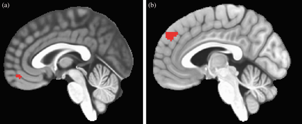
Analyses were conducted to examine the within-subject effects of RSA and SCR on neural activity controlling for the effects of trial block (i.e., detrending) and whether adolescents rated faces on subjective sadness or nose width during the block. Within-subject increases and decreases in RSA across the faces task were found to be inversely related to activity in a 50-voxel cluster located in the ventral medial frontal gyrus (i.e., vmPFC; Center of Mass = 1, 51, –14; Figure 1a). Within-subject increases and decreases in the number of SCRs across the faces task were found to be inversely related to activity in a 95-voxel cluster in the dorsal medial frontal gyrus (i.e., dmPFC; Center of Mass = –4, 49, 38; Figure 1b). No significant differences in activation or coupling with RSA or SCR were observed in vmPFC or dmPFC, respectively, between blocks where the question was “How sad does this person make you feel?” compared to blocks where the question was “How wide is the nose?” (see Weissman, Guyer, et al., Reference Weissman, Guyer, Ferrer, Robins and Hastings2018, for more details). Therefore, coupling across the entire task regardless of question was used in all further analyses to maximize the number of trials. Average activation beta values within each of these ROIs over the 12 blocks of the task were extracted to be used in further analyses.
Brain activity in regions of interest associated with autonomic activity during emotion processing. (a) Ventromedial prefrontal cortex region where activity is inversely related to respiratory sinus arrhythmia during emotion processing (50 voxels; Center of Mass = 1, 51, –14). (b) Dorsomedial prefrontal cortex region where activity is inversely related to number of skin conductance.

Multilevel structural equation modeling
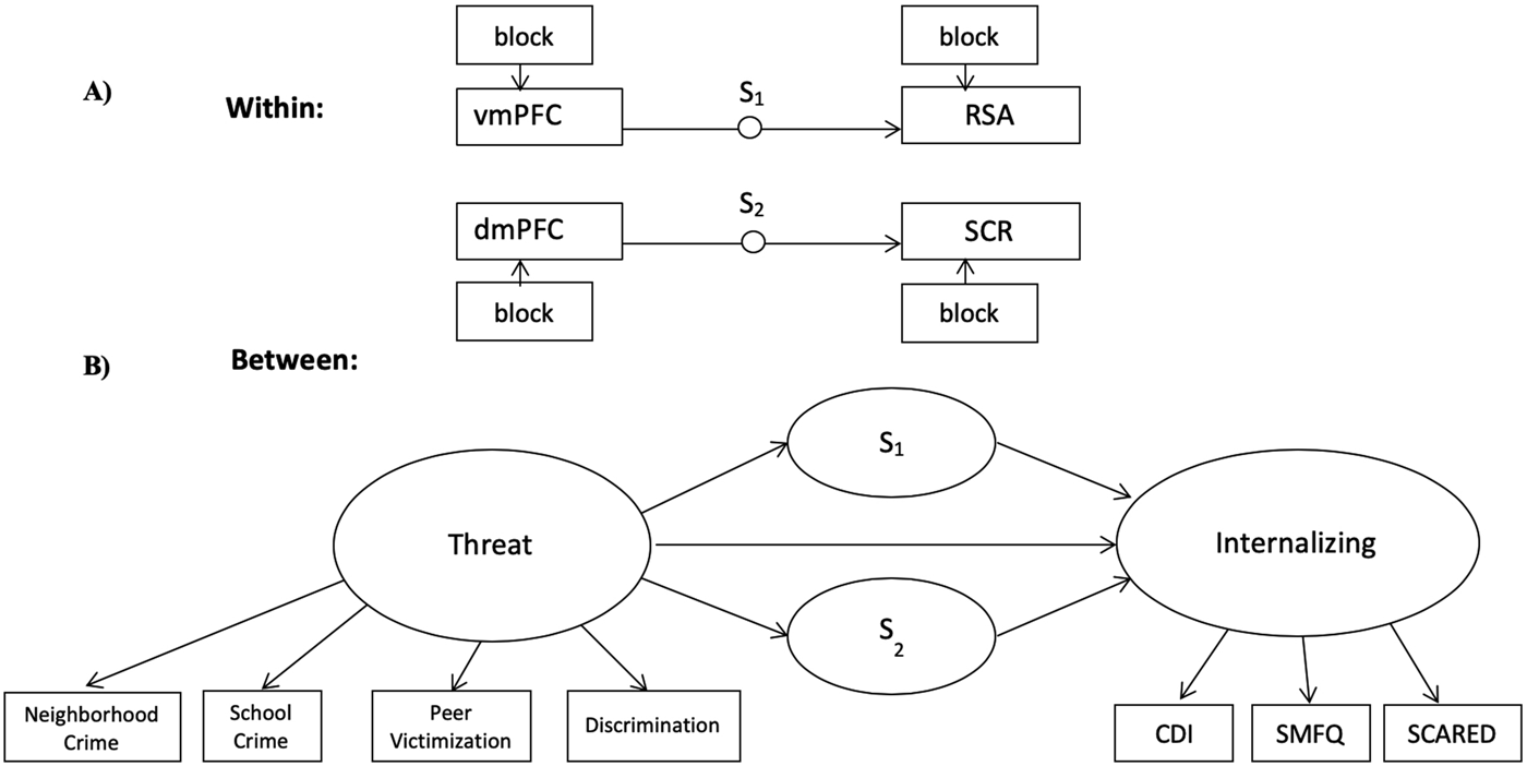
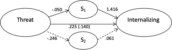
In order to investigate whether brain–ANS coupling mediated the link between threat exposure and internalizing problems, regression coefficients were extracted from the functional ROIs based on a significant relation between their activity and within-person fluctuations in RSA and number of SCRs. While the definition of the ROIs was based on adolescents with both ECG and skin conductance data (N = 133), betas from those ROIs were also extracted from adolescents who had fMRI data and at least one of the two ANS measures, including those with valid SCR but not ECG data (N = 28) and those with ECG but not SCR data (N = 18), bringing the final sample size for multilevel structural equation modelling analyses up to 179. Multilevel structural equation modelling in MPlus (Muthén & Muthén, Reference Muthén and Muthén2017) was then used to identify the random slope of the within-person relations (a) between mean-centered activity in vmPFC and RSA (S1) and (b) between mean-centered activity in dmPFC and the number of SCRs (S2; Figure 2a). These random slopes were then used as between-subject variables in a structural equation model. This approach to identifying variability in the degree of synchrony between two physiological signals is best suited for data in which the number of physiological observations per subject (12) is less than the number of subjects (179; Helm, Miller, Kahle, Troxel, & Hastings, Reference Helm, Miller, Kahle, Troxel and Hastings2018).
Multilevel structural equation model of the role of brain–autonomic coupling in the relation between past threat exposure and internalizing problems in late adolescence. (a) Within-subjects, random slopes of the relation between vmPFC activity and RSA (S1) and between dmPFC activity and SCR (S2) were calculated, controlling for the effect of task block (i.e., detrending). (b) Between-subjects, a latent variable was created for threat based on neighborhood crime, school crime, peer victimization, and discrimination, each averaged over four time points across adolescence. A latent variable was created for internalizing problems based on items from the Children's Depression Inventory (CDI), the Short Mood and Feelings Questionnaire (SMFQ), and the Screen for Child Anxiety-Related Emotional Disorders (SCARED). The effects of participant gender on S1, S2, and internalizing and the effect of Wave 1 internalizing on internalizing are not depicted, but were also included in the model.

At the between-subjects level, a latent variable representing threat exposure was defined by four indicators: mean scores on the neighborhood crime, school crime, peer victimization, and discrimination scales. A latent variable representing internalizing problems was defined by three indicators: mean scores on the CDI, SCARED, and SMFQ. The model tested the effect of threat exposure and participant gender on S1 and S2 and the effect of threat exposure, participant gender, S1, and S2 on internalizing problems (Figure 2b). Data and analysis code are available at https://github.com/dgweissman/adol-brain-ans-coupling.
Results
Descriptive statistics
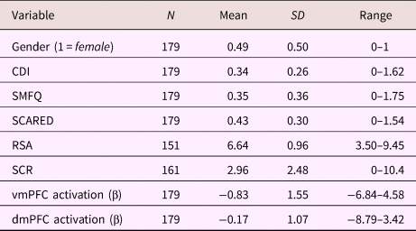
Adolescents’ reports of neighborhood crime, school crime, peer victimization, and discrimination were relatively low on average. With a possible range of 1 to 4, means for the ratings ranged from 1.06 for peer discrimination at the third and fourth assessments to 1.55 for school crime at the third assessment, but there was substantial variability within the sample (Table 1). Adolescents demonstrated mostly subclinical levels of internalizing problems, with mean ratings of 0.34, 0.35, and 0.43 out of 2 and 15, 22, and 43 adolescents meeting or exceeding recommended clinical cutoffs on the CDI, SMFQ, and SCARED, respectively (Table 2).
Descriptive statistics of threat variables over time

Descriptive statistics of internalizing problems and neurophysiological indices at 17 years

Note: CDI, Child's Depression Inventory. SMFQ, Short Mood and Feelings Questionnaire. SCARED, Screen for Child Anxiety-Related Emotional Disorders. RSA is mean respiratory sinus arrhythmia (in ln[ms2]) over the entire faces task. SCR is mean number of skin conductance responses per block (44 s) over the entire faces task. Standard deviations (SD) therefore indicate between-subjects variance only. vmPFC, ventromedial prefrontal cortex. dmPFC, dorsomedial prefrontal cortex.
Descriptive statistics for the neurobiological measures are in Table 2. Mean RSA (in ln[ms2]) during the faces task was 6.64 across the whole sample with a SD of 0.96 between-subjects, accounting for 72% of the total variance in RSA. Following subject mean-centering, the within-subjects SD was 0.60, accounting for 28% of the total variance in RSA. On average, participants had 2.96 SCRs per block, with a SD of 2.48, accounting for 65% of the total variance in SCR. Following subject mean-centering, the within-subjects SD was 1.83, accounting for 35% of the total variance in SCR. On average, the vmPFC had an activation beta of –0.83 during each block of the faces task with an SD of 1.55, accounting for 40% of the total variance in vmPFC activation. Following subject mean-centering, the within-subjects SD was 1.90, accounting for 60% of the total variance in vmPFC activation. On average, the dmPFC had an activation beta of –0.17 during each block of the faces task with an SD of 1.07, accounting for 38% of the total variance in dmPFC activation. Following subject mean-centering, the within-subjects SD was 1.37, accounting for 62% of the total variance in dmPFC activation.
Test of the mediation model
Confirmatory factor analysis on the measurement model of the threat and internalizing problems latent variables had good model fit, χ2 = 19.012, df = 13, p = .123, root mean square error of approximation = .051, standard root mean square residual = .039, comparative fit index =.987.
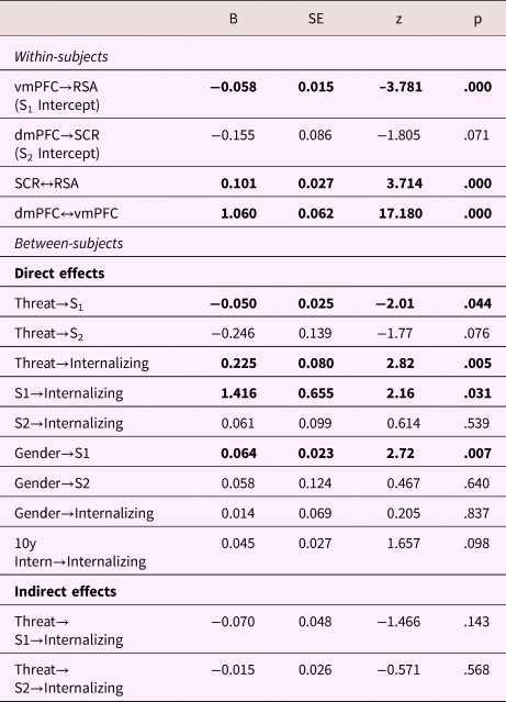
The within-subjects relations between vmPFC and RSA, and between dmPFC and SCR, were negative (Table 3). However, unlike what has been previously reported (Weissman, Guyer, et al., Reference Weissman, Guyer, Ferrer, Robins and Hastings2018), the negative relation between dmPFC activity and SCR was only marginally significant. This discrepancy from the original report was due to including the 46 participants with valid data for only one of the two ANS measures in the current analysis, who were not included in analyses used to localize the dmPFC ROI. The residual covariance between vmPFC–RSA coupling (S1) and dmPFC–SCR coupling (S2) was close to zero and nonsignificant (β = 0.001, SE = 0.008, p = .89), and so was constrained to be 0 in the final model. Female participants had significantly less negative vmPFC–RSA coupling than males. Females and males did not differ in the strength of their dmPFC–SCR coupling (Table 3).
Multilevel structural equation modeling results

Note: N = 179. All coefficients are unstandardized. Significant effects are in bold. → regression; ↔ covariance; x→y→z indirect path. S1, random slope of the within-subject relation between the ventromedial prefrontal cortex (vmPFC) activity and resting sinus arrhythmia (RSA). S2, random slope of the within-subject relation between the dorsomedial prefrontal cortex (dmPFC) and skin conductance responses (SCR). 10y Intern, Diagnostic Interview Schedule for Children anxiety and depression symptom counts in 5th grade. Internalizing, mean internalizing problem score at 17 years. Gender: female = 1, male = 0.
Neighborhood crime, school crime, peer victimization, and discrimination loaded on a common factor representing threat exposure across adolescence (Table 3). All three scales of adolescent-reported internalizing problems loaded on a common factor. Higher threat exposure was significantly related to higher levels of internalizing problems (Table 3, Figure 3).
Multilevel structural equation model results (between subjects). All coefficients are unstandardized. Solid lines indicate significant paths. Dotted lines indicate nonsignificant paths. The effects of participant gender on S1, S2, and internalizing and the effect of Wave 1 (5th grade) internalizing on internalizing are not depicted, but were also included in the model.

Higher threat exposure was significantly associated with more negative vmPFC–RSA coupling and marginally related with more negative dmPFC–SCR coupling (Table 3, Figure 3).
More positive (or less negative) vmPFC–RSA coupling during emotion processing was associated with significantly higher levels of internalizing problems (Table 3, Figure 3). However, because the indirect effect of threat on internalizing problems through vmPFC–RSA coupling was not significant (Table 3), this model did not meet the evidentiary criteria for suppression or inconsistent mediation (MacKinnon, Krull, & Lockwood, Reference MacKinnon, Krull and Lockwood2000). Internalizing problems were not significantly related to dmPFC–SCR coupling (Table 3).
Discussion
Exposure to threat in the environment during adolescence, a period of development characterized by change in the structure and function of neurophysiological mechanisms involved in the processing of emotion, may calibrate these mechanisms with consequences for future adjustment. The present study investigated how functional calibration of the interface of brain and body through the ANS in relation to prior experiences of adversity may convey risk to and resilience from internalizing problems in Mexican-origin adolescents. The results of this study suggest that (a) exposure to threats in community and peer contexts during adolescence increases risk for later internalizing problems, (b) negative vmPFC–RSA coupling is enhanced in adolescents who experienced more threatening environments, potentially reflecting an adaptation to a less safe environment, and (c) negative vmPFC–RSA coupling is associated with lower levels of internalizing problems, accounting for prior threat exposure. As hypothesized, threat exposure was associated with internalizing problems. However, mPFC–ANS coupling did not mediate this association as predicted. Instead, vmPFC–RSA coupling was independently associated with both higher threat and lower internalizing problems, while dmPFC–SCR coupling was associated with neither threat nor internalizing problems.
Contrary to our prediction, greater threat exposure was associated with significantly more negative vmPFC–RSA coupling and marginally more negative dmPFC–SCR coupling, suggesting that greater threat exposure was associated with an enhancement of mPFC–ANS coupling, and not a reduced magnitude of coupling, as was expected. It is plausible that more frequent activation of the stress response system provides the brain with more opportunities to reduce prediction errors between mPFC visceromotor output and interoceptive feedback from visceral responses mediated by the ANS. Conversely, as was predicted, more negative vmPFC–RSA coupling predicted fewer internalizing problems. Tighter regulation of PNS activity may allow for better emotion regulation, and fewer internalizing problems, while less coordinated activity of vmPFC and RSA, suggests persistent unresolved prediction error between visceromotor activity in the vmPFC and actual RSA. This decoupling has been hypothesized to contribute to the emergence of internalizing problems (Barrett & Simmons, Reference Barrett and Simmons2015). Because the indirect effect of threat on internalizing problems via vmPFC–RSA coupling was not significant, these should be interpreted as independent effects. Thus, threat increases risk for internalizing problems, while also being associated with more negative vmPFC–RSA coupling. More negative vmPFC–RSA coupling was associated with lower levels of internalizing problems, but this association was consistent, whether or not more negative vmPFC–RSA coupling occurred in the context of higher threat exposure.
Beauchaine (Reference Beauchaine2015a) proposed that emotion regulation is coordinated through PFC control of somatic arousal via the PNS, with breakdowns in that brain–body neural circuitry potentially underlying emotion dysregulation. Our observation that less coordinated activity of the vmPFC and RSA, as reflected by less negative coupling, was associated with higher internalizing problems could be seen as consistent with this proposal, and adds complexity to the idea that RSA may be a peripheral index of emotion regulation by the prefrontal cortex (Beauchaine, Reference Beauchaine2015a, Reference Beauchaine2015b). Our results demonstrated that adolescents differed in the extent to which their RSA activity corresponded with prefrontal cortex activity, and these individual differences related to adolescents’ contextual experiences and had consequences for their mental health. These findings also may contribute to an understanding of why, despite both RSA and childhood adversity demonstrating consistent associations with emotion dysregulation, they do not tend to be significantly related to one another (Chida & Hamer, Reference Chida and Hamer2008; Mclaughlin, Alves, et al., Reference McLaughlin, Alves and Sheridan2014; Skowron, Cipriano-Essel, Gatzke-Kopp, Teti, & Ammerman, Reference Skowron, Cipriano-Essel, Gatzke-Kopp, Teti and Ammerman2014). It is possible that while adversity does not influence baseline RSA, it does influence mPFC–RSA coupling, thereby enhancing the link between the dysregulated prefrontal cortex activity that contributes to psychopathology and low baseline RSA. Thus, low baseline RSA would be a better indicator of emotion dysregulation among adolescents, who through repeated threat exposure, had calibrated their ANS responding to be more tightly coupled with mPFC activity.
Female adolescents in this study demonstrated less negative vmPFC–RSA coupling than males. Females tend to have higher resting heart rate variability than males (Koenig & Thayer, Reference Koenig and Thayer2016), suggesting higher levels of tonic PNS activity, which may result from differences in the regulation of PNS activity. Despite females having higher baseline RSA, baseline RSA has been found to only moderate the association between childhood adversity and internalizing problems among male adolescents (McLaughlin et al., Reference McLaughlin, Rith-Najarian, Dirks and Sheridan2015). As with youth who experience higher levels of threat, low baseline RSA may be more of an indicator of emotion dysregulation among male adolescents, for whom prefrontal cortex activity is more tightly coupled with RSA. Moreover, less negative vmPFC–RSA coupling is associated with higher levels of internalizing problems and may therefore contribute directly to well-documented gender differences in internalizing problems (Hartung & Widiger, Reference Hartung and Widiger1998).
Our findings suggest two parallel paths to internalizing problems in adolescence. One is neurophysiological decoupling of vmPFC and RSA, indicating emotion dysregulation. The other is increased exposure to threat. It is probable that more negative vmPFC–RSA coupling among threat-exposed adolescents may serve as a compensatory regulatory mechanism for heightened emotional reactivity. Similarly, children who experience early neglect demonstrate more anxiety and more negative mPFC–amygdala connectivity than typically developing children (Gee, Gabard-Durnam, et al., Reference Gee, Gabard-Durnam, Flannery, Goff, Humphreys, Telzer and Tottenham2013). However, among the neglected children, more negative mPFC–amygdala connectivity actually predicts fewer anxiety symptoms (Gee, Gabard-Durnam, et al., Reference Gee, Gabard-Durnam, Flannery, Goff, Humphreys, Telzer and Tottenham2013), suggesting that prefrontal inhibition of emotional arousal serves as a compensatory regulatory mechanism. Although the timing and nature of the adversity experienced by the adolescents in this study is very different, there are clear parallels in the findings. Just as the absence of caregiver support during the sensitive window of infancy is then adaptively compensated for by an enhancement of mPFC–amygdala coupling, exposure to threats in the peer and community context during the sensitive window of adolescence may contribute to an enhancement of mPFC–RSA coupling to compensate for emotional responding that is heightened or dysregulated through other mechanisms.
To our knowledge, this is the first study to investigate how brain–ANS coupling may be calibrated by threat exposure across early through middle adolescence. However, our results can be interpreted through a synthesis of findings from studies of brain–ANS regulation in adults and adolescents, the development of mPFC–amygdala connectivity, and the relation between mPFC–amygdala connectivity and adversity. Studies of adults have typically found vmPFC activity to be positively associated with heart rate variability (Gianaros, Van der Veen, & Jennings, Reference Gianaros, Van der Veen and Jennings2004; Lane et al., Reference Lane, McRae, Reiman, Chen, Ahern and Thayer2009; Thayer et al., Reference Thayer, Åhs, Fredrikson, Sollers and Wager2012; Thayer & Lane, Reference Thayer and Lane2009) and negatively associated with skin conductance level (Nagai et al., Reference Nagai, Critchley, Featherstone, Trimble and Dolan2004; Zhang et al., Reference Zhang, Hu, Chao, Ide, Luo, Farr and Li2014). However, in the current sample of adolescents, vmPFC activity was negatively coupled with RSA (Weissman, Guyer, et al., Reference Weissman, Guyer, Ferrer, Robins and Hastings2018). This apparent developmental shift may be related to a similar shift in amygdala–mPFC connectivity.
Amygdala–mPFC connectivity during emotion processing has been found to undergo a developmental transition from positive to negative during adolescence (Gee, Humphreys, et al., Reference Gee, Humphreys, Flannery, Goff, Telzer, Shapiro and Tottenham2013; Silvers et al., Reference Silvers, Insel, Powers, Franz, Helion, Martin and Ochsner2017). Amygdala activity is thought to have an excitatory influence on the SNS via the rostral ventrolateral medulla, and inhibits the PNS activity through the nucleus ambiguus (Thayer & Lane, Reference Thayer and Lane2009). Thus, if the amygdala's inhibitory influence on the PNS is consistent from adolescence to adulthood, the positive to negative shift in amygdala–mPFC connectivity would correspond to a negative to positive shift in vmPFC–RSA coupling from adolescence to adulthood. The developmental trajectory of amygdala–mPFC connectivity has been found to be calibrated by childhood and adolescent adversity with consequences for internalizing problems (Colich et al., Reference Colich, Williams, Ho, King, Humphreys, Price and Gotlib2017; Gee, Gabard-Durnam, et al., Reference Gee, Gabard-Durnam, Flannery, Goff, Humphreys, Telzer and Tottenham2013; Herringa et al., Reference Herringa, Birn, Ruttle, Burghy, Stodola, Davidson and Essex2013, Reference Herringa, Burghy, Stodola, Fox, Davidson and Essex2016; Park et al., Reference Park, Leonard, Saxler, Cyr, Gabrieli and Mackey2018; Wolf & Herringa, Reference Wolf and Herringa2016). Higher amygdala-mPFC connectivity and, in this study, more negative mPFC–RSA coupling are associated with lower levels of internalizing problems in the context of prior adversity, potentially reflecting more effective regulation of emotional responding.
Coupling of dmPFC with the SNS was not significantly associated with either threat or internalizing problems, although dmPFC–SCR coupling was marginally associated with greater threat exposure. This suggests that inhibition of sympathoexcitatory brain regions by dmPFC and/or deactivation of dmPFC and other default mode network regions in response to salience cues indicated by SCRs are not mechanisms through which exposure to threat in adolescence conveys vulnerability to internalizing problems. Differences in vmPFC regulation of RSA between adolescents (Weissman, Guyer, et al., Reference Weissman, Guyer, Ferrer, Robins and Hastings2018) and adults (Gianaros et al., Reference Gianaros, Van der Veen and Jennings2004; Lane et al., Reference Lane, McRae, Reiman, Chen, Ahern and Thayer2009; Thayer et al., Reference Thayer, Åhs, Fredrikson, Sollers and Wager2012; Thayer & Lane, Reference Thayer and Lane2009) suggest a developmental transition, while dmPFC regulation of the skin conductance was similar in adolescents (Weissman, Guyer, et al., Reference Weissman, Guyer, Ferrer, Robins and Hastings2018) to what has been observed in adults (Nagai et al., Reference Nagai, Critchley, Featherstone, Trimble and Dolan2004; Zhang et al., Reference Zhang, Hu, Chao, Ide, Luo, Farr and Li2014). In addition, RSA in particular has been associated with individual differences in emotion dysregulation (Beauchaine, Reference Beauchaine2001, Reference Beauchaine2015b, Reference Beauchaine2015a) and with risk for and resilience to internalizing problems following exposure to threat and early life stress (Mclaughlin, Alves, et al., Reference McLaughlin, Alves and Sheridan2014; McLaughlin et al., Reference McLaughlin, Rith-Najarian, Dirks and Sheridan2015). The transition in vmPFC–RSA coupling may make this system particularly sensitive to exposure to threats and important for the development of internalizing problems in adolescence.
Limitations and future directions
The present study has many strengths, including a large, ethnic-minority sample of adolescents, longitudinal assessments of threat across adolescence, and simultaneous measures of RSA, SCR, and fMRI to evaluate brain–ANS coupling. However, several limitations merit consideration. First, although the results suggest that earlier threat exposure shapes neurophysiological functioning during emotion processing with consequences for internalizing problems, with only a single time point of neurophysiological data and concurrent measures of internalizing problems, the causal nature of these relations cannot be determined. Future work with multiple time points of neurophysiology, threat exposure, and mental health assessments could more robustly test their causal associations. Second, although the stimuli used in this study were emotional in nature, the faces adolescents viewed were presented in relatively short epochs. We therefore were not able to distinguish patterns of mPFC–ANS coupling to specific emotions with the current task design. Neurophysiological responses to negative and threatening stimuli (e.g., viewing fear faces) have been found to be specifically related to prior adversity (e.g., Gee, Humphreys, et al., Reference Gee, Humphreys, Flannery, Goff, Telzer, Shapiro and Tottenham2013; Herringa et al., Reference Herringa, Burghy, Stodola, Fox, Davidson and Essex2016). Future work comparing the nature of brain–ANS coupling in response to positive versus negative or threatening versus nonthreatening stimuli may contribute to a more complete understanding of the brain's regulation of the stress response and their relation to threat exposure and internalizing problems. Third and finally, the 400-Hz sampling frequency for ECG recording used in this study is below current standard recommendations of 512 Hz or above (Beauchaine et al., Reference Beauchaine, Bell, Knapton, McDonough-Caplan, Shader and Zisner2019; Berntson et al., Reference Berntson, Thomas Bigger, Eckberg, Grossman, Kaufmann, Malik and Van Der Molen1997). The lower sampling frequency was due to the technical limitations of the fMRI-compatible hardware used. However, empirical tests of this issue suggest that while higher resolution is better, sampling frequencies above 250 Hz are likely adequate for quantifying RSA (Berntson et al., Reference Berntson, Thomas Bigger, Eckberg, Grossman, Kaufmann, Malik and Van Der Molen1997; Merri, Farden, Mottley, & Titlebaum, Reference Merri, Farden, Mottley and Titlebaum1990; Riniolo & Porges, Reference Riniolo and Porges1997).
Conclusion
Our findings suggest that exposure to threats in the peer and neighborhood contexts puts Mexican-origin adolescents at risk for increased internalizing problems. However, in adolescence, the nervous system may adapt to threat exposure through stronger negative coupling of the vmPFC with RSA, which is associated with decreased internalizing problems. These results indicate that prefrontal cortex inhibition of physiological activity during emotion processing is tuned by experiences of threat and contributes to emotion dysregulation and risk for and resilience to internalizing problems. As these mechanisms are better understood, interventions that may influence prefrontal regulation of RSA, such as mind–body therapies (Muehsam et al., Reference Muehsam, Lutgendorf, Mills, Rickhi, Chevalier, Bat and Gurfein2017), may improve resiliency, and reduce emotion dysregulation among adolescents in high-threat environments. At a broader level, these findings support the idea that adolescence is a developmental period in which neurophysiological functioning is still being calibrated by experience, making it a period not only of risk but also of opportunity.
Acknowledgments
We thank Nicole Welindt, Timothy Bell, Elizabeth Kim, Lucia Leon, and Nilsen Gomez-Tabal for assisting with recruitment and data collection and processing.
Financial support
This work was supported by National Institutes of Health Grants R01-MH098370 (to A.E.G. and P.D.H.), R01-DA017902 (to R.W.R., Conger, and Widaman), and 5T32MH020006-19 (to Johnson and Gross), the National Science Foundation Grant IBSS 1327768 (to Page, P.D.H., Conger, and A.E.G.), and a US Department of Health and Human Services Poverty Research Center grant (to Stevens and Page).