Introduction
Anatomical variations of the bony skeleton of the nose and paranasal sinuses are common in adults. The importance of these variations in the sinonasal skeleton lies both in their potential relationship with chronic rhinosinusitis and in the surgical challenges they pose during endoscopic sinus surgery. In view of the increasing number of reports of chronic rhinosinusitis in children, the prevalence, significance and management of such anatomical variations in children have recently become the subject of much debate.Reference Kim, Cho, Lee, Kim, Kahng and Kim1
Concha bullosa is the term coined by Zuckerladl in 1862 to describe pneumatisation of the middle turbinate.Reference Nouraei, Elisay, DiMarco, Abdi, Majidi and Madani2 It is the commonest anatomical variation in adults, and has been implicated as a contributory factor for chronic rhinosinusitis by blockage of the ostiomeatal unit.
Haller cells are infraorbital ethmoid cells which, due to their position, increase the risk of damage to the orbit during ethmoidectomy.Reference Arslan, Aydinhoglu, Bozhurt and Egeli3
Kuhn cells are ethmoid cells in the frontal recess above the agger nasi, and can be divided into four types as follows: I, a single frontal recess cell above the agger nasi; II, a tier of cells in the frontal recess above the agger nasi; III, a single, massive frontal recess cell that pneumatises superiorly into the frontal sinus; and IV, a single cell contained entirely within the frontal sinus.Reference Bent, Cuilty-Siller and Kuhn4 The latter may well be an artefact of previous computed tomography (CT) technology; now, imaging quality has improved and thinner slices are used.
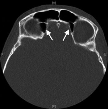
Supraorbital ethmoid cells pneumatise forward to displace the posterior wall of the frontal sinus, and are also thought to encroach on its drainage pathway.Reference Owen and Kuhn5
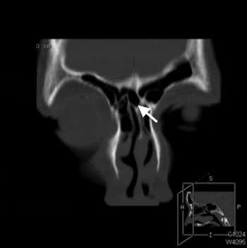
A further type of cell implicated in obstructing the frontal sinus drainage is the intersinus septal cell. This cell represents a pneumatisation within bony lamella between the frontal sinuses.Reference Bent, Cuilty-Siller and Kuhn4
In this report, we describe the case of an adolescent with chronic rhinosinusitis who exhibited all these anatomical variations, and who required endoscopic sinus surgery for chronic rhinosinusitis.
Case report
A 15-year-old male adolescent was referred to the ENT clinic with a 3-year history of hyposmia and nasal obstruction which was more pronounced on the right side. He had a past medical history of mild asthma rarely requiring salbutamol inhalers, and had no known drug allergies. His general practitioner had treated him with fluticasone nasal spray and cetirizine, with some symptomatic relief.
On the patient's initial visit, his Sino-Nasal Outcome Test 22 score was 43. Notably, he scored highly for the ‘need to blow nose’ and ‘runny nose’ items.
On examination, the patient had large inferior turbinates with rhinitic mucosa and evidence of septal deviation to the left.
Nasendoscopy revealed grade III polyps on the right (polyps touching the floor of the nose) and grade II polyps on the left (polyps extending beyond the middle meatus but not reaching the floor of the nose).
At the initial out-patient appointment, the patient was prescribed Pulmicort nasal douches (AstraZeneca, Luton, UK; 0.5 mg in 2 ml nebules diluted in 250 ml saline solution, twice daily) and a short course of oral prednisolone (40 mg daily for two weeks, reducing by 5 mg daily over a further week). A skin prick test and sweat test were also requested, and a follow-up appointment was arranged for two months hence.
The allergen test was positive for timothy grass, house dust mite and cat hair. The sweat test was borderline positive; however, genetic testing was negative for all common cystic fibrosis mutations.
At his follow-up appointment, the patient reported minimal benefit from the prescribed topical and systemic steroid therapy (his Sino-Nasal Outcome Test 22 score at this time was 47). Thus, arrangements were made for him to undergo surgery.
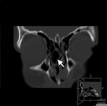
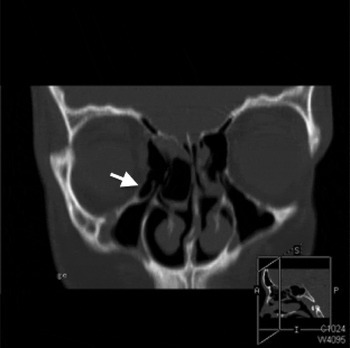
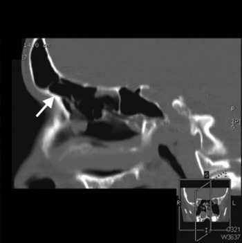
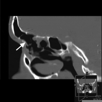
Computed tomography, performed as part of pre-operative investigation, revealed concha bullosa (Figure 1) and Haller cell on the right side (Figure 2), bilateral type III Kuhn cells (Figures 3 and 4) and supraorbital ethmoid cells (Figure 5), and an intersinus septal cell (Figure 6).
Coronal computed tomography image showing right-sided concha bullosa (arrow). S = superior; A = anterior; P = posterior; I = inferior

Coronal computed tomography image showing a right-sided Haller cell (arrow). S = superior; A = anterior; P = posterior; I = inferior

Sagittal computed tomography image showing a left-sided, type III Kuhn cell (arrow). S = superior; R = right; L = left; I = inferior

Sagittal computed tomography image showing a right-sided, type III Kuhn cell (arrow). S = superior; R = right; L = left; I = inferior

Axial computed tomography image showing supraorbital ethmoid cells (arrows). A = anterior; R = right; L = left; P = posterior

Coronal computed tomography image showing an intersinus septal cell (arrow). S = superior; A = anterior; P = posterior; I = inferior

The patient underwent endoscopic sinus surgery aided by the Fusion ENT navigation system (Medtronic, Minneapolis, Minnesota, USA), together with sinus lavage, endoscopic septoplasty and submucosal reduction of the inferior turbinates. He suffered no post-operative complications and tolerated the procedure well.
At two months' follow up, the patient reported almost complete resolution of his symptoms and had a Sino-Nasal Outcome Test 22 score of 10.
After 10 months' follow up, he continued to do well on once-daily Pulmicort irrigations.
Discussion
The anatomical variations observed in the nose and paranasal sinuses of adults have been well described, as have their associations with chronic rhinosinusitis and their impact on operative safety.Reference Nouraei, Elisay, DiMarco, Abdi, Majidi and Madani2, Reference Arslan, Aydinhoglu, Bozhurt and Egeli3, Reference Bolger, Butzin and Parsons6, Reference Meyers and Valvassori7 In children, the prevalence of these variations and their relationship with the observed pathology are less well documented. There is a distinction between adolescents over the age of 12 years, whose sinuses are already fully formed, and younger children, with varying degrees of development of the frontal, sphenoid and maxillary sinuses.
There is no clear consensus as to the optimal management strategy, and especially the timing of surgery. According to current literature, the indications for endoscopic sinus surgery include failure of maximal medical therapy, anatomical variations contributing to obstruction of sinonasal drainage pathways, sinonasal polyposis, and cases in which rhinosinusitis has led to orbital or intracranial complications.Reference Lieser and Derkay8 Even though evidence suggests that functional endoscopic sinus surgery is an effective treatment for paediatric chronic rhinosinusitis refractory to medical treatment, some authors have raised concerns regarding its possible interference with sinus and midfacial development.Reference Criddle, Stinson, Savliwala and Cottichia9
• Sinonasal anatomy variants are common in adults and increasingly reported in children
• The prevalence of paediatric chronic rhinosinusitis is increasing
• Neuronavigation can improve sinus surgery efficacy and safety in children with chronic rhinosinusitis refractory to medication
Other aspects that should prompt extra caution in the management of chronic rhinosinusitis in children include the radiation exposure associated with CT scans, the semi-invasive nature of the surgery, and the need for immobility, necessitating anaesthesia in the younger subpopulation.Reference Bhattacharrya, Jones, Hill and Shapiro10 In particular, when using the neuronavigation system, thinner cuts and a greater number of slices are required. Finally, there is a well-known risk associated with systemic glucocorticoids in growing children, as regards their effect on growth; however, topical administration has been shown to be safe.Reference Maspero, Rosenblut, Finn, Lim, Wu and Philpot11 All these factors need to be considered when assessing these patients with a view to surgery.
Conclusion
We report the case of an adolescent boy whose chronic rhinosinusitis was successfully managed with endoscopic sinus surgery despite multiple anatomical variations. Endoscopic sinus surgery in the paediatric population offers its own set of unique challenges. With the reported increasing prevalence of chronic rhinosinusitis in children, and emerging evidence that endoscopic sinus surgery is beneficial in this group of patients, it is expected that this management option will become increasingly popular. Given our limited knowledge of paediatric sinonasal variations, computer-assisted sinus surgery can aid intra-operative decision-making and improve surgical safety.