Introduction
Bacterial mastitis is a significant welfare and financial problem in sheep flocks (Gelasakis et al. Reference Gelasakis, Mavrogianni, Petridis, Vasileiou and Fthenakis2015). Various approaches are employed for diagnosis of mastitis in ewes (recently reviewed by Fragkou et al. Reference Fragkou, Boscos and Fthenakis2014). These more often include clinical, bacteriological and/or cytological methodologies. Clinical mastitis can be readily detected by clinical examination. For subclinical mastitis, the combination of bacteriological and cytological examinations is considered to be the most reliable method. In recent years, ultrasonographic examination of the udder has been used, as it can provide images of mammary glands and can yield useful information regarding the condition of mammary parenchyma (Barbagianni et al. Reference Barbagianni, Mavrogianni, Vasileiou, Fthenakis and Petridis2017).
Contrast-enhanced ultrasonographic examination is a novel imaging technique that can be faster and more convenient for evaluation of medical abnormalities throughout the body; its use can lead to application of fewer redundant, unnecessary examinations (International Contrast Ultrasound Society, 2017). A second generation contrast agent consisting of microbubbles, containing sulphur hexafluoride, which is an inert and hydrophobic gas, stabilised by a thin and flexible monolayer shell of phospholipids (Sono Vue, Bracco, Milano, Italy) is licenced for use (Schneider, Reference Schneider1999). The sulphur hexafluoride dissolves in the blood and is subsequently exhaled. After a single intravenous injection of 1- or 10-fold the maximum clinical dose to humans, the sulphur hexafluoride is cleared rapidly. The mean terminal half-life is 12 min (range: 2–33 min). Over 80% of the administered sulphur hexafluoride is recovered in exhaled air within 2 min post-injection and almost 100% after 15 min (European Medicines Agency, 2006). The properties of the microbubbles slow down gas diffusion into the blood, increasing stability/persistence in bloodstream and resistance to external pressures, thus preventing bubbles to dissolve, burst or coalesce forming larger ones (Bouakaz et al. Reference Bouakaz, Frinking, de Jong and Bom1999).
This research communication describes the use of contrast-enhanced ultrasonographic examination for diagnosis of long-standing mastitis in ewes. To the best of our knowledge, this is the first report of the use of this modality in diagnostic imaging of mammary glands of ruminants.
Materials and methods
Lacaune × breed ewes (bodyweight: 57·0–63·5 kg) in late lactation (n = 2; described henceforward as A and B) from a private flock were included in the study for the assessment of their mammary glands. Two ewes of similar age and stage of lactation, with no history of mastitis, were used as controls. Ewes A and B had previously developed clinical mastitis of staphylococcal aetiology, which had been treated with antibiotic administration with apparent clinical cure (no samples had been collected for confirmation).
Clinical examination of the mammary glands of the ewes was performed (see Fthenakis (Reference Fthenakis1994) and Mavrogianni et al. (Reference Mavrogianni, Fthenakis, Brooks, Papaioannou, Cripps, Taitzoglou, Brellou and Saratsis2005) for details). Milk samples were collected from each mammary gland of all animals for bacteriological and cytological examination by conventional techniques, as described previously (Fragkou et al. Reference Fragkou, Mavrogianni, Cripps, Gougoulis and Fthenakis2007, Reference Fragkou, Boscos and Fthenakis2014).
B-mode examination was performed by an ultrasound scanner (MyLab® 30; ESAOTE SpA, Genova, Italy), with a linear transducer, using a frequency of 12·0 MHz and scanning depth of 50 mm (Barbagianni et al. Reference Barbagianni, Mavrogianni, Vasileiou, Fthenakis and Petridis2017). The supramammary lymph nodes were also examined (Barbagianni et al. Reference Barbagianni, Mavrogianni, Vasileiou, Fthenakis and Petridis2017). Images were processed by means of ImageJ software (National Institutes of Health, Rockville Pike, MD, USA) (Petridis et al. Reference Petridis, Gouletsou, Barbagianni, Amiridis, Brozos, Valasi and Fthenakis2014; Barbagianni et al. Reference Barbagianni, Gouletsou, Valasi, Petridis, Giannenas and Fthenakis2015), which took into account the image's overall pixel grey-scale intensity values (Ojala et al. Reference Ojala, Pietikaeinen and Maeenpaea2002) and results were expressed on a 0 (black) to 255 (white) scale.
Doppler measurements were taken at the external pudendal artery (before its branching) with an ultrasound scanner (MyLab® 30), with linear transducer, using a frequency of 6·6 MHz and scanning depth 50 to 60 mm (Petridis et al. Reference Petridis, Gouletsou, Barbagianni, Amiridis, Brozos, Valasi and Fthenakis2014, Reference Petridis, Barbagianni, Ioannidi, Samaras, Fthenakis and Vloumidi2017; Barbagianni et al. Reference Barbagianni, Gouletsou, Valasi, Petridis, Giannenas and Fthenakis2015, Reference Barbagianni, Mavrogianni, Vasileiou, Fthenakis and Petridis2017). A ‘Doppler angle’ of 60° was employed in the examination (Petridis et al. Reference Petridis, Gouletsou, Barbagianni, Amiridis, Brozos, Valasi and Fthenakis2014; Barbagianni et al. Reference Barbagianni, Gouletsou, Valasi, Petridis, Giannenas and Fthenakis2015). Images of cross-sections of external pudendal artery and spectral waveforms of external pudendal artery were processed by the MyLab software (ESAOTE SpA); the following haemodynamic parameters were calculated: resistance index, pulsatility index, systolic:diastolic velocity ratio, general and mean pressures, mean velocity, systolic acceleration and blood input (Ginther, Reference Ginther2007; Petridis et al. Reference Petridis, Barbagianni, Ioannidi, Samaras, Fthenakis and Vloumidi2017).
Contrast-enhanced ultrasonographic (CEUS) examination was performed using an ultrasound scanner (Vivid-I; General Electric, Tirat Carmel, Israel), with a convex transducer (4C RS) of varying frequencies (1·8–6·0 MHz). B-mode sections were taken using a frequency of 5·0 MHz and a scanning depth of 120 mm, eventually switching the imaging settings to a preset coded phase inversion mode. Frequency, mechanical index and power were automatically set to lower values (i.e., 2·0/4·0 MHz, 0·09 and 22 dB, respectively). One focal zone was used at a scanning depth of 70 mm.
A volume of 2·5 ml of the contrast agent (20 µl of sulphur hexafluoride in microbubbles, equivalent to 112·5 mg; excipients: macrogol 4000, distearoylphosphatidylcholine, dipalmitoylphosphatidylglycerol sodium, palmitic acid; solvent: sodium chloride 9 mg ml−1) was injected into the jugular vein, followed by intravenous injection of 10 ml of normal saline. In the absence of a licenced dose for sheep, the dose licenced for humans has been used (European Medicines Agency, 2006), as animals weighed 11 to 19% less than average humans (Walpole et al. Reference Walpole, Prieto-Merino, Edwards, Cleland, Stevens and Roberts2012), thus no risk of unclear imaging of lesions during examinations was taken. The imaging plane remained unchanged during the examination. Real-time images of the contrast agent uptake (‘wash-in’) and clearance (‘wash-out’) were taken for up to 120 s post-injection.
Video images were analysed in sequence of frames (JPG format; first frame at time 0 and then one frame every 2 s) using the Free Studio (v. 6.6.35.323) multimedia software developed by DVDVideoSoft (Digital Wave Ltd, London, United Kingdom). The frames were opened as a stack with ImageJ software. Four regions of interest were used in the evaluation: external pudendal artery, mammary parenchyma, lactiferous ducts and teat tissues for calculation of total gray-scale intensity of signals. Image enhancement in each region was measured in linear arbitrary enhancement units (AEU). A time–intensity curve was generated for each region of interest and for each examination the below parameters were calculated.
-
• Peak enhancement (expressed in AEU): enhancement curves were produced after measurement of intensity by means of Vivid-I software (General Electric) and dividing by the maximum value of intensity.
-
• Time to peak (s): calculated from injection of contrast agent to peak intensity.
-
• Time to wash-out (s): calculated from injection of contrast agent to return to baseline.
-
• Total enhancement time (s): calculated from beginning of enhancement to return to baseline.
-
• Wash-in time (s): calculated from beginning of enhancement to peak intensity.
-
• Wash-out time (s): calculated from peak intensity to return to baseline.
For all ultrasonographic findings, one set of data was calculated for [a] three mammary glands of ewes A and B, which had been found with no clinically evident abnormalities and [b] four mammary glands of the two healthy control ewes. Comparisons of results of grey-scale intensity values obtained by B-mode ultrasonographic examination and results of haemodynamic parameters between [a] and [b] were made by means of Mann-Whitney test for small sample numbers. For CEUS results, repeated measures mixed effect linear regression models were used to study outcomes over the measurement period. The effect of animals was included as random effect in the model, which was adjusted for repeated measures within animals and comparisons were made between [a] and [b]. An electronic data management tool was employed (Lowry, Reference Lowry2012, Reference Lowry2015).
Results and discussion
In palpation, mammary glands of ewes A and B were found to be smaller in size relative to those of the control animals. The udder of ewe A was soft, symmetrical and with no palpable abnormalities; in the udder of ewe B, there was a palpable hard mass, occupying most of the parenchyma in the right gland, whilst the left gland was soft and with no palpable abnormalities. No clinical abnormalities were evident in mammary glands of the control animals. Milk somatic cell counts of all four ewes were below 0·4 × 106 cells ml−1, with only scattered macrophages identified in milk films.
During B-mode examination, the mammary parenchyma of both ewe A glands, the mammary parenchyma of ewe B left gland and the mammary parenchyma of both control ewes’ glans were imaged as an homogeneous, granular structure with mildly increased echogenicity with no abnormal structures (Barbagianni et al. Reference Barbagianni, Mavrogianni, Vasileiou, Fthenakis and Petridis2017). Anechoic structures identified therein corresponded to lactiferous ducts and vessels imaged as those observed in the control ewes, with no abnormal structures therein (online Supplementary material 1). Mammary parenchyma of the right gland of ewe B was characterised by the presence of an encapsulated round structure with hypoechoic capsule and a hyperechoic content. Results of grey-scale evaluation (online Supplementary material 2) indicated a significant difference (P = 0·05) between mammary glands of ewes A and B and those of control animals. Mammary lymph nodes in all ewes were imaged with homogeneously hypoechoic parenchyma; the hilar area was imaged as a highly echogenic linear structure, with no changes in grey-scale evaluation or dimensions observed.
During Doppler examination, only differences in total blood input (P = 0·05) were significant between mammary glands of ewes A and B and those of control animals (online Supplementary material 2). The two most frequently used indices (resistance index and pulsatility index) in ewes A and B were within the reference range (Petridis et al. Reference Petridis, Gouletsou, Barbagianni, Amiridis, Brozos, Valasi and Fthenakis2014). Blood velocity, acceleration and total input were outside the reference range (online Supplementary material 3).
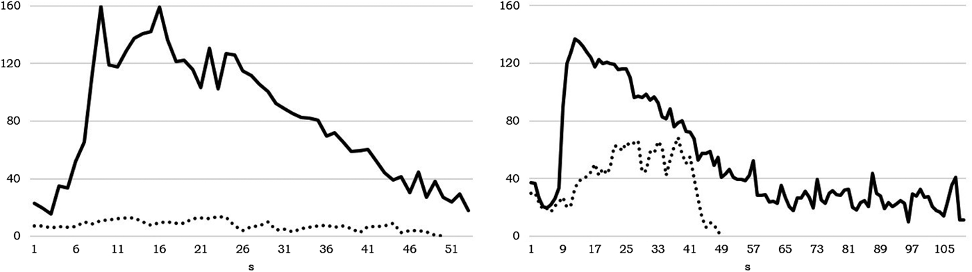
No adverse effects were observed clinically in any animal after administration of the contrast agent. The dose administered allowed clear imaging of mammary structures in all cases. In healthy mammary glands, CEUS examination revealed a steady biphasic pattern of contrast agent kinetics, characterised by initial uptake (wash-in phase) within 15 to 40 s post-injection, at which time intensity peaked with strong enhancement (130–200 AEU), followed by a gradual wash-out phase (Fig. 1). In contrast, in mammary glands of ewes A and B, the pattern was particularly inconsistent and unclear, with weak enhancement (<100 AEU; P < 0·01) lasting for a short period (Fig. 1).
Patterns of image detection enhancement in mammary glands of ewes after contrast-medium administration; left: external pudendal artery (up to 50 s post-injection) – right: mammary parenchyma (up to 104 s post-injection) (straight line: mammary glands of healthy ewes, dotted line: mammary glands of ewes with history of mastitis).

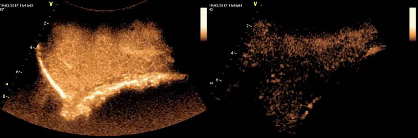
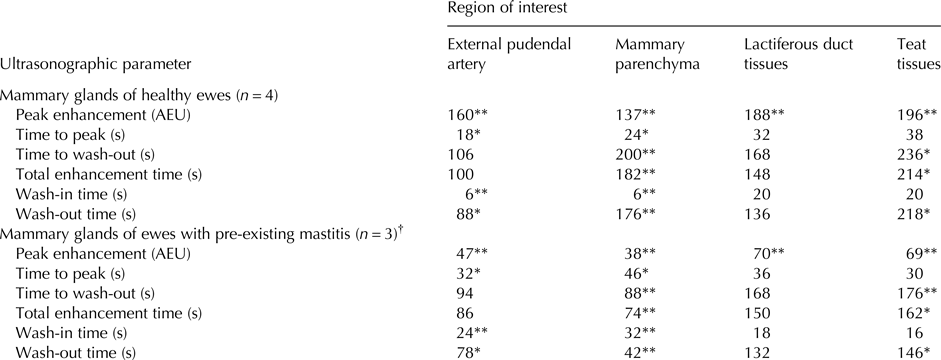
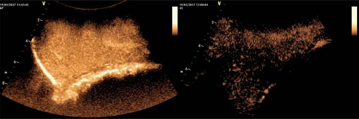
Enhancement and clearance were evident initially in the external pudendal artery. Enhancement in mammary parenchyma started with a delay, but lasted longer. Enhancement of the lactiferous ducts started shortly after that of mammary parenchyma and lasted longer. The lactiferous duct was the last structure where enhancement was observed (Table 1). Enhancement allowed clear visualisation of the entire parenchyma of the mammary glands of healthy ewes; in the three mammary glands of ewes A and B, mammary parenchyma could be visualised weakly and only regionally (Fig. 2, online Supplementary material 4 and 5).
Contrast-enhanced ultrasonographic presentation of mammary parenchyma; image taken at the 6th month of lactation period. Along the long axis of the udder, left: imaging of mammary gland of a healthy ewe, with peak enhancement of mammary parenchyma in 24 s – right: imaging of mammary gland of a ewe with history of mastitis, with reduced enhancement of mammary parenchyma in 46 s (images taken and processed on a Vivid-I ultrasonography system (General Electric) with convex transducer, imaging frequency: 2·0/4·0 MHz – mechanical index: 0·09 – power: 22 dB – scanning depth: 60 mm – contrast agent: 20 µl sulphur hexafluoride in microbubbles).
Quantitative results (median) of contrast-enhanced ultrasonographic examination of mammary glands of ewes

*P < 0·05, **P < 0·01 between respective parameters in mammary glands of healthy ewes and of ewes with pre-existing mastitis.
† Results for the right gland of ewe B have not been included, as clinically evident abnormalities were recorded.
The contrast agent took longer time to perfuse the udder tissues in ewes A and B than in control animals, and enhancement lasted for a shorter period. Similar results were observed in measurements performed in the external pudendal artery, in the mammary parenchyma, in the lactiferous duct tissues and in the teat tissues of all ewes (Table 1).
CEUS provided a definitive indication that both mammary glands of ewe A and the left gland of ewe B were not fully functional. Decreased enhancement indicated reduced perfusion of contrast agent into the mammary parenchyma. In chronic mastitis, destruction of alveolar integrity and mammary epithelium and proliferation of fibrous tissue are well-documented features (Fthenakis & Jones, Reference Fthenakis and Jones1990; Tzora et al. Reference Tzora, Fthenakis and Linde1998). Therefore, it can be suggested that the reduced amount of functional mammary tissue would have contributed to the decreased perfusion of the contrast agent. Hence, the reduced enhancement reflected tissue damage, which, in turn, would have resulted in smaller milk yield during the subsequent lactation period.
In this case, conventional techniques (e.g., clinical examination, examination of milk samples) could not fully support a diagnosis regarding condition of mammary glands of ewes A and B. Clinical abnormalities were observed immediately only in one mammary gland of ewe. B-mode examination provided an initial suspicion, but the results were treated cautiously, given that in mammary glands at the end of a lactation period and at early involution, significant variations have been reported (Petridis et al. Reference Petridis, Gouletsou, Barbagianni, Amiridis, Brozos, Valasi and Fthenakis2014). Doppler examination then corroborated the initial findings. Doppler ultrasonographic examination of mammary glands of sheep has been described repeatedly in recent reports (e.g., Petridis et al. Reference Petridis, Gouletsou, Barbagianni, Amiridis, Brozos, Valasi and Fthenakis2014; Barbagianni et al. Reference Barbagianni, Gouletsou, Valasi, Petridis, Giannenas and Fthenakis2015, Reference Barbagianni, Mavrogianni, Vasileiou, Fthenakis and Petridis2017). However, it is noteworthy that interpretation of such findings requires specific training and cannot be achieved readily under clinical conditions.
CEUS is a non-invasive imaging modality, which provided immediate and easy to interpret results. Nevertheless, its cost of 35 € per examination (i.e., 70 € per animal) might be a limiting factor. As the contrast agent is not licenced for sheep, long withdrawal periods should also be maintained. This will be a significant problem in cases where culling of animals would be recommended.
No previous reports of CEUS in the mammary gland of ruminants have been found in the international literature. In female dogs, Feliciano et al. (Reference Feliciano, Uscategui, Maronezi, Simões, Silva, Gasser, Pavan, Carvalho, Canola and Vicente2017) have described the use of CEUS for diagnosis of neoplastic lesions in mammary glands and have also confirmed association of imaging findings with histological evidence. This can be of value, as it lends support to our findings that the limited functionality of the mammary glands of ewes A and B (with history of mastitis) was related to the reduced acoustic enhancement in the same mammary glands, as observed in the current report. Further, in women, Jiang et al. (Reference Jiang, Liu, Zhu, Sun and Chang2007) and Liu et al. (Reference Liu, Jiang, Liu, Zhu, Sun and Chang2009) have each reported diagnosis of one case of mastitis by means of the method. More recently, Xiao et al. (Reference Xiao, Ou, Yang, Wu and Luo2014) have indicated that, although this imaging modality was useful in diagnosing abnormalities (including mastitis) in the mammary glands of women, it could not differentiate between mastitis and malignant lesions.
In conclusion, this imaging modality could be useful in ruminants, particularly in cows. In those animals, the large size of the udder and smaller financial constraints would make the modality more applicable. Use of this imaging modality may contribute to improved diagnosis of mastitis cases, especially on occasions when abnormalities cannot be easily confirmed by more conventional methods. Certainly, further investigations should be performed before the modality may be used readily.
Supplementary material
The supplementary material for this article can be found at https://doi.org/10.1017/S002202991800002X.