INTRODUCTION
Declarative, episodic, and working memory have long been recognized as being dependent on the function of an intact hippocampus, a medial temporal lobe structure necessary for encoding and consolidating new memories. Schizophrenia is a disorder characterized by both episodic memory deficits and hippocampus abnormality. Because selective memory impairment has been observed in never-medicated patients experiencing first-episode schizophrenia, episodic memory has been posed as a core deficit of the disorder rather than an effect of chronicity or antipsychotic medication (Saykin et al., Reference Saykin, Shtasel, Gur, Kester, Mozley, Stafiniak and Gur1994). Episodic memory deficits appear to be present in schizophrenia before the onset of frank psychosis. In a study of patients at high risk of developing psychosis (predominantly schizophrenia), impairments in the Visual Reproduction and Logical Memory subtests of the Wechsler Memory Scale-Revised (WMS-R) differentiated patients who went on to develop psychosis within the next 12 months from those who did not (Brewer et al., Reference Brewer, Francey, Wood, Pantelis, Phillips, Yung, Anderson and McGorry2005). While it is possible for lower scores on neuropsychological tests of memory to be secondary to any number of environmental or individual attributes, episodic memory impairment in schizophrenia is not commensurate with global impairment, nor is it attributable to lack of motivation, attention, or cooperation (Gold et al., Reference Gold, Randolph, Carpenter, Goldberg and Weinberger1992; Saykin et al., Reference Saykin, Gur, Gur, Mozley, Mosley, Resnick, Kester and Stafaniak1991; Tamlyn et al., Reference Tamlyn, McKenna, Mortimer, Lund, Hammond and Baddeley1992).
Functional neuroimaging has linked hippocampus dysfunction to impaired performance on memory tasks in schizophrenia. In a positron emission tomography (PET) study, Heckers et al. (Reference Heckers, Rauch, Goff, Schacter, Fischman and Alpert1998) identified lesser hippocampus activity in patients than controls during attempts to recall studied words. In a functional magnetic resonance imaging (fMRI) study, controls exhibited greater activation than patients in left anterior hippocampus during encoding and in hippocampus bilaterally during recognition (Jessen et al., Reference Jessen, Scheef, Germeshausen, Tawo, Kockler, Kuhn, Maier, Schild and Heun2003). Functional impairment related to the hippocampus extends to the ability to comprehend relationships and draw inferences, as demonstrated by a selective deficit in discrimination accuracy when cognitive flexibility is required, which has been observed in schizophrenia using fMRI (Öngur et al., Reference Öngür, Cullen, Wolf, Rohan, Barreira, Zalesak and Heckers2006). Using a similar relational-memory task, Hanlon et al. (Reference Hanlon, Weisend, Yeo, Huang, Lee, Thoma, Moses, Paulson, Miller and Cañive2005) directly assessed hippocampus activity with magnetoencephalography (MEG), reporting abnormal right hemisphere processing of nonverbal stimuli accompanied by a possibly compensatory, left-lateralized activation in schizophrenia.
Structural abnormalities of the medial temporal lobe, which includes the hippocampus, are among the most robust findings in schizophrenia research. A number of studies have reported smaller volume of the medial temporal lobe in schizophrenia (Bogerts et al., Reference Bogerts, Lieberman, Ashtari, Bilder, Degreef, Lerner, Johns and Masiar1993; Gur et al., Reference Gur, Turetsky, Cowell, Finkelman, Maany, Grossman, Arnold, Bilker and Gur2000; McCarley et al., Reference McCarley, Wible, Frumin, Hirayasu, Levitt, Fischer and Shenton1999; Wright et al., Reference Wright, Rabe-Hesketh, Woodruff, David, Murray and Bullmore2000), particularly in the left hemisphere (DeLisi et al., Reference DeLisi, Hoff, Schwartz, Shields, Halthore, Gupta, Henn and Anand1991; Honea et al., Reference Honea, Crow, Passingham and Mackay2005; Shenton et al., Reference Shenton, Kikinis, Jolesz, Pollak, LeMay, Wible, Hokama, Martin, Metcalf, Coleman and McCarley1992). However, while hippocampus abnormality is consistently found, the precise nature of the results has varied. Meta-analyses have posited that smaller hippocampus volume is equivalent across hemispheres (Nelson et al., Reference Nelson, Saykin, Flashman and Riordan1998), even when limited to studies of first-episode patients (Steen et al., Reference Steen, Mull, McClure, Hamer and Lieberman2006). However, other first-episode studies have resulted in findings of significant left-less-than-right hippocampus asymmetry (Bogerts et al., Reference Bogerts, Ashtari, Degreef, Alvir, Bilder and Lieberman1990; Hirayasu et al., Reference Hirayasu, Shenton, Salisbury, Dickey, Fischer, Mazzoni, Kisler, Arakaki, Kwon, Anderson, Yurgelun-Todd, Tohen and McCarley1998). In a longitudinal study, Velakoulis et al. (Reference Velakoulis, Wood, Wong, McGorry, Yung, Phillips, Smith, Brewer, Proffitt, Desmond and Pantelis2006) demonstrated hippocampus volume deficits bilaterally in chronic schizophrenia, in the left hemisphere only in first-episode schizophrenia, and not at all in schizophreniform psychosis. It is also important to note that there have been scattered reports of null findings with regard to hippocampus volume in schizophrenia. For example, Marsh et al. (Reference Marsh, Harris, Lim, Beal, Hoff, Minn, Csernansky, DeMent, Faustman, Sullivan and Pfefferbaum1997) reported no group difference in hippocampal volume in an inpatient sample with severe and chronic schizophrenia. Because of the variability in group differences across reports, the etiological role of hippocampal anomaly in schizophrenia and its association with subsequent symptoms and course of the disorder remains a critical area for research (White et al., Reference White, Cullen, Rohrer, Karatekin, Luciana, Schmidt, Hongwanishkul, Kumra, Schulz and Lim2008).
With some degree of hippocampal volume deficits in schizophrenia well established but the nature and mechanisms of its functional consequences unclear, attention is turning to localizing hippocampus volume decrements along the anterior-posterior axis. One study confirmed the presence of overall hippocampus volume deficits in schizophrenia but maintained that the loss was diffuse rather than topographically specific (Weiss et al., Reference Weiss, DeWitt, Goff, Ditman and Heckers2005). However, Narr et al. (Reference Narr, Thompson, Szezsko, Robinson, Jang, Woods, Kim, Hayashi, Asunction, Toga and Bilder2004) made a strong argument for factoring regional specificity into hippocampus measurements in a study that demonstrated volume deficits localized to midbody and anterior hippocampus in first-episode schizophrenia. Consistent with Narr et al.’s finding, smaller anterior hippocampus volumes have been observed in both first-episode and chronic schizophrenia (Lieberman et al., Reference Lieberman, Chakos, Wu, Alvir, Hoffman, Robinson and Bilder2001; Pegues et al., Reference Pegues, Rogers, Amend, Vinogradov and Deicken2003; Szeszko et al., Reference Szeszko, Goldberg, Gunduz-Bruce, Ashtari, Robinson, Malhotra, Lencz, Bates, Crandall, Kane and Bilder2003). Other studies have found deficits localized to posterior hippocampus in chronic schizophrenia (Narr et al., Reference Narr, Thompson, Sharma, Moussai, Blanton, Anvar, Edris, Krupp, Rayman, Khaledy and Toga2001) and in first-episode patients (Hirayasu et al., Reference Hirayasu, Shenton, Salisbury, Dickey, Fischer, Mazzoni, Kisler, Arakaki, Kwon, Anderson, Yurgelun-Todd, Tohen and McCarley1998).
The relationship of neuropsychological function to structural abnormality in the hippocampus is under debate. Lack of a relationship between hippocampus volume and episodic memory in people with schizophrenia or controls has been reported by some studies (DeLisi et al., Reference DeLisi, Hoff, Schwartz, Shields, Halthore, Gupta, Henn and Anand1991; Torres et al., Reference Torres, Flashman, O’Leary, Swayze and Andreasen1997). Gur et al. (Reference Gur, Turetsky, Cowell, Finkelman, Maany, Grossman, Arnold, Bilker and Gur2000), on the other hand, found a positive correlation between gray matter volume of the hippocampus and episodic memory scores across patients and controls of both sexes. This finding is supported by other studies reporting similar correlations between verbal memory and left hippocampus volume (Goldberg et al., Reference Goldberg, Torrey, Berman and Weinberger1994; Seidman et al., Reference Seidman, Faraone, Goldstein, Kremen, Horton, Makris, Toomey, Kennedy, Caviness and Tsuang2002) or amygdala-anterior hippocampus volume (O’Driscoll et al., Reference O’Driscoll, Florencio, Gagnon, Wolff, Benkelfat, Mikula, Lal and Evans2001) in people with schizophrenia, their relatives, and controls. However, some investigations of structure-function relationships have found differential patterns of correlations between hippocampus volume and verbal IQ or verbal or visual memory in controls and people with schizophrenia (Kuroki et al., Reference Kuroki, Kubicki, Nestor, Salisbury, Park, Levitt, Woolston, Frumin, Niznikiewicz, Westin, Maier and Shenton2006; Sachdev et al., Reference Sachdev, Brodaty, Cheang and Cathcart2000; Sanfilipo et al., Reference Sanfilipo, Lafargue, Rusinek, Arena, Loneragan, Lautin, Rotrosen and Wolkin2002; Toulopoulou et al., Reference Toulopoulou, Grech, Morris, Schulze, McDonald, Chapple, Rabe-Hesketh and Murray2004). Findings of differential patterns of correlations for patients and controls are thought to indicate a loss of normal structure-function relationships, possibly arising from aberrant neurodevelopment.
One reason for discrepancies among volumetric studies may be that critical hippocampus subregions are not being discriminated, as few studies have combined neuropsychological assessment with hippocampus subregion measurements as discussed earlier. Anterior hippocampus volume deficits have been correlated with decrements in executive and motor function, but not memory, in first-episode schizophrenia (Bilder et al., Reference Bilder, Bogerts, Ashtari, Wu, Alvir, Jody, Reiter, Bell and Lieberman1995), with the effect present for men but not for women in a similar study (Szeszko et al., Reference Szezsko, Strous, Goldman, Ashtari, Knuth, Lieberman and Bilder2002). Given the substantial evidence for episodic memory dysfunction and hippocampus volume deficits in schizophrenia, further investigation taking subregional measurements into account seems warranted.
The present study was conceived to investigate how measures of cognitive function track structural abnormalities in specific hippocampus subregions. Based on prior research, it was expected that (1) episodic memory would be impaired and (2) hippocampus volume would show deficits in the schizophrenia group. It was also predicted that (3) there would be positive correlations between hippocampus volumes and episodic memory scores in the control group, establishing an assemblage of correlational relationships that represent normal brain-behavioral functioning. It was further predicted that (4) correlations between regional hippocampus volumes and episodic memory scores would differentiate the groups, supporting the presence of abnormal brain-behavioral relationships in schizophrenia. Demographic variables that are known to affect hippocampus volume, such as age and gender, were also investigated.
METHOD
Participants
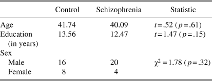
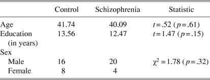
Participants in this study were 24 schizophrenia patients and 24 healthy control subjects matched for age, education, and sex (see Table 1). Group membership was determined with the Structured Clinical Interview for DSM-IV Axis I Disorders, Clinician Version (SCID-CV; First et al., Reference First, Spitzer, Gibbon and Williams1996). No subject had a history of head injury, neurological disorder, or severe medical illness.
Sample demographics
The schizophrenia group consisted of volunteers and referrals who were relatively stable patients, well known to their providers. They met predetermined criteria for clinical stability, as they had been treated with the same antipsychotic medications for at least 3 months and had had no inpatient stays during the past year. Absence of other current psychiatric disorders was determined using the SCID-CV. Patients with schizophrenia were all taking antipsychotic medications, either haloperidol (n = 6), olanzapine (n = 4), risperidone (n = 5), clozapine (n = 4), aripiprazole (n = 2), or quetiapine (n = 3).
The control group consisted of healthy volunteers recruited via advertisements in local newspapers. Control subjects were screened for the presence of Axis-I psychopathology via SCID-CV with a licensed clinical psychologist (Thoma) or with a psychology graduate student under his direct supervision. Axis II psychopathology was ruled out with a diagnostic clinical interview with a licensed clinical psychologist (Thoma) if necessary. If there was a question of the presence of schizotypal or schizoid symptoms, scores on the Social Anhedonia Scale (Chapman et al., Reference Chapman, Chapman, Kwapil and Eckblad1994) and the Magical Ideation Scale (Eckblad & Chapman, Reference Eckblad and Chapman1983) were reviewed. During the clinical interview, potential control subjects were asked if they had a first-degree relative with schizophrenia or other psychotic disorder, and if so, they were not included in the study.
Institutional Review Board approval was obtained prior to running subjects, and subjects were informed that they could leave the study at any time. Appropriate informed consent was obtained from all subjects, and data was obtained in compliance with institutional regulations.
Hippocampus volume data from these subjects was previously published elsewhere as part of an analysis of psychophysiological anomalies (Thoma et al., Reference Thoma, Hanlon, Petropoulos, Miller, Moses, Smith, Parks, Lundy, Sanchez, Jones, Huang, Weisend and Cañive2008).
Procedures
Magnetic resonance images
Three-dimensional structural magnetic resonance images (MRI), T1 weighted, were collected with a 1.5 Picker Edge Imager at the NMVAHCS Magnetic Source Imaging center. A gradient echo 3-D sagittal sequence with parameters TR = 15 ms; TE = 4.4 ms; FOV = 256 mm; 192 x 256 matrix, flip angle = 25; slice thickness = 1.5 mm, with no gap, was used.
Structural MRI analysis
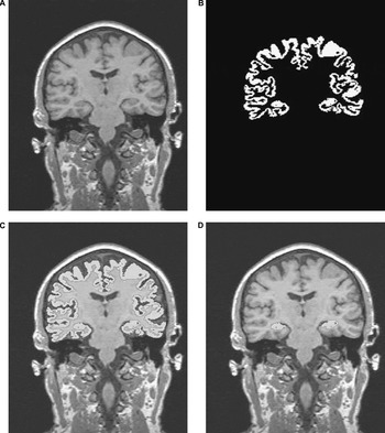
The method used for this hippocampus volumetric analysis was first described in detail elsewhere (Thoma et al., in press). MRIs were first resliced into coronal images of 1.0 mm thickness (Figure 1a). The skull was then stripped from each MRI image using Brain Extraction Tool software (BET: fMRIB Image Analysis Group, Oxford, UK). Intracranial volume (ICV) was calculated from the mask produced from this program. Images were then segmented using an automated k-means clustering segmentation algorithm, and the volumes of gray matter (GM; not including cerebellum; see Figure 1b), white matter (WM; not including cerebellum), cerebrospinal fluid (CSF), and total ICV (including cerebellum) were determined by the number of pixels in each of their respective clusters (Petropoulos et al., Reference Petropoulos, Sibbitt and Brooks1999). Pixels that could not be assigned exclusively to GM or CSF were considered partial volume (PV). The number of PV pixels was divided in half and then added to the GM for a final count.
A pictorial representation of the steps involved in the hippocampus quantification routine. (1a) Sagittal MRI series is resliced into 1-mm coronal images. (1b) An example of cortical gray matter segmentation. (1c) Cortical mask “overlay” on T1 image. (1d) Hippocampus tissue extracted by trained rater utilizing manual editing to separate the body of the hippocampus from the cortex.

The k-means algorithm segmented GM from WM to assist raters in selecting the hippocampus, and as a result hippocampus WM was excluded from the overall hippocampus volume measurement (Figure 1c). Two independent raters used interactive software (Driscoll, et al., Reference Driscoll, Hamilton, Petropoulos, Yeo, Brooks, Baumgartner and Sutherland2003) to conduct volumetric assessment of the hippocampus using the already k-means segmented coronal T1-weighted images. The software was designed to interact with the segmented imaging data for quantification and allows the user to select a segmented brain section. The user is able to magnify the area of interest to remove sections of segmented data from the volumetric analysis and allow the user to specify brain structures and easily quantify the selection via a pixel-counting algorithm (Figure 1d).
Raters followed the anatomical guidelines outlined by Watson et al. (Reference Watson, Andermann, Gloor, Jones-Gotman, Peters, Evans, Olivier, Melanson and Leroux1992) to measure hippocampus, except in regard to the posterior hippocampus definition. Watson et al. (Reference Watson, Andermann, Gloor, Jones-Gotman, Peters, Evans, Olivier, Melanson and Leroux1992) defined the most posterior slice as where the crux of the fornix separated from the hippocampus. This method, however, eliminates the most posterior slices of the hippocampus. Since active hippocampus MEG source dipoles have been previously found on these most posterior slices of the hippocampus (Hanlon et al., Reference Hanlon, Weisend, Yeo, Huang, Lee, Thoma, Moses, Paulson, Miller and Cañive2005), they were included in the present measurements. Consequently, the most posterior slice was defined in this study as the slice in which the hippocampus connects laterally to the lateral ventricle and medially to the midline. Hippocampus volume was determined for total, right, left, anterior (anterior 9 slices), and posterior (posterior 9 slices) (Maguire et al., Reference Maguire, Gadian, Johnsrude, Good, Ashburner, Frackowiak and Frith2000). Total hippocampus volume was the sum of the measurements collected for the images available, around 40 (1.0 mm) slices. The mean measurements from the two raters were used. Inter-rater reliability between two raters was established in a subset of 20 hippocampi (alpha = .82).
Neuropsychological battery
While the primary hypotheses focused on the relationship between hippocampus volume and episodic memory, a broad neuropsychological battery was administered to assess a wide array of cognitive functions, including intelligence, attention, working memory, and executive functioning, in addition to episodic memory function. Intelligence was assessed with the Shipley Institute for Living Scale (Shipley, Reference Shipley1940), attention with the Conners’ Continuous Performance Test (CPT; Conners, Reference Conners2000), working memory with the auditory consonant trigrams test (ACT), as well as Digit Span Forward (DSF) and Back (DSB), and executive functioning with the Trail Making Test (Trails A & B). For the episodic memory assessment, the Wechsler Memory Scale-Revised (WMS-R) Logical Memory I (LM I) and II (LM II) and Visual Reproduction I (VR I) and II (VR II) subtests were used (Wechsler, Reference Wechsler1987).
RESULTS
Hypothesis 1
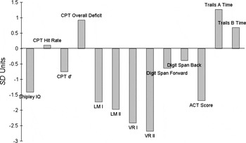
The schizophrenia group was impaired across all neuropsychological tests (p-values ranged from <.001 to .034) with two exceptions. There was a marginal group difference for digit span forward (p = .052), and no difference for digit span back (p = .187; see Figure 2).
Standardized representation of cognitive deficits in schizophrenia. Each bar represents mean schizophrenia group score as a z-score relative to normal control mean and standard deviation. For most tests, negative scores indicate a schizophrenia group deficit. Trails A, Trails B, and CPT Overall Deficit measures reflect schizophrenia group deficit in terms of increased time on task.

Hypothesis 2
Group differences in hippocampus substructure volumes were assessed using a mixed-model multivariate analyses of variance (MANOVA), with Hemisphere (right and left) x Region (anterior and posterior hippocampus) entered as repeated measures. Overall hippocampus volume was smaller in the schizophrenia group than in controls [Group main effect, F(1, 46) = 6.689, p = .013]. Across groups, anterior hippocampus volume was larger than posterior hippocampus volume [Region main effect, F(1, 46) = 254.554, p < .001], and left hippocampus was larger than right [Hemisphere main effect, F(1, 46) = 4.195, p = .046].
The Group x Region interaction was marginally significant, F(1, 44) = 3.122, p = .084, two-tailed test, and investigation of this predicted interaction using simple-effects tests demonstrated a trend toward smaller anterior hippocampus in the schizophrenia group than in controls, t(46) = 2.701, p = .010, with no group difference for posterior hippocampus (p = .821). No other main effects or interactions approached significance.
Hypotheses 3 and 4
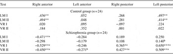
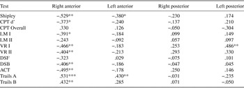
Table 2 shows partial correlations between hippocampus subregional volumes and neuropsychological memory test scores for each group, controlling for ICV. Groups did not differ in ICV, GM, or WM, so using ICV as a covariate is appropriate (Miller & Chapman, Reference Miller and Chapman2001). In the control group, LM I and LM II scores correlated positively with right anterior and left posterior hippocampus volumes. No significant brain-behavior correlations associated with VR I or VR II emerged for this group.
Correlations between neuropsychological tests and hippocampus subregions

Note.
Values represent partial correlations controlling for intracranial volume (ICV).
LM I, LM II = Wechsler Memory Scale-Revised (WMS-R) Logical Memory scores;VR I, VR II = WMS-R Visual Reproduction scores.
a Group difference in correlations, p < .05.
b Group difference in correlations, p < .10.
*p < .10. **p < .05. ***p < .01.
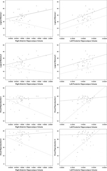
In the schizophrenia group, the pattern of correlations was markedly different. Only a single correlation was noted with LM: LM I was negatively correlated with right anterior hippocampus volume. VR I and VR II were also negatively correlated with right anterior hippocampus volume. VR I and VR II were positively correlated with bilateral posterior hippocampus volumes. See Figure 3 for scatterplots of relationships between episodic memory scores and hippocampus subregions.
The most dramatic correlational group differences were between memory test scores and right anterior and left posterior hippocampus. The scatterplots depict bivariate correlations between the variables (hippocampus volumes are in cc). Solid line = control group, dashed line = schizophrenia group.

Cohen’s statistical tables were used to test group differences between brain-behavior correlations (Cohen, Reference Cohen1988). Group correlational differences fell into two basic clusters. First, the schizophrenia group correlations between right anterior hippocampus volume and episodic memory test scores (i.e., LM I, VR I, VR II) were significantly different from those in the control group. In this cluster, the schizophrenia group correlations were strongly negative versus positive or absent in controls. Second, bilateral posterior hippocampus volume correlations with VR I and VR II were significantly different, in this case more positive in the schizophrenia group than those correlations in the control group. In addition, differences between correlations for left anterior volume and VR II and left posterior volume and LM II approached significance (p < .10).
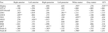
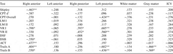
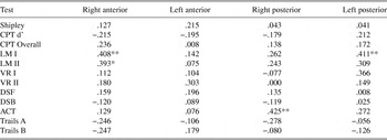
To provide some context for the findings presented in Table 2, Tables 3a and 3b show the zero-order correlations between most test scores derived from the neuropsychological battery and all brain regional volumetric data assessed for this study. Tables 4a and 4b show partial correlation coefficients between test scores and hippocampus regions controlling for gray matter volume. Bivariate correlations between neuropsychological tests and hippocampus regional volumes, overall WM and GM, and ICV for the control group (n = 24) ICV = intracranial volume, GM = gray matter, WM = white matter, CPT Overall = Continuous Performance Test Overall Deficit, CPT d’ = CPT detectability, DSF = Digit Span Forward, DSB = Digit Span Back, ACT = Auditory Consonant Trigrams, Trails A, B = Trail-Making Tests A & B. * p < .10. **p < .05. ***p < .01. Bivariate correlations between neuropsychological tests and hippocampus regional volumes, overall WM and GM, and ICV for the schizophrenia group (n = 24) *p < .10. **p < .05. ***p < .01. Partial correlations between neuropsychological tests and hippocampus regional volumes for the control group, controlling for GM (n = 24) *p < .10. **p < .05. ***p < .01. Partial correlations between neuropsychological tests and hippocampus regional volumes for the schizophrenia group, controlling for GM (n = 24) *p < .10. **p < .05. ***p < .01.
Note.

Note.

Note.

Note.
Demographic Variables
As a result of the nature of the population from which these samples were drawn (i.e., a VA population consisting largely of middle-aged veterans), only 13 women were included in the study. When sex was considered as a covariate in the mixed model MANOVA described earlier, there was no main effect (p = .381) or significant interactions between sex and hippocampus volume variables.
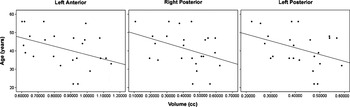
There was a group difference in how age related to hippocampus volume [Group x Age interaction, F(2, 45) = 4.513, p = .016]. Further correlational analysis indicated that there was no effect of age on hippocampus volumes for control subjects but that age was negatively correlated with left anterior hippocampus (r(24) = -.406, p = .049) and marginally correlated with right posterior (r(24) = -.382, p = .065) and left posterior (r(24) = -.383, p = .065) hippocampus volumes in the schizophrenia group (see Figure 4).
Scatterplots represent the significant correlations between age and hippocampus subregional volumes in the schizophrenia group.

Consistent with the prevailing literature, GM volume correlated negatively with age for both groups. In a regression, no effect was found for Group, or for the Group x Age interaction term, indicating that GM volume decreased equally with age for control and schizophrenia groups. A series of hierarchical regressions was used to investigate whether this generalized GM diminution might account for the age–hippocampus volume correlations in the schizophrenia group. With age used as a dependent variable, and GM as a lone predictor variable, the overall regression was significant for the schizophrenia group (R 2 = .386, p = .001). However, there was no additional variance accounted for when hippocampus volumes were added as predictors. Taken together, these analyses suggest that age–hippocampus volume correlations in schizophrenia are proportionate to age–GM correlations, whereas in controls, age predicts only smaller GM and there was no evidence of a correlation with hippocampus volume.
The mean number of years since schizophrenia symptom onset was 16.18 (SD = 8.34) and was considered as a demographic covariate for the schizophrenia group. To investigate an effect for time since first diagnosis, a mixed-model MANOVA was used with Region and Hemisphere as repeated measures and Years since symptom onset entered as a covariate. There was a three-way Hemisphere x Region x Years interaction, F(1, 19) = 7.233, p = .015. Post-hoc investigation with correlations suggested that Years was most significantly correlated with right hemisphere posterior hippocampus volume (r = –.505, p = .017).
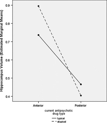
To investigate possible medication effects, Medication type (typical, atypical) was entered as a covariate for the schizophrenia group. Although there was no main effect of Medication type, there was a signification Medication type x Region interaction, F(1, 22) = 5.267, p = .032; schizophrenia subjects taking atypical antipsychotic medications had larger anterior hippocampus and smaller posterior hippocampus than those taking typical antipsychotic medications (see Figure 5). When Medication type was entered as a covariate in the correlation matrix relating neuropsychological tests and hippocampal subregions, there were no significant changes from Table 2 evident in the pattern or size of the correlations.
Schizophrenia patients taking atypical antipsychotic medications had larger anterior hippocampi and smaller posterior hippocampi than those taking typical antipsychotic medications.

DISCUSSION
Associations between hippocampus and memory are one of the core findings in neuropsychology (Milner, Reference Milner2005). Both hippocampus abnormality and normal variability in volume have been linked to episodic memory measures. Because hippocampus volume deficits are among the most consistent findings in schizophrenia, a key focus of this study was to better characterize the relationship between hippocampus subregion volume and episodic memory function in healthy control subjects and patients with schizophrenia.
The first hypothesis was that the schizophrenia group would be impaired on neuropsychological measures relative to controls. Across measures and presumed cognitive domains, with a single exception, neuropsychological function was impaired in the schizophrenia group, a finding consistent with the majority of studies investigating the extent of cognitive changes associated with schizophrenia (e.g., Goldberg et al., Reference Goldberg, Torrey, Gold, Ragland, Bigelow and Weinberger1993; Gruzelier et al., Reference Gruzelier, Seymour, Wilson, Jolley and Hirsch1988; Saykin et al., Reference Saykin, Gur, Gur, Mozley, Mosley, Resnick, Kester and Stafaniak1991; 1994). The exception was for the digit span backward portion of the digit span test. Further analysis indicated that a single, high-scoring subject in the schizophrenia group accounted for the lack of group difference, as he achieved the highest score of either group. With his data removed from the analysis, patients scored significantly worse than controls on the digit span backward test.
The second hypothesis was that hippocampus volume would be smaller in the schizophrenia group. This hypothesis was supported, but of particular interest was the finding that the group difference was specific to bilateral anterior (not posterior) hippocampus. Overall intracranial, GM, and WM volumes were statistically equivalent across groups, although these three measures were smaller in mean size. With increased power, that is, larger sample size, group differences may have been found. Certainly, such a finding would not be unusual and would be well supported by the prevailing literature (Andreone et al., Reference Andreone, Tansella, Cerini, Rambaldelli, Versace, Marrella, Perlini, Dusi, Pelizza, Balestrieri, Barbui, Nosé, Gasparini and Brambilla2007; Sanfilipo et al., Reference Sanfilipo, Lafargue, Rusinek, Arena, Loneragan, Lautin, Feiner, Rotrosen and Wolkin2000; Reference Sanfilipo, Lafargue, Rusinek, Arena, Loneragan, Lautin, Rotrosen and Wolkin2002; Steen et al., Reference Steen, Mull, McClure, Hamer and Lieberman2006; Zipursky et al., Reference Zipursky, Lambe, Kapur and Mikulis1998). In any case, however, none of these measures accounted for the group difference in anterior hippocampus when considered as statistical covariates, indicating that smaller hippocampus volume is not part of a more general effect for GM or WM volume, but is specific to anterior hippocampus.
Because of the importance of an intact hippocampus for episodic memory, the third hypothesis predicted that hippocampus volumes would positively correlate with episodic memory scores. Again, there was support for this hypothesis, but the answer was more complicated than initially supposed. Correlations between hippocampus and episodic memory scores were first evaluated in healthy control subjects to establish a frame of reference. In the control group, strong positive correlations between the size of hippocampus and episodic memory test performance were observed only for LM I and LM II, tests of immediate and delayed verbal memory. Verbal memory is often thought to be lateralized to the left hemisphere, yet positive correlations with both left posterior and right anterior hippocampus volumes indicate that current models of episodic memory may be oversimplified. Certainly, frontotemporal interaction and intrahippocampus circuitry are critical to the encoding, storage, and retrieval of relevant information. VR I and VR II, tests of memory for nonverbal information, evinced no correlations with any measure of hippocampus volume in controls.
A fourth prediction was that the pattern of correlations established in controls would not hold for the schizophrenia group, and this hypothesis was supported. Patterns of correlations between episodic memory scores and hippocampus volumes differed markedly according to diagnosis. Bilateral posterior hippocampus volumes correlated positively with performance on VR I and VR II in the schizophrenia group. Since mean posterior hippocampus volumes did not differ by group, this finding suggests that hippocampus organization in service to episodic memory is quite different in schizophrenia. Perhaps most damaging to a simple “bigger-is-better” model was the finding that right anterior hippocampus volume was negatively correlated with both verbal and nonverbal memory functions (i.e., LM I, VR I, and VR II) in schizophrenia. That these partial correlations were weaker and generally nonsignificant (but still negative) in bivariate correlations suggests that the effect is partially, but not entirely, accounted for by overall brain size in schizophrenia. It may be that smaller size reflects more efficient design or some developmental anomaly associated with schizophrenia. For example, anomalous schizophrenia neurodevelopment is thought to involve neurodevelopmental delays interacting with a fixed “developmental window” (see Yeo et al., Reference Yeo, Gangestad and Thoma2007). To the extent that a “smaller-is-better” model applies to hippocampus function in normal childhood (Van Petten, Reference Van Petten2004), it is possible that the present negative correlations simply reflect a straightforward schizophrenia neurodevelopmental delay. Of course, the current negative association between hippocampal volume and episodic memory score runs contrary to the typical findings in this literature and requires replication.
Beyond the main hypotheses, the effects of demographic variables were considered. The age of subjects in this study ranged from 20 to 62 years. There was a strong negative association between overall GM volume and age that was equivalent for both groups. In controls, however, there was no effect for age on hippocampus volume, which appeared to remain relatively constant across the lifespan (at least ages 20–62). In contrast, hippocampus volume in the schizophrenia group correlated with age to the same extent as overall cortical volume. The cross-sectional design and limited sample size in the present study allow only for speculation as to the cause of this finding. Other researchers have demonstrated similar effects among controls (Sullivan et al., Reference Sullivan, Marsh, Mathalon, Lim and Pfefferbaum1995) and subjects with schizophrenia (Chakos et al., Reference Chakos, Schobel, Gu, Gerig, Bradford, Charles and Lieberman2005). These findings invite further investigation into possibly differential aging of neocortical versus limbic cortical tissue. Another demographic variable effect investigated was gender. This sample was drawn from the population of a veteran’s hospital, and the small number of women included reflects the representation of women in that population. Because of the small sample size, this study was not an ideal test of population-typical gender differences. When sex was considered as a covariate in the analysis, there was no evidence of an effect.
In brain-behavioral studies of schizophrenia, the effect of medication must be considered. When included as a covariate, medication type had no effect on the hippocampus–memory correlations, suggesting that medication does not mediate this relationship. Rather, schizophrenia subjects taking atypical antipsychotic medications were found to have larger anterior hippocampi and smaller posterior hippocampi than those taking typical antipsychotic medications. This group was a chronic population and had been on medication for years or decades. Medication decisions for these patients were based on clinical criteria of maximizing good effects and limiting side effects, using the standard VA formulary. It may be that this pattern of hippocampal structure is an indicator of who will derive the best outcome from each medication type; however, this result was unexpected and will require replication.
Hippocampus volume is consistently and positively associated with episodic memory performance across both the normal lifespan and a host of disorders, including temporal lobe epilepsy, neurodegenerative disease, depression, post-traumatic stress disorder, dementia, and Alzheimer’s disease (Griffith et al., Reference Griffith, Pyzalski, Seidenberg and Hermann2004; Hickie et al., Reference Hickie, Naismith, Ward, Turner, Scott, Mitchell, Wilhelm and Parker2005; Kramer et al., Reference Kramer, Schuff, Reed, Mungas, Du, Rosen, Jagust, Miller, Weiner and Chui2004; Reference Kramer, Rosen, Du, Schuff, Hollnagel, Weiner, Miller and Delis2005; Mungas et al., Reference Mungas, Harvey, Reed, Jagust, DeCarli, Beckett, Mack, Kramer, Weiner, Schuff and Chui2005; Nemeroff et al., Reference Nemeroff, Bremner, Foa, Mayberg, North and Stein2006; Petersen et al., Reference Petersen, Jack, Xu, Waring, O’Brien, Smith, Ivnik, Tangalos, Boeve and Kokmen2000; Reminger et al., Reference Reminger, Kaszniak, Labiner, Littrell, David, Ryan, Herring and Kaemingk2004; Walhovd et al., Reference Walhovd, Fjell, Reinvang, Lundervold, Fishl, Quinn and Dale2004). In none of these disorders has the relationship between hippocampus volume and episodic memory function differed notably from that for normal samples. Hence, growing evidence for a divergent pattern of correlations between hippocampus volumes and episodic memory scores in schizophrenia suggests the presence of multiple processes in this disorder. These processes may be more complex than the hippocampus atrophic changes found in dementia or epilepsy that result in a relatively linear correspondence between reduced hippocampus volume and impaired episodic memory. Although the present study did identify an inverse correlation of hippocampus volume and age in the schizophrenia group, this effect was not associated with decrements in episodic memory performance. It may be that the abnormal structure-function relationship between hippocampus and episodic memory in schizophrenia precedes the onset of the disorder and catalyzes an abnormal aging process throughout the lifespan.
Several weaknesses in this research remain to be addressed. The generalizability of the present conclusions regarding hippocampus volume and episodic memory relationships is limited by the relatively small sample size. The use of a longitudinal design in the future would allow for stronger conclusions regarding aging and hippocampus–memory relationships in schizophrenia. Further, the investigation of sex differences was weakened by the gender bias in this VA sample. Moreover, very recent research by Squire and colleagues (i.e., Bayley et al., Reference Bayley, O’Reilly, Curran and Squire2008) suggests that certain aspects of semantic memory processing traditionally assumed to be based in the hippocampus actually involve neocortical structures while minimally engaging the hippocampus. To the extent that this is true, some of the variance unaccounted for in episodic memory scores might be explained by abnormalities in distal neocortical regions. It may be that schizophrenia hippocampus–memory relationships are best considered in the context of a distributed brain network (Eyler et al., Reference Eyler, Jeste and Brown2008; Nestor et al., Reference Nestor, Kubicki, Kuroki, Gurrera, Niznikiewicz, Shenton and McCarley2007), and future research will be necessary to investigate this possibility. Our current results require replication prior to drawing any strong conclusions, but the data serve as an incremental step in our understanding of brain–behavioral relationships in schizophrenia.
ACKNOWLEDGMENTS
This research was supported by the National Institutes on Alcohol Abuse and Alcoholism grants 1K23AA016544-01 and 1R21AA017313-01 to Dr. Thoma and by the National Institute of Mental Health grant 1R01MH65304-01 and VA MERIT grant 0104 to Dr. Cañive. None of the authors have commercial interests constituting potential conflict of interest. Information in this article was previously published only where noted.