INTRODUCTION
Toxoplasma gondii, one of the most widespread obligate intracellular protozoan parasites, belongs to the phylum Apicomplexa and has been recognized as an important opportunistic pathogen for an immunocompromising individual and a significant cause of congenital infection and abortion when vertically transmitted during pregnancy (Tenter et al. Reference Tenter, Heckeroth and Weiss2000; Montoya and Remington, Reference Montoya and Remington2008). The life cycle of Toxoplasma has multiple differentiation stages that are vital for parasites to survival in intermediate and final hosts. During the infection of intermediate hosts, including human and a large number of mammalian hosts, Toxoplasma can invade a wide range of nucleated cells and occur on the developmental interconvert between tachyzoite and bradyzoite stages. Chronic infection with latent bradyzoite cysts is asymptomatic in immunocompetent individuals. Upon host immunosuppression (due to chemotherapy, organ transplantation or AIDS), however, reconversion of bradyzoites into tachyzoites might occur and then the rapidly dividing tachyzoites can cause severe tissue damage resulting in organ failure and death (Sullivan and Jeffers, Reference Sullivan and Jeffers2012). Hence, it is critical to understand that the molecular mechanisms underpinning the conversion of these life cycle stages, which may identify suitable novel targets for the treatment of toxoplasmosis.
Autophagy is a self-catabolic process conserved in eukaryotic cells, by which cellular components include defunct proteins and organelles that are degraded by delivering them into mammalian cell lysosomes or vacuoles in plant and yeast (Levine and Klionsky, Reference Levine and Klionsky2004). As it is widely known, the fundamental cellular process is associated with a variety of cellular functions such as regulation of cell death and differentiation, recycling of the degraded proteins and organelles as energy sources or building blocks for the synthesis of new cellular constituents, and encystation of protozoan parasites under nutrient-limiting or stress stimulus conditions (Levine and Klionsky, Reference Levine and Klionsky2004; Gozuacik and Kimchi, Reference Gozuacik and Kimchi2007; Picazarri et al. Reference Picazarri, Nakada-Tsukui and Nozaki2008; Kourtis and Tavernarakis, Reference Kourtis and Tavernarakis2009; Kroemer et al. Reference Kroemer, Marino and Levine2010). There are three major classes of autophagy, such as macroautophagy, microautophagy and chaperone-mediated autophagy. However, there is no evidence of microautophagy or chaperone-mediated autophagy in Apicomplexa. In this paper, the term ‘autophagy’ will indicate macroautophagy unless otherwise specified.
In the autophagic process, cytosolic or organelle components are captured and sequestered for destruction in a unique double-membrane vesicle called the autophagosome (Mizushima et al. Reference Mizushima, Noda, Yoshimori, Tanaka, Ishii, George, Klionsky, Ohsumi and Ohsumi1998; Weidberg et al. Reference Weidberg, Shvets and Elazar2011). As the central event of autophagy, the formation of autophagosome is governed by a series of autophagy-related (Atg) proteins (Nakatogawa et al. Reference Nakatogawa, Suzuki, Kamada and Ohsumi2009; Mizushima et al. Reference Mizushima, Yoshimori and Ohsumi2011). To date, although many Atg proteins associated with the autophagic process are not found in T. gondii, database searches and bioinformatics have illustrated that the parasite appears to contain the core conjugation system involved in the attachment of Atg8 to the lipid phosphatidylethanolamine (PE), including TgAtg3, TgAtg7 and TgAtg8. Moreover, the autophagic vesicles have been observed in both extracellular and intracellular dividing tachyzoites of Toxoplasma RH strain under the conditions of starvation via using TgAtg8 as an autophagosomal marker to detect and quantify autophagy (Besteiro et al. Reference Besteiro, Brooks, Striepen and Dubremetz2011; Besteiro, Reference Besteiro2012; Ghosh et al. Reference Ghosh, Walton, Roepe and Sinai2012). Nevertheless, very little is known about the variation of Atg proteins in Toxoplasma tachyzoite to bradyzoite transformation.
The objectives of the present study were to assess the relative protein expression abundance of three TgAtg proteins, TgAtg3, TgAtg7 and TgAtg8, between tachyzoite and bradyzoite stages of Pru (Prugniaud) TgAtg7-HA transgenic strains. Our results indicate that autophagy is downregulated in Toxoplasma bradyzoite in vitro. Furthermore, we modulated autophagy with two kinase inhibitors and determined that Toxoplasma autophagy may interfere with the transformation of tachyzoite to bradyzoite in vitro.
MATERIALS AND METHODS
Host cells and parasite culture
The type II avirulent Pru (Prugniaud) ΔKu80 strain tachyzoite of T. gondii was maintained in human foreskin fibroblast (HFF) cells (Roos et al. Reference Roos, Donald, Morrissette and Moulton1994). To induce bradyzoite in vitro, ~1 × 10⁵ PruΔKu80 tachyzoites were inoculated onto confluent HFF monolayers with the normal culture media. After 2–3 h of invasion, the medium was replaced with an alkaline medium (pH 8·2), which was changed daily. After 10 days of being cultivated in the alkaline stress medium, intracellular parasites were separated from host cells by passage through 23- and 25-gauge syringe needles. Then, the parasites were purified from host cell debris using 3·0-μ m Nuclepore filters (Whatman). The purified parasites were washed twice with cold sterile phosphate-buffered saline (PBS).
Generation of transgenic parasite lines
To stably express TgAtg7 (TGME49_229690) fused at its C-terminus with the 3× haemagglutinin (HA) epitope in parasite, ~1·5-kb fragment of the TgAtg7 gene 3′ end was amplified from PruΔKu80 strain genomic DNA using sense primer: 5′- TACTTCCAATCCAATTTAATGCCGTGCTCACGTTCACATTATC-3′ and antisense primer: 5′-TCCTCCACTTCCAATTTTAGCCTTGGTCTCTGCTGCATCCT-3′ (the sequences underscored represented the ligation-independent cloning sequences). The amplified fragments were cloned into the pLIC-HA × 3-DHFR-Ts (Suvorova et al. Reference Suvorova, Radke, Ting, Vinayak, Alvarez, Kratzer, Kim, Striepen and White2013) endogenous tagging vector such that the TgAtg7 coding sequence was fused in frame with the epitope coding region. Plasmids were linearized using RsrII (NEB) prior to the transfection into parasites by electroporation as previously described (Roos et al. Reference Roos, Donald, Morrissette and Moulton1994). The transfected cultures were supplemented with 1·0 µ m pyrimethamine (Sigma) for drug selection.
Modulation of autophagy with either rapamycin or 3-methyladenine (3-MA) in vitro
Autophagy was modulated with kinase inhibitors as described previously with modifications (Besteiro et al. Reference Besteiro, Brooks, Striepen and Dubremetz2011). Briefly, the purified extracellular tachyzoites of Pru TgAtg7-HA were incubated in a complete Dulbecco's modified Eagle medium (DMEM) with either 5 µg mL−1 of rapamycin (Sigma) or 10 mm of 3-MA (Sigma) for 8 h. Afterwards, the treated tachyzoites were allowed to infect confluent HFFs, and then induced into bradyzoites in the alkaline medium as described above. After 10 days, the induced bradyzoites from each group were obtained to evaluate the transcription and protein levels of both TgAtg8 and BAG1. Parasites which are not treated with drugs were used as control.
Western blot and antibodies
Parasites were lysed in ice-cold PBS with Complete protease inhibitor (Roche) followed by sonication. Equal amounts of the parasite lysates, 20 µg, were used for immunoblotting as described previously (Li et al. Reference Li, Hu, Wan, Xie, Li, Chen, Cheng, Yi, Liang and Tan2014). For identification of the presence of the lipidated TgAtg8, parasite lysates were detected by Western blot after separation by 6 m urea sodium dodecyl sulphate–polyacrylamide gel electrophoresis (SDS–PAGE) as described previously (Besteiro et al. Reference Besteiro, Brooks, Striepen and Dubremetz2011).
Primary antibody mouse anti-HA antibodies (1:1000, Roche) were used for the analysis of TgAtg7-HA transgenic strain and protein expression of TgAtg7. For observation of protein expression of both TgAtg3 and TgAtg8, rabbit polyclonal antibodies against anti-TgAtg3 (1:1000) (Hua et al. Reference Hua, Li, Wan, Chen, Zhao, Jiang, Hu, Pan and Tan2014) or anti-TgAtg8 (1:1000) (Tan et al. Reference Tan, Hua, Li, Li and Liang2014) were used as the primary antibody, respectively. For monitoring the bradyzoite formation and normalization, both rabbit polyclonal antibodies against Toxoplasma BAG1 (1:500) (Raygene Custom Antibody Service, Shanghai) and rabbit polyclonal antibodies against Toxoplasma β-tubulin (1:1000) (Mao et al. Reference Mao, Hua, Li, Yao and Tan2014) were used, respectively. Corresponding horseradish peroxidase (HRP)-conjugated anti-mouse or anti-rabbit antibodies were used as secondary antibodies at 1:5000. The density of bands detected in Western blot was analysed by ImageJ software and normalized with the β-tubulin loading control as the ratio of TgAtg/β-tubulin.
Semiquantitative RT–PCR
Total RNA was extracted using the Trizol Reagent (Ambion) and cDNA was synthesized using RevertAid First Strand cDNA Synthesis Kit (Thermo Scientific). Semiquantitative RT–PCR was performed using SYBR Green PCR Master Mix (Applied Biosystems). Primers used in this study were as follows: β-tubulin (for normalization): sense, 5′-ATGTTCCGTGGTCGCATGT-3′, antisense, 5′-TGGGAATCCACTGAACGAAGT-3′; BAG1: sense, 5′-TGAGCGAGTGTCCGGTTATTT-3′, antisense, 5′-TAGAACGCCGTTGTCCATTG-3′. PCR reactions were carried out in triplicate using the StepOne Real-time PCR Systems and analysed with relative quantification software (StepOne Software Version 2.1, Applied Biosystems).
Indirect immunofluorescent assay (IFA)
All incubations were performed at room temperature unless otherwise stated. Parasites were fixed in 4% paraformaldehyde/PBS for 20 min and permeabilized in PBS containing 0·2% Triton X-100 plus 3% bovine serum albumine (BSA) for 10 min. After blocked in PBS with 3% BSA for 1 h, the parasites were first probed with mouse anti-HA antibodies (1:200) for the analysis of TgAtg7-HA transgenic strain or rabbit anti-TgAtg8 (1:500) for the detection of TgAtg8 puncta. Secondary antibodies were Alexa Fluor 488 goat anti-mouse or rabbit IgG (1:1000, Invitrogen). Fluorescent images were obtained with a Nikon ECLIPSE Ci-L epifluorescence microscope and the number of parasites bearing fluorescently labelled puncta was quantified. At least 200 cells were counted in each TgAtg8 puncta experimental set.
Statistical analyses
These results were expressed as the mean ± s.d. of at least three independent experiments. Data from these experiments were analysed with either Student's t-test or one-way ANOVA (SPSS13.0 software) followed by the Student–Newman–Keuls (SNK) test. Significant differences were established at P < 0·05.
RESULTS
Identification of transgenic strains Pru TgAtg7-HA in vitro
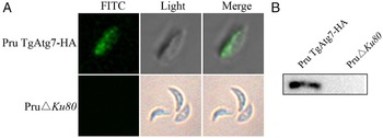
IFA showed that TgAtg7 fusion protein distributes in the cytoplasm of parasites, whereas this protein was not detected in the parental PruΔKu80 strains (Fig. 1A). Meanwhile, a specific protein band of 150 kDa in parasite lysate was revealed by Western blot using the anti-HA antibodies. There is consistent with the predicted size of the TgAtg7-HA fusion protein, whereas no protein band was observed in parental parasites (Fig. 1B).
Identification of the expression of TgAtg7-HA fusion protein in transgenic strains. (A) Subcellular localization of TgAtg7 in Toxoplasma by IFA. PruΔKu80 strain was used as a HA-negative control. (B) Confirmation of the C-terminal HA × 3-tagging of the endogenous TgAtg7 locus in Pru TgAtg7-HA strains with Western blot using the anti-HA antibody. Lysate from parental PruΔKu80 strain was included as a HA-negative control.

Induction of bradyzoite downregulates Toxoplasma autophagy in vitro
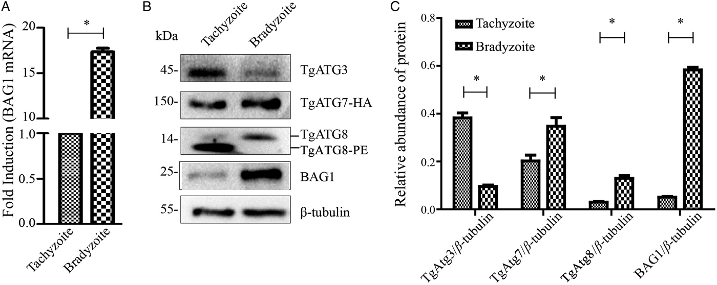
As a bradyzoite stage-specific protein, both the transcript and protein level of BAG1 were significantly higher at bradyzoites in comparison with the tachyzoites (Fig. 2A and B), demonstrating that the bradyzoites were induced successfully in vitro. Contrary to BAG1, the ratio of the TgAtg3/β-tubulin protein level was significantly decreased in cultures enriched with bradyzoites (0·10 ± 0·01) compared with tachyzoites (0·38 ± 0·02) (P < 0·05, Fig. 2B and C). Unlike TgAtg3, however, TgAtg7 expression was significantly higher in bradyzoites (0·35 ± 0·04) in comparison with tachyzoites (0·20 ± 0·03) (P < 0·05, Fig. 2B and C).
Identification of the protein abundance for TgAtg3, 7 and 8 in tachyzoite and bradyzoite, respectively. (A) The abundance for BAG1 transcript was quantified by semiquantitative RT–PCR in both tachyzoite and bradyzoite from Pru TgAtg7–HA strain. (B) Western blot confirms the relative expression abundance for TgAtg3, 7, 8 and BAG1 protein in both tachyzoite and bradyzoite from Pru TgAtg7-HA strain; (C) The density of bands detected in Western blot was analysed by ImageJ software and normalized with the β-tubulin loading control as the ratio of TgAtg/β-tubulin. Data are mean ± s.d. values obtained from one of three independent experiments. *P < 0·05.

To further assess the autophagic process in tachyzoite to bradyzoite transformation, urea SDS–PAGE followed by Western blot was performed using anti-TgAtg8 antibodies. The results showed that TgAtg8 is present in both lipidated (TgAtg8-PE) and non-lipidated forms in tachyzoites. In contrast, TgAtg8 was present only as a single non-lipidated form in bradyzoites (Fig. 2B). Furthermore, IFA showed the proportion of parasites containing TgAtg8 puncta reduced significantly in bradyzoite (31·0 ± 3·39%) than in tachyzoite (55·9 ± 3·82%) (P < 0·01).
Toxoplasma autophagy involved in the transformation of tachyzoite to bradyzoite in vitro
To modulate Toxoplasma autophagy in vitro, Pru TgAtg7-HA tachyzoites were treated with two kinase inhibitors, rapamycin or 3-MA, before induction of bradyzoite. As shown in Fig. 3A, compared with the control group (30·15 ± 3·23), the treatment with rapamycin resulted in significantly higher proportions of parasites containing TgAtg8 puncta (57·30 ± 5·09, P < 0·05) and 3-MA treatment reduced significantly the proportion of parasites containing TgAtg8 puncta (13·00 ± 5·66, P < 0·01). These results were further confirmed by the Western blot.
Effect of modulation of autophagy on the transformation of tachyzoite to bradyzoite. (A) The proportions of parasites containing fluorescence-labelled TgAtg8-PE puncta from Pru TgAtg7-HA strain. (B) The relative levels for BAG1 transcript were quantified by semiquantitative RT–PCR in rapamycin-treated bradyzoites, 3-MA-treated bradyzoites or untreated control. (C,D) The relative protein expression abundancesfor TgAtg8 and BAG1 were detected by Western blot in rapamycin-treated bradyzoites, 3-MA-treated bradyzoites or untreated control. Data are mean ± s.d. values obtained from one of the three independent experiments. *P < 0·05.

To evaluate the regulation of autophagy in tachyzoites to bradyzoites transformation, the levels of transcript and protein for BAG1 were observed. In comparison with the control group, BAG1 transcript level was significantly lower in rapamycin-treated bradyzoites and higher in 3-MA-treated bradyzoites (Fig. 3B). Consistent with these results, Western blots revealed that the ratio of BAG1/β-tubulin protein level was significantly decreased in rapamycin-treated bradyzoites (0·74 ± 0·01) and increased in 3-MA-treated bradyzoites (1·14 ± 0·02) compared with control bradyzoites (0·93 ± 0·03) (P < 0·01, Fig. 3C and D), which suggests that induction of Toxoplasma autophagy suppresses the transformation of tachyzoite to bradyzoite in vitro.
DISCUSSION
During the formation of autophagosome, Atg8 requires the activity of two ubiquitin-like conjugation systems to bind the autophagosomal membrane, involving Atg7–Atg3 and Atg12–Atg5, respectively (Ohsumi and Mizushima, Reference Ohsumi and Mizushima2004; Yorimitsu and Klionsky, Reference Yorimitsu and Klionsky2005; Geng and Klionsky, Reference Geng and Klionsky2008). To date, however, it has been shown that the Atg12–Atg5 conjugation system is dispensable in vitro (Ichimura et al. Reference Ichimura, Imamura, Emoto, Umeda, Noda and Ohsumi2004) and seems to be absent from the most of Apicomplexan parasites, including T. gondii (Besteiro et al. Reference Besteiro, Brooks, Striepen and Dubremetz2011), as well as other protists (Ichimura et al. Reference Ichimura, Imamura, Emoto, Umeda, Noda and Ohsumi2004; Ponder and Bogyo, Reference Ponder and Bogyo2007; Rigden et al. Reference Rigden, Michels and Ginger2009). On the contrary, genes encoding orthologues of a complete set of the Atg7–Atg3 conjugation system, including TgAtg3, TgAtg4, TgAtg7 and TgAtg8, have been shown in T. gondii genome (Besteiro et al. Reference Besteiro, Brooks, Striepen and Dubremetz2011). However, TgAtg4 is not involved in regulating the autophagosomal formation although this plays a role in the recycling of membrane-bound TgAtg8 (Kong-Hap et al. Reference Kong-Hap, Mouammine, Daher, Berry, Lebrun, Dubremetz and Besteiro2013). In this study, we tagged firstly the C-terminus of the endogenous TgAtg7 in PruΔKu80 parasite strain with a 3 × HA tag via LIC strategy. Stable Pru TgAtg7-HA transgenic strains were confirmed successfully by the IFA and Western blot and showed no apparent difference in growth or morphology (data not shown). Based on this strain, the expression levels for three TgAtg proteins (TgAtg3, TgAtg7 and TgAtg8) were compared between tachyzoite and bradyzoite in order to evaluate the autophagy level in tachyzoite to bradyzoite transformation.
Atg8 is usually recognized as the most reliable marker for autophagosomes since it is a key and conserved protein for the autophagy (Nakatogawa et al. Reference Nakatogawa, Ichimura and Ohsumi2007; Xie et al. Reference Xie, Nair and Klionsky2008; Shpilka et al. Reference Shpilka, Weidberg, Pietrokovski and Elazar2011; Weidberg et al. Reference Weidberg, Shvets and Elazar2011). There are two forms of Atg8 in most organisms, including cytosolic unconjugated Atg8 and membrane-bound Atg8-PE, which can be separated by urea SDS–PAGE (Ichimura et al. Reference Ichimura, Kirisako, Takao, Satomi, Shimonishi, Ishihara, Mizushima, Tanida, Kominami, Ohsumi, Noda and Ohsumi2000; Besteiro et al. Reference Besteiro, Brooks, Striepen and Dubremetz2011). In the present study, two separated bands, TgAtg8 and TgAtg8-PE, were observed using urea-containing SDS–PAGE and Western blot analysis. There is consistent with previously issued that a significant proportion of native TgAtg8 appears to be already conjugated with PE in basal conditions (Besteiro et al. Reference Besteiro, Brooks, Striepen and Dubremetz2011; Kong-Hap et al. Reference Kong-Hap, Mouammine, Daher, Berry, Lebrun, Dubremetz and Besteiro2013). In contrast, only a single band corresponding to free TgAtg8 was detected in induced bradyzoite lysates, suggesting that there is declined in autophagy activity in the bradyzoite stage of Toxoplasma. To further verify this finding, IFA using the anti-TgAtg8 antibody was performed to quantify the fluorescent puncta because the fluorescence signal dispersed throughout the cytoplasm of parasite was generally assembled as the punctate structure, while TgAtg8 was relocalized from the cytoplasm to autophagic vesicular structures (Besteiro et al. Reference Besteiro, Brooks, Striepen and Dubremetz2011). The results showed that the number of parasites containing fluorescence-labelled puncta was significantly decreased from 56 ± 0·57% in tachyzoite to 30·8 ± 0·28% in bradyzoites.
In most eukaryotes, Atg3 accepts Atg8 from the Atg8/Atg7 intermediate to form an Atg8/Atg3 thioester intermediate, which promotes the transfer of Atg8 from the intermediate to PE for the efficient formation of Atg8–PE conjugates (Yamaguchi et al. Reference Yamaguchi, Noda, Nakatogawa, Kumeta, Ohsumi and Inagaki2010). Hence, Atg3 is another essential element for the association of Atg8 with the autophagosome membrane and plays crucial role in autophagic function. Likewise, in Toxoplasma, it has revealed that the ability of TgAtg8 lipidation and autophagosome formation was seriously impaired in TgAtg3-depleted strains (Besteiro et al. Reference Besteiro, Brooks, Striepen and Dubremetz2011). Recently, we have determined the TgAtg3 can bind directly to TgAtg8 (Di Chen et al. Reference Chen, Lin, Liu, Li, Chen, Hua, Nie, Hu and Tan2016). Accordingly, the relative abundance for TgAtg3 protein was also detected in this study. Similar to the TgAtg8, a signification decline level of TgAtg3 was identified. Combined with these observations, it suggests that the induction of bradyzoite leads to a reduced capacity in TgAtg8-conjugation to the autophagosomes and thus likely downregulates the autophagic function in Toxoplasma.
As an obligate intracellular protozoan parasite, Toxoplasma tachyzoites firstly establish themselves into a parasitophorous vacuole (PV), which constructs a niche that offers protection from hostile cytosolic surveillance of innate immunity and allows parasite to consume host nutrients for intracellular growth and multiplication (Laliberte and Carruthers, Reference Laliberte and Carruthers2008). Thus, it is unlikely that the intracellular parasite is experiencing nutrient limiting. However, once parasites have egressed from their host cells, the fate of extracellular tachyzoite is highly reliant on parasites’ motility and the time of parasite invasion into their neighbouring cells. Although little is known about the exact circumstances confronted within their hosts, autophagy could also be seen as a mechanism of improving the ability of Toxoplasma to survive during the extracellular journey of the parasite within the host body. In this study, the upregulation of the autophagic function in Toxoplasma tachyzoites supports the hypothesis.
The activation of Atg8 through Atg7 is first event during autophagosome formation in which a thioester intermediate is formed between Atg7 and Atg8 (Tanida et al. Reference Tanida, Mizushima, Kiyooka, Ohsumi, Ueno, Ohsumi and Kominami1999). In the present research, however, contrarily to the decreased expression of TgAtg8 and TgAtg3 in the bradyzoite stage, TgAtg7 was more abundantly expressed in this stage at the protein level, suggesting that TgAtg7 has a dual role in T. gondii. On the one hand, it functions in the catabolic processes involved in activation of TgAtg8 in Toxoplasma autophagy, but it also associates with tachyzoite–bradyzoite differentiation of Toxoplasma. These ideas could be verified in future experiments using genetic strategies.
The observation of downregulation of autophagy in bradyzoite prompts us to further investigate whether the transformation of tachyzoite to bradyzoite is associated with Toxoplasma autophagy. Autophagy is a multistep pathway regulated by several upstream kinases. The ‘target of rapamycin’ (TOR) kinase possesses a central position in regulating cellular growth by repressing autophagy. As TOR inhibitor, therefore, rapamycin is well known to increase the autophagy activity in several eukaryotes (Diaz-Troya et al. Reference Diaz-Troya, Perez-Perez, Florencio and Crespo2008). Conversely, 3-MA has been shown to reduce autophagy activity by specifically inhibiting the class III phosphoinositide 3-kinase (PI3K) which has been identified to promote autophagy (Petiot et al. Reference Petiot, Ogier-Denis, Blommaart, Meijer and Codogno2000). Importantly, both rapamycin and 3-MA have been identified to inhibit their corresponding target kinase activity in T. gondii (Besteiro et al. Reference Besteiro, Brooks, Striepen and Dubremetz2011; Ghosh et al. Reference Ghosh, Walton, Roepe and Sinai2012). Nevertheless, both rapamycin and 3-MA have to be used with higher concentration than those used for mammalian and yeast cells. In considerations of the impact by both drugs on the host cells, the tachyzoites were treated at first with two kinase inhibitors prior to infection of host cells. Under such conditions, the levels of transcript and protein for BAG1 were significantly decreased at rapamycin-treated bradyzoites and increased at 3-MA-treated bradyzoites, which imply the autophagy involved in regulating the transformation of tachyzoite to bradyzoite in vitro. Nevertheless, future work is necessary to elucidate the spatial and temporal regulation, as well as the molecular regulation mechanism.
In conclusion, we have shown here that the level of autophagy was downregulated in Toxoplasma bradyzoite and the transformation of tachyzoite to bradyzoite was strongly influenced via modulation of autophagy, which presents an attractive strategy for the development of drugs for treatment of infections caused by T. gondii.
ACKNOWLEDGEMENTS
We are grateful to Dr William J. Sullivan, Jr. (Indiana University School of Medicine, IN) for providing the pLIC-HA × 3-DHFR-Ts vector, and Dr Louis Weiss (Albert Einstein College of Medicine, NY) and Dr Xiaoguang Chen (Southern Medical University, Guangzhou, China) for the PruΔKu80 parasite strain.
FINANCIAL SUPPORT
This work was supported by grants from the Natural Science Foundation of Zhejiang Province (no. LY12H19004) and the Commonweal Technology Application Project Program of Zhejiang Province (grant no. 2014C33161) to FT, Natural Science Foundation of Zhejiang Province (grant no. LQ14H190003) to XH, Zhejiang Xinmiao talent plan (grant no. 2014R413082) to XL, as well as Student Scientific Research Project of Wenzhou Medical University (grant no. wyx2015101098) to YL.