


Introduction: vertebrate middle ears
Sound transduction in vertebrates is performed by the hair cells of the inner ear, which are surrounded by fluid. This presents a problem for the detection of airborne sound because the acoustic input impedance of the inner ear is much higher than that of air, which would tend to result in the reflection of acoustic energy at the interface. A tympanic middle ear, found in amphibians, reptiles, birds and mammals, consists of a tympanic membrane overlying an air-filled middle-ear cavity, vibrations of the membrane being somehow coupled to the oval window, the entrance to the inner ear. The tympanic middle ear represents an impedance-matching device, allowing a far greater proportion of incident sound energy to be transmitted through to the inner ear than would be the case in its absence. It has been theoretically and clinically demonstrated in humans that ossicular interruption in the presence of an intact drum results in a conductive hearing loss of around 60 dB.Reference Merchant, Ravicz, Voss, Peake and Rosowski 1
Among vertebrates in general, the tympanic membrane is coupled to the oval window by means of a chain of bony or cartilaginous structures, generically referred to here as ‘conducting elements’. In mammals, the conducting elements are the three auditory ossicles: the malleus, incus and stapes. Non-mammalian tetrapods lack a malleus and incus: their stapes (sometimes referred to as the columella) is coupled to the tympanic membrane via a cartilaginous extrastapes (also known as the extracolumella).
The tympanic middle ear is believed to have evolved several times independently in different vertebrate groups.Reference Clack 2 – Reference Clack, Allin, Manley, Popper and Fay 6 Although details are debated among palaeontologists, it is generally agreed that a tympanic ear evolved once in the ancestors of living amphibians,Reference Lombard, Popper and Fay 7 , Reference Bolt and Lombard 8 and probably several times within amniotes, including independent origins within the turtle, diapsid (a group including lizards, crocodiles and birds) and mammalian lineages.Reference Clack 2 The mammalian three-ossicle ear is thought to have evolved from ancestors which used their malleus (equating to the articular and prearticular bones) and incus (equating to the quadrate) as part of the jaw joint; it is still unclear if and where a tympanic membrane was present in these ancestors, and to what extent they used their stapes to conduct sound vibrations.Reference Clack, Allin, Manley, Popper and Fay 6 , Reference Maier 9 – Reference Meng, Wang and Li 11 Whether or not a three-ossicle ear actually evolved more than once among mammals remains controversial.Reference Ji, Luo, Zhang, Yuan and Xu 12 – Reference Rougier, Forasiepi and Martinelli 14
Given that a tympanic ear appears to have evolved multiple times within vertebrates, it is instructive to look for convergently derived features which might represent necessary design constraints. Flexibility within the chain of conducting elements, manifested either as relative movement at an articulation between two components or as bending of one particular component, appears to be one such feature. This paper will review the existence and anatomical basis of flexibility within the ears of living vertebrate groups, with a view to gaining insights into its functional and clinical implications for humans.
Amphibian middle ears
Among amphibians, there is a wide diversity of pathways by which vibrations, travelling initially in air, through a solid substrate or through water, can get to the inner ear.Reference Mason, Narins, Feng, Fay and Popper 15 Frogs are the only living amphibian group to possess tympanic ears, but many frog species show reduction or loss of various components. Of those frogs with ‘complete’ tympanic ears, the best-studied examples are found within the family Ranidae.
The tympanic membrane of ranid frogs is located on the side of the head. The air-filled middle-ear cavity beneath the membrane communicates with the mouth cavity through a wide and typically patent eustachian tube. Tympanic membrane vibrations are communicated to the inner ear via a cartilaginous extrastapes and a largely bony stapes (Figure 1a). Two recent studies on ranid frogs have shown that the stapes is hinged where it articulates with the skull along the ventral rim of the oval window, such that an inwards movement of the tympanic membrane results in an outwards movement of the stapes footplate.Reference Jørgensen and Kanneworff 16 , Reference Mason and Narins 17 The conducting elements therefore work as a first-order lever (like a see-saw or crowbar). Furthermore, given that the ratio of lengths from the tip of the extrastapes to the fulcrum and from the fulcrum to the centre of the stapes footplate exceeds one, force amplification is expected at the oval window relative to the tympanic membrane, at the cost of velocity reduction. This is presumably part of the impedance-matching function of the middle ear. However, in both cited studies the anatomical lever ratios were found to be much lower than the velocity ratios obtained using laser vibrometry, over the measured frequency range of up to 2–3 kHz which largely encompasses the hearing range of these animals. Although velocity ratios were approximately flat over these frequencies, a phase lag was observed between tympanic membrane and footplate which increased with frequency. These observations imply some kind of flexibility within the chain of conducting elements.
Diagrammatic representations of the middle-ear structures of vertebrates (not to scale): (a) frog; (b) lizard; (c) bird; and (d) mammal (freely mobile morphology). Some key elements of the conducting apparatus, referred to in the text, are labelled. Numbers indicate regions believed to represent points of flexibility in each group: 1 = attachment of frog extrastapes to tympanic membrane; 2 = articulation between extrastapes and stapes; 3 = reptile extrastapes pars inferior; 4 = manubrium of malleus; 5 = malleo-incudal articulation; 6 = pedicle of lenticular apophysis; 7 = incudo-stapedial articulation. TM = tympanic membrane
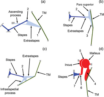
The cartilaginous extrastapes in the American bullfrog (Lithobates catesbeianus, formerly Rana catesbeiana) is tethered to the skull via its thin ascending process, which bends to generate a rocking movement of the extrastapes when the tympanic membrane is pushed inwards.Reference Mason and Narins 17 This requires some movement of the distal extrastapes relative to the tympanic membrane, to which it is only loosely attached. The resulting motion of the extrastapes is then coupled to the bony stapes shaft, to which it is fused. Although there is some relative movement at the stapes–extrastapes articulation, which acts as a hinge-joint, researchers have found no evidence of bending of the shaft of the extrastapes itself, up to at least 2 kHz. Although unossified, the extrastapes in the bullfrog therefore works in effect as a second ‘ossicle’ coupled via a flexible articulation to the stapes proper, a conclusion reached independently by Werner.Reference Werner 18
The stapes footplate of frogs typically shares the oval window with a cartilaginous operculum; vibrations of the footplate in response to airborne sound are conveyed to the operculum in bullfrogs.Reference Mason and Narins 19 The articulation between these two coupled elements represents another source of flexibility within the amphibian middle ear.
Reptile middle ears
Living reptiles are commonly divided into four groups: Testudines (turtles and tortoises), Squamata (lizards, snakes and amphisbaenians), Rhynchocephalia (the tuatara) and Crocodylia (crocodilians). Of these, snakes, amphisbaenians, tuatara and some lizards (e.g. chameleons) lack tympanic ears as defined above, and will not be considered here.
The middle-ear structures of lizards (Figure 1b) have been reviewed by Baird,Reference Baird, Gans and Parsons 20 WeverReference Wever 21 and Saunders et al. Reference Saunders, Duncan, Doan, Werner, Dooling, Fay and Popper 22 Lizard tympanic membranes are convex externally, unlike those of mammals, and are found at the bottom of short external ear canals. As in frogs, the middle-ear cavities are in free communication with the pharynx by means of short, wide, patent eustachian tubes, and vibration transmission from tympanic membrane to oval window involves a cartilaginous extrastapes and bony stapes. The extrastapes terminates in a number of radiating processes which attain different degrees of prominence in different species. Of these processes, the pars superior and pars inferior together form a bar which inserts along a radius of the tympanic membrane. The ligamentous attachment of the pars superior to the bony frame surrounding the tympanic membrane forms a fulcrum. In some lizards, the articulation between extrastapes and stapes has some degree of mobility and thus is considered a flexible joint, while in others the joint appears to be stiff and is supported by a dense connective tissue sheath.
The extrastapes in lizards is considered to act as a second-order lever (like a wheelbarrow), one lever-arm being the distance between the tip of the pars inferior to the fulcrum at the tip of the pars superior, the other the distance from the point of origin of the extrastapes shaft to the same fulcrum, which equates to the length of the pars superior only.Reference Wever and Werner 23 Experimental studies suggest that this kind of lever motion prevails at least up to around 1 kHz in the alligator lizard Gerrhonotus multicarinatus.Reference Rosowski, Peake, Lynch, Leong and Weiss 24 Studies on the tokay gecko (Gekko gecko), using the Mössbauer technique,Reference Manley 25 have enabled a comparison of the vibrations of the tip of the pars inferior with those of the point of origin of the extrastapes shaft. It was found that the amplitude ratio varied between 6 and 14 dB at low frequencies but increased at frequencies greater than 4 kHz, and exceeded 20 dB at 10 kHz. This increase was attributed to flexion of the pars inferior at higher frequencies, which would increase the relative velocity of its tip.Reference Manley 26 Using laser vibrometry, Werner et al. Reference Werner, Montgomery, Safford, Igic and Saunders 27 found an increase in velocity ratio between the same two points at frequencies over 4 kHz in the gekkonoid Oedura marmorata; however, results from another study, in which the Mössbauer technique was used to compare eardrum and stapes footplate movement in two other species of lizards, were less consistent with this general picture.Reference Saunders and Johnstone 28
The middle ear of crocodiles has not been extensively investigated, but it appears to resemble that of birds (see below). The cartilaginous extrastapes has a number of processes radiating from its distal end.Reference Baird, Gans and Parsons 20 , Reference Wever 21 The connection between extrastapes and stapes is described as ‘a more or less flexible joint’.Reference Wever 29
Although considered to possess a tympanic ear,Reference Clack, Allin, Manley, Popper and Fay 6 testudines lack the thin tympanic membrane usually found in vertebrates specialised for hearing in air. The extrastapes of the terrestrial box turtle Terrapene carolina is expanded distally into a cartilaginous plate, lying underneath largely undifferentiated skin and separated from it by ‘a loose network of fibrous and fatty tissue’.Reference Wever and Vernon 30 The extrastapedial plate is hinged through a stiff ligamentous connection to the skull at its posterior edge, resulting in a lever action which increases force (slightly) at the stapes footplate.Reference Wever 21 No flexibility has been described in the turtle ear, and the structure of the extrastapes appears simpler than that of other non-mammalian vertebrates. However, the connective tissue layer between the skin and the extrastapedial plate would be expected to show some compliant behaviour, and one might expect some kind of bending between the conducting elements if a rocking movement of the extrastapedial plate was to be translated into a piston-like motion of the stapes within the oval window, as Wever and Vernon observed. Marine turtle middle ears are essentially similar to what is described above, except that more fat is present between extrastapes and skin.Reference Lenhardt, Klinger and Musick 31 , Reference Ridgway, Wever, McCormick, Palin and Anderson 32 The ear morphology of testudines in general has been regarded as a compromise between aquatic and terrestrial hearing.Reference Wever 33
Bird middle ears
Birds emerged from within the reptile group. They are most closely related to crocodiles, among living reptiles, which explains the similarities in their auditory structures. Like reptiles, birds have a long, bony stapes which is attached to the externally convex tympanic membrane via a cartilaginous extrastapes (Figure 1c), although the extrastapes may show partial ossification in some adult birds.Reference Saunders 34 , Reference Gaudin 35 The extrastapes possesses a number of radiating processes which vary in morphology and orientation according to species. Of these, the infrastapedial process extends towards the edge of the tympanic membrane and in some species articulates with its supporting bony rim.Reference Saunders, Duncan, Doan, Werner, Dooling, Fay and Popper 22 , Reference Norberg 36
Based on a consideration of the anatomy and micromanipulation of the structures involved, the extrastapes of birds is generally believed to undergo a rocking movement. The exact location of the axis or fulcrum about which the extrastapes rocks differs according to different studies on different species, but the tip of the infrastapedial process is commonly implicated, either as the position of a fulcrumReference Starck 37 or as one point on a rotatory axis.Reference Norberg 36 , Reference Gummer, Smolders and Klinke 38 The rotation of the extrastapes would then work as a second-order lever, increasing force at the footplate. Vibration velocities recorded using the Mössbauer technique suggest that simple, rigid-body rotation of the extrastapes occurs at low frequencies up to approximately 1 kHz.Reference Gummer, Smolders and Klinke 38 At higher frequencies, a rise in the velocity ratio and an increasing phase-lag was observed between the extrastapes and the stapes footplate, thought to indicate a change in vibratory mode which might, at some frequencies at least, involve flexion somewhere within the chain of conducting elements.Reference Gummer, Smolders and Klinke 38 In anatomical studies and those employing static pressure changes to displace the tympanic membrane, the flattened extrastapes–stapes junction region has been identified as a point of flexibility within the avian middle ear.Reference Norberg 36 , Reference Starck 37 , Reference Pohlman 39 – Reference Mills and Zhang 41
Mammalian middle ears
Anatomy of the mammalian ossicular chain
Ossicular morphology in mammals varies considerably, the most extreme adaptations being found, as would be expected, in those species which inhabit atypical acoustic environments, e.g. cetaceans (which are modified to hear underwater)Reference Nummela, Reuter, Hemilä, Holmberg and Paukku 42 and golden moles (which appear to hear ground vibrations through inertial bone conduction).Reference Mason 43 As FleischerReference Fleischer 44 observed, however, the majority of living mammals fall somewhere on a spectrum ranging between the ‘freely mobile’ ossicular morphology seen in humans and guinea pigs (Figures 1d and 2a) and the ‘microtype’ morphology characteristic of bats, shrews and mice (Figure 2b). Anatomical observations suggest that the malleus–incus complex should rotate about an axis extending between the main points of its attachment to the surrounding tympanic cavity walls, i.e. from the anterior process of the malleus to the short process of the incus.Reference Dahmann 45 The tip of the manubrium of the malleus, which inserts into the tympanic membrane pars tensa, is typically further from this ‘anatomical axis’ than is the articulation of the stapes at the end of the long process of the incus (Figure 1d), resulting in a second-order lever action which is widely believed to contribute to middle-ear impedance matching. However, the ligamentous attachments of ossicles to the walls of the tympanic cavity tend to allow more than one degree of freedom of movement: rotation about the ‘anatomical axis’ may be a reasonable approximation only at low frequencies.Reference Decraemer, Khanna and Funnell 46 , Reference Decraemer and Khanna 47
Light micrographs showing internal views of the tympanic membrane, malleus and incus of two mammalian species. In both, the malleus and incus are fused and the line of articulation between the two ossicles is barely visible. (a) Guinea pig (Cavia porcellus), left structures of a stillborn neonate from a laboratory colony (scale bar = 2 mm); the ossicles are ‘freely mobile’ in this species. (b) Common shrew (Sorex araneus), structures in an adult found dead in Cambridgeshire, UK. This species has ‘microtype’ ossicles (the original image of right structures has been laterally inverted to facilitate comparison) (scale bar = 1 mm). 1 = tympanic membrane pars tensa; 2 = head of malleus; 3 = long process of incus; 4 = manubrium of malleus; 5 = orbicular apophysis

The human malleo-incudal articulation has been described in detail.Reference Marquet 48 , Reference Sim and Puria 49 Among mammals in general, this articulation is typically synovial,Reference Hinchcliffe and Pye 50 the joint surface usually being described as saddle-shaped.Reference Fleischer 44 The articular surfaces can be flattened and the inter-ossicular connection very loose in some mammals, notably subterranean species,Reference Mason 51 – Reference Segall 54 but in certain other mammals the malleus and incus may actually be fused. This seems to be the normal condition among most rodents of the Ctenohystrica clade,Reference Burda, Bruns and Hickman 53 , Reference Wible, Wang, Li and Dawson 55 – Reference Mason 58 a group including guinea pigs and chinchillas. In the guinea pig, the malleo-incudal articulation starts to form in the embryo but never cavitates.Reference Amin and Tucker 59
Contrary to the belief that only ‘freely mobile’ species such as guinea-pigs have fused ossicles,Reference Fleischer 44 , Reference Amin and Tucker 59 the malleo-incudal articulation also appears to be fused in shrews of the genus Sorex (as noted by BurdaReference Burda 60 and M.J. Mason, unpublished observations), which have a ‘microtype’ ossicular morphology (Figure 2b). The malleus and incus are said to be fused in the echidna and adult platypus too,Reference Doran 56 , Reference Zeller, Szalay, Novacek and McKenna 61 although in the case of the latter this is disputed.Reference Gates, Saunders, Bock, Aitkin and Elliott 62 There are contradictory reports of malleo-incudal fusion in various other species of mammals, which remain to be confirmed; some of these claims may arise from stiffened capsular ligaments in dried or preserved specimens.
The anatomy of the incudo-stapedial articulation, formed between the lenticular apophysis of the incus and the head of the stapes, has been best described in humans and cats.Reference Chien, Northrop, Levine, Pilch, Peake and Rosowski 63 – Reference Karmody, Northrop and Levine 65 This articulation is synovial in mammals, with the possible exception of some bat speciesReference Hinchcliffe and Pye 50 and the platypus.Reference Zeller, Szalay, Novacek and McKenna 61 A fibrocartilaginous disc (meniscus) has been described in humans, separating the joint cavity into two compartments.Reference Chien, Northrop, Levine, Pilch, Peake and Rosowski 63 , Reference Karmody, Northrop and Levine 65 The lenticular apophysis is usually attached to the long process of the incus by means of a thin, bony pedicle, which in many species appears to be flexible.Reference Mason 51 , Reference Chien, Northrop, Levine, Pilch, Peake and Rosowski 63 , Reference Mason, Lai, Li and Nevo 66 A finite-element model of the cat incudo-stapedial articulation prompted the conclusion that the pedicle may in fact be more flexible than the articulation between the lenticular apophysis and the stapes itself.Reference Funnell, Heng Siah, McKee, Daniel and Decraemer 64
Experimental evidence for flexibility within the mammalian middle ear
From experiments using a capacitive probe, MøllerReference Møller 67 concluded that the malleus and incus of the cat are rigidly coupled at frequencies up to 2.5 kHz, at ‘reasonably low’ sound pressure levels. Examining ossicular motion visually using stroboscopic illumination, Guinan and PeakeReference Guinan and Peake 68 found that the cat incus lagged in phase relative to the malleus at sound frequencies over approximately 3 kHz, which they interpreted as evidence of movement at the malleo-incudal articulation. Decraemer and KhannaReference Decraemer, Khanna, Gyo, Wada, Hato and Koike 69 also reported slippage, increasing with frequency, at the feline malleo-incudal joint. Using the Mössbauer technique in guinea pigs, Manley and JohnstoneReference Manley and Johnstone 70 found an increase in the malleus–incus lever ratio together with an increasing incudal phase lag at frequencies over 25 kHz; it was suggested that this might arise from energy loss at the malleo-incudal articulation, despite this being fused. The source of flexibility within the guinea pig ear merits further investigation.
Guinan and PeakeReference Guinan and Peake 68 found no significant differences in amplitude or phase between the end of the incudal long process and the stapes in cats at the tested frequencies up to 15.5 kHz at moderate sound pressure levels; however, they did observe movement at the incudo-stapedial joint at sound pressure levels of more than 150 dB.
However, movement at the ossicular articulations is not the only possible source of flexibility within the mammalian middle ear. Experimental evidence from mouse, bat (Rhinolophus ferrumequinum), cat and gerbil suggests manubrial bending at high frequencies, the frequencies in question being species-specific but exceeding a few kilohertz.Reference Decraemer, Khanna and Funnell 46 , Reference Decraemer and Khanna 47 , Reference Saunders and Summers 71 – Reference de la Rochefoucauld and Olson 73 However, significant manubrial bending was not observed in the guinea pig ear, over the measured range of up to 45 kHz.Reference Manley and Johnstone 70
Flexibility within the human middle-ear apparatus
Although movement at the human malleo-incudal joint at ‘physiological’ sound pressure levels has long been regarded as negligible,Reference Gyo, Aritomo and Goode 74 – Reference Elpern, Greisen and Andersen 77 recent results obtained using more sophisticated measurement systems show that this is not the case.Reference Decraemer, Khanna, Gyo, Wada, Hato and Koike 69 , Reference Sim, Puria, Steele, Gyo, Wada, Hato and Koike 78 – Reference Willi, Ferrazzini, Huber, Gyo, Wada, Hato and Koike 80 Using scanning laser Doppler vibrometry of human temporal bone preparations, Willi et al. Reference Willi, Ferrazzini and Huber 79 demonstrated movement at this joint even at moderate sound pressure levels (i.e. 75–90 dB SPL). A 90 dB SPL stimulus results in a transmission loss of approximately −10 to −15 dB at frequencies greater than 2 kHz.Reference Willi, Ferrazzini, Huber, Gyo, Wada, Hato and Koike 80 This malleo-incudal flexibility appears to be sufficient to explain the fact that tympanic membrane (umbo) velocity changes little with stapes fixation in humans, such that stapes fixation cannot be reliably diagnosed using tympanometry.Reference Ravicz, Peake, Nakajima, Merchant, Rosowski, Gyo, Wada, Hato and Koike 81
Flexibility elsewhere in the human ear in response to acoustic stimulation has been less extensively examined. Elpern et al. Reference Elpern, Greisen and Andersen 77 found that experimental fixation of the incudo-stapedial articulation made essentially no difference to the transmission of sound through the middle ear, as measured via vibrations of the round window, but these experiments have been criticised.Reference Willi, Ferrazzini and Huber 79 By introducing experimental fixations of the ossicles within a human temporal bone preparation exposed to a chirp stimulus (containing frequencies from 0.025 to 20 kHz, at 105–114 dB SPL), Nakajima et al. Reference Nakajima, Ravicz, Merchant, Peake and Rosowski 82 demonstrated that flexibility of the human ossicular chain over the measured frequency range (0.1 to 4–6 kHz) includes a significant incudo-stapedial component. Of the total compliance of the system seen at the umbo, Nakajima et al. estimated that 55 per cent arises from the annular ligament around the footplate of the stapes, 27 per cent from the incudo-stapedial joint, 13 per cent from the malleo-incudal joint and 4 per cent from manubrial bending, although certain simplifying assumptions were made in obtaining these values.
Although the idea of significant malleo-incudal flexibility in humans at normal sound pressure levels has only recently gained general acceptance, it has long been recognised that greater pressures applied to the ear result in joint movement. Such pressures include ‘non-physiological’ sound pressuresReference Gundersen and Høgmoen 83 and the results of physical probing.Reference Cancura 84 HüttenbrinkReference Hüttenbrink 85 found that static air pressures of up to ±400 mmH2O in the external ear canal resulted in a gliding movement at both inter-ossicular joints, the result of which was a reduction of stapes displacement relative to the displacement of the tympanic membrane. Hüttenbrink found that stapes displacement was 21 times less than manubrium displacement in the normal ear, but only 4.2 times less following experimental fixation of the malleo-incudal joint.
Effects of flexibility in vertebrate ears
Rather than working as simple pistons, the evidence reviewed above suggests that the ‘columellar’ conducting elements of frogs, lizards and birds actually rotate about an axis. The resulting lever arrangement is believed to increase force at the oval window, in an analogous manner to what is achieved by the three-ossicle system of mammals. Some form of flexibility, often occurring at a joint between conducting elements, also appears to be universal within tympanic ears: the supposedly ‘single ossicle’ ears of non-mammalian vertebrates are functionally ‘two ossicle’ ears. Testudines may represent an exception, but these animals appear adapted to some extent towards aquatic sound reception, for which different rules apply.
Some level of flexibility must be inevitable given stimuli of high enough amplitude or frequency. It may in some cases be geometrically necessary, for example in order to convert rotation of the extrastapes into a piston-like motion of the stapes shaft in birds.Reference Starck 37 , Reference Pohlman 39 Whether an inevitable by-product or an adaptive feature in its own right, flexibility will have an effect on the function of the middle ear.
Predictions from a simple model
In order to assess the likely effects of flexibility within a chain of conducting elements, we shall first consider the implications of a very simple model (Figure 3a), based on that of Nakajima et al. Reference Nakajima, Ravicz, Merchant, Peake and Rosowski 82 The model consists of two ossicular elements (ossicle 1 and ossicle 2, consisting of masses L 1 and L 2 moving at velocities v 1 and v 2, respectively) connected together by a spring (compliance C 1) representing a flexible inter-ossicular joint; ossicle 2 is articulated with the skull by means of a second spring, C 2. The motion of any real middle-ear apparatus is clearly far more complex than the one-dimensional vibration considered in our model, especially at high frequencies, but our purpose here is only to outline the general effects that flexibility between the ossicles might be expected to have, in the simplest case.
(a) A simple mechanical model of a middle ear containing two ossicular conducting elements separated by a compliant articulation. (b) An electrical analogue of model (a). See text for details.
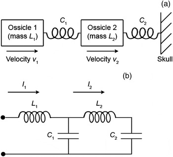
An electrical analogue to the mechanical model of Figure 3(a) is represented as Figure 3(b). Here, velocities are represented as currents: current I 1 (passing through inductor L 1) represents the velocity of ossicle 1, while I 2 (passing through L 2) represents the velocity of ossicle 2. We shall assume for the moment that the system is driven by a sinusoidal pressure source (a voltage, in the electrical analogue) with no source impedance. It can be shown that:
 $${I_2\over I_1}={1\over 1-\omega^2 C_1L_2+{C_1\over C_2}}$$
where ω is the radian frequency of vibration. As frequency tends to zero, the denominator of equation 1 tends to 1 + (C 1/C 2), the result obtained by Nakajima et al. Reference Nakajima, Ravicz, Merchant, Peake and Rosowski 82 This means that ossicle 2 moves with a lower velocity than ossicle 1, for all non-zero values of C 1. When frequency is very high, the relative velocity of ossicle 2 tends to zero. Without a concomitant increase in force as might be expected of a stiff lever mechanism, a reduced velocity of ossicle 2 reflects a loss of power.
It is instructive also to compare the difference in absolute velocity of each ossicle, between the conditions of inter-ossicular flexibility (C 1 > 0) and total ossicular fusion (C 1 = 0). The ratio of the two velocities of ossicle 1 can be calculated as follows:
 $${I_{1\lpar C_1\gt 0\rpar }\over I_{1\lpar C_1=0\rpar }}={\lsqb C_1+C_2-\omega^2C_1C_2L_2\rsqb \lsqb 1-\omega^2C_2\lpar L_1+L_2\rpar \rsqb \over C_2\lsqb 1-\omega^2C_2\lpar L_1+L_2\rpar -\omega^2C_1L_1+\omega^4C_1C_2L_1L_2\rsqb }$$
As ω tends to zero, the right-hand side of equation 2 tends to (C 1 + C 2)/C 2, while at high frequencies it tends to (L 1 + L 2)/L 1. A flexible ossicular joint therefore increases the velocity of ossicle 1 at the lowest frequencies, and it does the same at the highest frequencies for all non-zero values of L 2.
Turning to ossicle 2, the equivalent ratio of velocities in the flexible and fused states is:
 $${I_{2\lpar C_1\gt 0\rpar }\over I_{2\lpar C_1=0\rpar }}={\lsqb 1-\omega^2C_2\lpar L_1+L_2\rpar \rsqb \over \lsqb 1-\omega^2C_2\lpar L_1+L_2\rpar -\omega^2C_1L_1+\omega^4C_1C_2L_1L_2\rsqb }$$
As ω tends to zero, the right-hand side of equation 3 tends to one. At high frequencies, the ratio tends to zero, the result of the velocity of ossicle 2 tending to zero faster with a flexible inter-ossicular joint than it would if fused to ossicle 1.
Overall, this simple model suggests that inter-ossicular joint flexibility will have the following effects. (1) The relative velocity of ossicle 1 exceeds that of ossicle 2 at both very low and very high frequencies. (2) The absolute velocity of ossicle 1 is increased at both very low frequencies (compliance-dominated; both springs flex) and very high frequencies (in which case it becomes substantially easier to move one mass than two). (3) Although unaffected at very low frequencies, at very high frequencies the absolute velocity of ossicle 2 declines to zero faster with a mobile inter-ossicular joint. (4) At certain intermediate frequencies (when the denominators of equations 2 and 3 are equal to zero), resonance will occur within the system.
These conclusions are essentially the same if the model is extrapolated to three ossicles. In general terms, the velocity of ‘upstream’ ossicles at both the lowest and highest frequencies is increased by ‘downstream’ compliance, while the velocity of ‘downstream’ ossicles is reduced by ‘upstream’ compliance, but at high frequencies only. A reduced velocity of ossicle 2 will translate into reduced power transmission to the cochlea at these frequencies. Resonance will increase the velocity of ossicle 2 at intermediate frequencies, but high amplitudes and sharp tuning could be detrimental to hearing. Resonance can be reduced by resistance in the inter-ossicular joint(s), at the cost of reduced power transmission to the cochlea; resistive source or load impedances can also reduce resonance in the system. If we introduce a high source impedance, the limiting case being a pure velocity source driving the system (a current source, in the electrical analogue), the absolute velocity of ossicle 1 is less affected by ossicular fusion, but the above conclusions 1, 3 and 4 still hold.
In a comparison of power flow from middle to inner ears between humans, cats and guinea pigs, at frequencies from approximately 0.2 to 10 kHz, guinea pigs were calculated to have a higher power transfer efficiency than the other two species across nearly the whole frequency range.Reference Rosowski, Carney, Lynch, Peake, Allen, Hall, Hubbard, Neely and Tubis 86 Of these species, only the guinea pig has a fused malleo-incudal articulation, consistent with the above model's predictions that ossicular flexibility is generally detrimental to the transmission of sound energy.
Given that ossicular flexibility is predicted to result in reduced velocity of ‘downstream’ ossicular elements at high frequencies, we would expect it to be particularly detrimental to high-frequency hearing. Hearing tends to be limited to the sonic range in non-mammalian vertebrates,Reference Saunders, Duncan, Doan, Werner, Dooling, Fay and Popper 22 , Reference Manley 87 , Reference Megela-Simmons, Moss and Daniel 88 although Feng et al. Reference Feng, Narins, Xu, Lin, Yu and Qiu 89 report an exception. Sound localisation at low frequencies is made possible in these animals through the use of their acoustically coupled ears as pressure-difference receivers.Reference Christensen-Dalsgaard 90 Flexibility-induced reduction of sound transmission at higher frequencies may therefore be of little consequence.
The hearing ranges of mammals extend to much higher frequencies than those of other vertebrates, exceeding 100 kHz in some echo-locating bats (for an example, see Heffner et al.).Reference Heffner, Koay and Heffner 91 It has been argued that such high-frequency hearing was made possible by the evolution of the mammalian ossicular chain, which, being ossified, has greater stiffness than the equivalent conducting elements in non-mammalian tetrapods.Reference Manley 5 However, only a minority of living mammals (not including bats) possess fused mallei and incudes, despite the fact that fusion would appear to benefit sound transmission still further. Why do ossicular articulations remain flexible in most species?
Could flexibility ever improve acoustic transmission?
Ossicular fusion is clearly achievable among mammals and, according to our simple model, should improve sound transmission at high frequencies at least. Most mammals, however, retain mobile ossicular articulations, which suggests that flexibility confers some kind of adaptive advantage. Three proposals from the literature suggest that flexibility could under certain circumstances improve sound transmission through mammalian middle ears.
‘Transmission line’ hypothesis
The controversial theory that the mammalian middle ear works as a ‘transmission line’Reference Wilson and Bruns 72 , Reference Overstreet and Ruggero 92 , Reference Puria and Allen 93 requires that the conducting elements be regarded as a series of masses coupled by springs, each mass-stiffness pair being matched to the load impedance of the cochlea. The flexible joints between the ossicles then act as shunting stiffnesses which counteract the effect of the ossicular masses, the result of which is wide-band and potentially ‘lossless’ sound transmission at the cost of acoustic delay.Reference Overstreet and Ruggero 92 , Reference Puria and Allen 93 Such a mechanism would explain not just the presence of the inter-ossicular articulations but also the otherwise paradoxical capability of the mammalian middle ear to transmit acoustic energy at very high frequencies. A phase lag has indeed been measured in human middle-ear transmission from umbo to stapes, and this is reduced by experimental fixation of the ossicular joints.Reference Hato, Gyo, Stenfelt, Welsh, Goode, Gyo, Wada, Hato and Koike 94 However, the predictions of the transmission line hypothesis are only upheld in the gerbil middle ear at frequencies from 5 to 35 kHz, not at lower or higher frequencies,Reference Ravicz, Cooper and Rosowski 95 suggesting that things are not this simple.
Change in ossicular rotatory modes
Based on calculations of ossicular rotatory moments of inertia, Puria and SteeleReference Puria and Steele 96 have argued that the mallei of humans and cats adopt different rotatory axes in response to high-frequency sound, which would reduce their moments of inertia and so improve sound transmission. The new mode of malleus vibration could be communicated through to the incus by means of the flexible malleo-incudal articulation, working as a type of gear. Puria and Steele have suggested that this change in rotatory mode is not necessary in small mammals such as guinea pigs and chinchillas because their ossicular moments of inertia are already small, hence their fused malleo-incudal articulations. Given that ossicular fusion is actually very unusual among small mammals, fusion of the malleo-incudal joint in guinea pigs and chinchillas is more likely to relate to something specific to these animals rather than just to size,Reference Mason, Willi and Narins 97 perhaps their unusual malleus morphology.Reference Lavender, Taraskin and Mason 98
Reduction in tympanic membrane constraint
The simple model described above predicts that inter-ossicular flexibility will increase the absolute velocity of motion of ossicle 1, at both the lowest and the highest frequencies. Could this make it easier for the tympanic membrane, to which this ossicle is attached, to vibrate? A related idea has been proposed by Funnell et al.,Reference Funnell, Khanna and Decraemer 99 considering just the manubrial process of the malleus. Although a completely flexible manubrium will fail to couple vibrations through to the malleus body, a rigid manubrium might significantly constrain membrane movement and add mass loading if rigidity is conferred through making it more robust. This being the case, a light manubrium with some limited flexibility might, at least in principle, represent the optimal design.
Any or all of these three proposals might help to explain the prevalence of ossicular flexibility in mammals. However, it should be noted that there is as yet only limited evidence supporting the transmission line hypothesis, and that the other two proposals (i.e. altered ossicular rotatory modes and reduced tympanic membrane constraint) await experimental verification.
Flexibility and pressure-buffering
The high pressures found within the middle-ear cavities of frogs, associated both with ‘force-pump’ ventilation and vocalisation, result in sometimes visible bulging of the tympanic membranes outwards.Reference Purgue and Narins 100 – Reference Narins, Lewis, Purgue, Bishop, Minter and Lawson 103 The externally located membranes are also vulnerable to external insult. Flexibility within the frog middle ear must confer some protection against such ‘quasi-static’ pressure changes, which would otherwise result in damagingly high-amplitude movements of the stapes footplate within the oval window.Reference Mason and Narins 17 , Reference Mason and Narins 19 Contraction of the opercularis muscle might contribute to this protection,Reference Mason and Narins 19 as might pressure-release features of the inner ear.Reference Purgue and Narins 100 , Reference Purgue and Narins 104
In other terrestrial vertebrates, ventilation does not involve the high pressures expected of a force-pump mechanism, and the tympanic membrane is protected by an external meatus. However, a slow decrease in middle-ear pressure has been recorded in birds, reset probably by opening of the eustachian tubes and pressure changes during vocalisation.Reference Larsen, Dooling, Ryals, Lewis, Long, Lyon, Narins, Steele and Hecht-Poinar 105 The avian middle ear might also be subject to significant pressure changes when flying or (in some species) diving.Reference Mills and Zhang 41 Middle-ear pressure changes in birds affect the frequency response of the tympanic membrane through their effects on membrane displacement.Reference Larsen, Dooling, Ryals, Lewis, Long, Lyon, Narins, Steele and Hecht-Poinar 105 Flexibility within the chain of conducting elements is believed to protect the avian inner ear against the effects of such pressure changes.Reference Pohlman 39 , Reference Mills and Zhang 41 Contraction of the stapedius muscle might contribute to this protective mechanism by increasing the amount of flexion that is possible between stapes and extrastapes.Reference Mills and Zhang 41
Flexibility within the mammalian ossicular chain is also thought to protect the annular ligament and inner ear against excessive stapes displacement caused by high static pressures.Reference Kirikae 75 , Reference Hüttenbrink 85 , Reference Kobrak 106 Middle-ear pressure changes exceeding 1 kPa are said to occur with events such as gushes of wind, sneezing and diving in humans,Reference Hüttenbrink 85 and pressures of up to 5 kPa can occur during the Valsalva manoeuvre.Reference Tideholm, Carlborg, Jönsson and Bylander-Groth 107 When middle-ear pressure was increased by 1.6 kPa in a human temporal bone preparation, the umbo was found to be displaced by over 300 µm, a value three orders of magnitude greater than the umbo displacement measured by Tonndorf and KhannaReference Tonndorf and Khanna 108 in response to a 121 dB SPL acoustic stimulus at 525 Hz.Reference Dirckx and Decraemer 109 Positive pressures inside the middle-ear cavity give rise to a greater umbo displacement than negative pressures,Reference Murakami, Gyo and Goode 110 which may relate to the amount of flexion possible at the inter-ossicular joints in each direction.Reference Ladak, Decraemer, Dirckx and Funnell 111 Given that mammalian middle-ear pressure fluctuates slowly throughout the day,Reference Tideholm, Jönsson, Carlborg, Welinder and Grenner 112 the flexibility of the articulations might also contribute to accommodation of the middle-ear apparatus, allowing efficient transmission at all times.
It is unknown to what extent static pressure changes represent a problem for reptiles, and whether they too use flexibility as a protective mechanism.
Middle-ear muscles and ossicular joints
Middle-ear muscles have been described in all tympanic middle ears except, apparently, the unusual testudine ear. The literature on middle-ear muscle function in mammals is very extensive, and there are significant differences between species. We shall restrict our discussion here to the possible relationships between the muscles and ossicular chain flexibility in mammals.
Although the most familiar role of the mammalian middle-ear muscles is in the acoustic reflex in response to loud sounds, muscle activity in humans has been found to occur in many circumstances which might involve changes in middle-ear pressure relative to atmospheric pressure, including vocalising, laughing, coughing, eructation, swallowing, yawning, voluntary contractions of the soft palate or base of the tongue, and a general startle arising from an air-jet to the eye.Reference Djupesland 113 – Reference Ingelstedt and Jonson 116 When middle-ear pressure is high, the tensor tympani could be used to restrain outwards movement of the malleus.Reference Hüttenbrink 85 This circumstance might also arise in acute otitis media. It has been proposed that hearing thresholds are affected by intracochlear static pressures,Reference Bell 117 in which case restraining the movement of the ossicular chain could be useful in maintaining optimal pressure within the inner ear. Conversely, if tensor contraction indirectly pushes the stapes footplate further into the oval window and thereby increases inner-ear pressure, this could form part of a protective reflex.Reference Bell 117 However, it is currently unclear what effect the tensor tympani actually has on ossicular movement; while some have suggested that contraction of this muscle tightens the ossicular joints among mammals in general,Reference Fleischer 44 others have found that traction applied to the tensor in humans appears to decouple malleus from incus.Reference Gundersen and Høgmoen 83
The effects of stapedius contraction are better understood. In both cats and humans, this results in a sliding motion of the incudo-stapedial joint, such that the stapes head moves caudally relative to the lenticular apophysis.Reference Kirikae 75 , Reference Pang, Peake, Allen, Hall, Hubbard, Neely and Tubis 118 , Reference Love and Stream 119 This is thought to pull the footplate outwards from the oval window, stiffening the annular ligament and reducing transmission of vibrations.Reference Pang, Peake, Allen, Hall, Hubbard, Neely and Tubis 118 Pulling the lenticular apophysis backwards in human temporal bone preparations also results in lateral (outward) movement of the manubrium of the malleus.Reference Hüttenbrink 85 , Reference Love and Stream 119 According to Hüttenbrink,Reference Hüttenbrink 85 the malleo-incudal articulation must be mobile for this to occur. Such an effect would allow the stapedius to act as the antagonist to the tensor tympani with respect to manubrial motion in humans, but whether this applies to mammals in general is not clear. No displacement of the malleus or incus was detected with stapes head displacements of up to 65 µm in the cat.Reference Pang, Peake, Allen, Hall, Hubbard, Neely and Tubis 118 Middle-ear muscles have also been suggested to ‘exercise the joints’, aiding the circulation of synovial fluid and thereby keeping them from stiffening.Reference Hüttenbrink 85
Clinical implications of ossicular joint flexibility in humans
Ossicular joint pathology in humans
The effects of otosclerosis, whereby the stapes becomes fixed within the oval window, are well-known. However, other pathological conditions are believed to affect flexibility within the ossicular chain itself. Malleo-incudal fusion has been shown to occur in Treacher Collins syndrome,Reference Jahrsdoerfer, Aguilar, Yeakley and Cole 120 , Reference Belal and Stewart 121 branchio-oto-renal syndrome,Reference Ceruti, Stinckens, Cremers and Casselman 122 trisomy 13–15121 and congenital aural atresia.Reference Schuknecht 123 Joint fusion usually coexists with many other abnormalities in these congenital syndromes, making interpretation of its effects difficult.
It should in principle be easier to identify effects specifically associated with ossicular joint pathology in cases of arthritis. Evidence that the synovial inter-ossicular joints develop increased stiffness and even fusion in osteoarthritis comes both from histological observationReference Lempert and Wolff 124 – Reference Etholm and Belal 126 and tympanometric and audiometric data.Reference Rawool and Harrington 127 ‘Moderate’ to ‘severe’ arthritic changes in the malleo-incudal joint were found in all 36 ears taken from individuals older than 70 years, and also in a significant proportion of younger ears, although the donors showed no clinical evidence of middle-ear disease.Reference Etholm and Belal 126 If flexibility decreases acoustic transmission, joint fusion may increase the sound intensities to which the cochlea is exposed; it is interesting in this regard to note that elderly people are at increased risk of noise-induced hearing loss.Reference Toppila, Pyykkö and Starck 128
A histological study of rheumatoid arthritis has also suggested malleo-incudal and incudo-stapedial joint involvement,Reference Gussen 129 and the expected increase in stiffness of the middle ear has been documented.Reference Ozturk, Yalcin, Kaygusuz, Sahin, Gok and Karlidag 130 – Reference Colletti, Fiorino, Bruni and Biasi 133 In contrast, other studies have found a decrease in stiffness,Reference Moffat, Ramsden, Rosenberg, Booth and Gibson 134 , Reference Raut, Cullen and Cathers 135 but this could relate to changes in other parts of the ear, such as the tympanic membraneReference Moffat, Ramsden, Rosenberg, Booth and Gibson 134 or the joint capsules and ligaments.Reference Colletti, Fiorino, Bruni and Biasi 133 Small conductive and sensorineural hearing losses have been reported in a proportion of patients with rheumatoid arthritis.Reference Takatsu, Higaki, Kinoshita, Mizushima and Koizuka 132 , Reference Özcan, Karakus, Gündüz, Tuncel and Sahin 136 – Reference Murdin, Patel, Walmsley and Yeoh 138 It has been suggested that the sensorineural hearing impairment may be due to an increase in compressive force on the labyrinth,Reference Salvinelli, Cancilleri, Casale, Luccarelli, Di Peco and D'Ascanio 131 perhaps due to a relative inability to buffer static pressure changes. However, alternative hypotheses include the effects of systemic neuritis, vasculitis or salicylate toxicity.Reference Colletti, Fiorino, Bruni and Biasi 133 The inter-ossicular joints may also be affected in the ears of patients with juvenile idiopathic arthritis.Reference Ikiz, Unsal, Kirkim, Erdag and Guneri 139
Flexibility in ossicular replacement prostheses
An extreme example of loss of joint flexibility in the human middle ear is reconstruction with a total ossicular replacement prosthesis (TORP). In ears reconstructed with a TORP, a 10-fold increase in pressure at the oval window (the inner ear having been removed) has been measured when pressure in the ear canal is varied.Reference Pau and Hüttenbrink 140 The potential for most severe damage arises following surgery for otosclerosis, when the restraining effect of the annular ligament is functionally eliminated.Reference Hüttenbrink 85 Rupture of the oval window membrane due to barotrauma on landing in an aeroplane has been reported in one patient with a prosthesis.Reference Marquet 141 In a study of patients who had undergone stapedectomy and ‘standard’ wire or Teflon wire prosthesis insertion 28–30 years previously, the initial gains in hearing following surgery were found later to have been lost. After Carhart correction, sensorineural function was found to be superior in non-operated, otosclerotic ears than in operated ears of the same patients,Reference Redfors and Möller 142 consistent with a hypothesis that loss of flexibility in a reconstructed middle ear increases the chance of hearing impairment.
MillsReference Mills 143 argued that efforts at human middle-ear reconstruction should be directed towards recreating something more similar to the three-ossicle arrangement. This author's incus–stapes replica achieved this to an extent, and included a flexible ‘joint’.Reference Mills, Zadrozniak and Jie 144 This prosthesis has been demonstrated to undergo less movement in the medial to lateral plane in response to static pressure variation, theoretically reducing the risk of cochlear damage.Reference Mills, Zadrozniak and Jie 144
Variation from optimal tension across the middle ear due to sub-optimal prosthesis length leads to impaired sound transmission.Reference Nishihara and Goode 145 – Reference Feenstra and Vlaming 147 Currently, selecting the correct prosthesis length is based solely on clinical experience, which places considerable demands on the surgeon and may be complicated by post-operative inwards movement of the tympanic membrane.Reference Goode and Hüttenbrink 148 It has been suggested that a more flexible prosthesis will lessen the deleterious effect of incorrect length selection.Reference Goode and Yamada 149 Attempts to produce such prostheses have met with varying success.Reference Goode and Yamada 149 – Reference Arechvo, Bornitz, Lasurashvili, Zahnert and Beleites 154 Whether the potential benefits would outweigh the loss of transmission introduced by the additional flexibility has been questioned.Reference Bornitz, Zahnert, Hüttenbrink, Hardtke, Gyo, Wada, Hato and Koike 155
Comparative anatomy has informed ossicular replacement prosthesis design, although most attention to date has been focussed on birds.Reference Mills 143 , Reference Arechvo, Bornitz, Lasurashvili, Zahnert and Beleites 154 A closer look at some of the universal features of tympanic ears among vertebrates in general, such as the apparent requirement for a flexible lever, may provide important insights.Reference Manley 156 Given the diversity of ear types both between and within major vertebrate groups, the species chosen as experimental models for human ear pathologies must be selected with care.
Conclusion
Tympanic middle ears have evolved multiple times independently among vertebrates. The chain of conducting elements typically forms a first- or second-order, force-amplifying lever. Flexibility may represent a universal feature of the conducting element chain in vertebrate tympanic ears, although this has yet to be demonstrated in testudines.
Simple models suggest that flexibility will generally reduce the transmission of sound energy through the middle ear, especially at high frequencies. The most likely adaptive role of such flexibility is to buffer the ear against high-amplitude static pressure changes.
Loss of flexibility could result in sensorineural hearing loss, due either to increased transmission of unbuffered pressure changes to the inner ear, or to an increase in transmitted sound intensity. Flexibility may theoretically improve acoustic transmission in some circumstances; if this is the case, then reduced flexibility may result in conductive hearing loss.
Acknowledgements
We should like to thank the Department of Physiology, Development and Neuroscience animal facility for the provision of the guinea pig specimen dissected in the course of this study, and the Midlands Institute of Otology for their kind provision of reference material. Special thanks go to Rebecca Jordan for her help with the preparation of Figure 1, and to Jon Gair and Heidi Nakajima for valuable comments relating to the model.



