Introduction
In the dementias there is widespread disruption of neuronal and synaptic integrity, involving multiple neurotransmitter systems. Advances in neuroimaging techniques over the past 20 years enable us to study several neurotransmitter systems in vivo, including cholinergic, serotonergic and dopaminergic systems. As research tools, these neuroimaging techniques can improve our understanding of the neural substrates of dementia, whilst potential clinical applications include diagnosis and monitoring of disease progression and treatment. At present, imaging of the dopaminergic system has the greatest clinical utility in dementia.
Dopaminergic pathways and neuroimaging
The best understood dopaminergic pathway is the one involved in the modulation of movement. It is also increasingly recognized that dopaminergic pathways play important roles in cognition and behavior. Dopaminergic neurons originate in the substantia nigra and ventral (anterior) tegmental area, and project as three main pathways:
1. Nigrostriatal pathway to striatum (caudate and putamen). This pathway plays an important role in the coordination and maintenance of movements.
2. Mesocortical pathway to prefrontal cortex (orbito-frontal cortex). This pathway is involved in attention, initiative, motivation, planning, working memory and other higher cortical functions.
3. Mesolimbic pathway to nucleus accumbens and amygdala. This pathway is believed to be involved in emotional behavior, motivation, drive and reward.
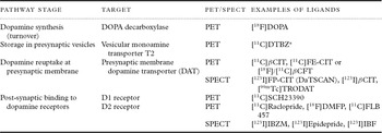
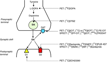
There are a number of components of dopaminergic neurotransmission that are amenable to neuroimaging. Figure 1 and Table 1 summarize the positron emission tomography (PET) and single photon emission computed tomography (SPECT) ligands that target dopamine synthesis, storage, presynaptic dopamine transporter and post-synaptic D1 and D2 dopamine receptors.
PET and SPECT ligands for dopaminergic imaging

*The early stage of [11C]DTBZ-PET scanning can be used to assess perfusion.
Nigrostriatal dopaminergic synapse and targets of PET and SPECT ligands. Tyrosine is converted to L-DOPA by tyrosine hydroxylase. L-DOPA is then converted to dopamine (DA), which is gathered into vesicles by vesicular monoamine transporter (VMAT T2) and from which it is released into the synaptic cleft. Dopamine binds to D1 and D2 receptors on the post-synaptic membrane. Some dopamine re-enters the presynaptic terminal via dopamine transporters (DAT).

PET and SPECT scanning
In both PET and SPECT a ligand that is attached to a radionuclide (or tracer), e.g. 11C or 123I, is administered by intravenous injection. The ligand is a biologically active molecule that attaches to a specific target (e.g. a dopamine transporter).
SPECT is an imaging technique that uses gamma rays to provide three-dimensional (3D) information. The gamma rays emitted by the injected radionuclide + ligand are detected by a gamma camera to yield 2D images of ligand concentration. Multiple 2D images are then reconstructed to yield a 3D dataset. This dataset may then be manipulated to show slices along any chosen axis of the brain, similar to those obtained from other tomographic techniques, e.g. magnetic resonance imaging (MRI) and computed tomography (CT).
PET radionuclides emit positrons that travel a short distance before hitting an electron (annihilation), producing a pair of photons moving in opposite directions. Detection of these pairs of photons enables production of 3D images that are of higher resolution than those produced by SPECT.
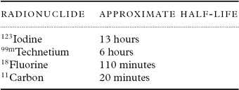
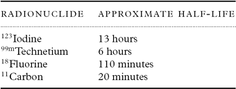
The time window during which the PET or SPECT scan can be acquired depends largely on the affinity of the ligand for its target. In [123I]FP-CIT-SPECT imaging, this window is about 3–6 hours from the time of injection of the ligand. The shelf-life of the ligand is determined by the half-life of the radionuclide (Table 2). Radionuclides used in PET scanning typically have a short half-life and must therefore be produced using a cyclotron close to the imaging facility. SPECT scanning is consequently more widely available and significantly less expensive than PET. [123I]FP-CIT-SPECT is the most widely used form of dopaminergic imaging.
Approximate half-lives of common PET and SPECT radionuclides
Dopaminergic imaging in dementia
Dementia with Lewy bodies and Parkinson's disease dementia
Dementia with Lewy bodies (DLB) is part of the Lewy body spectrum of disorders that are all characterized by dopaminergic loss. It is the second most common cause of neurodegenerative dementia. The clinical picture is of a progressive dementia accompanied by some or all of the following features: fluctuations in cognition and alertness, visual hallucinations, spontaneous motor parkinsonism, neuroleptic sensitivity and rapid eye movement (REM) sleep behavior disorder (McKeith et al., Reference McKeith2005).
Patients with Parkinson's disease (PD) frequently go on to develop PD dementia (PDD) (Aarsland et al., Reference Aarsland, Andersen, Larsen, Lolk and Kragh-Sorensen2003). DLB and PDD may represent different manifestations of the same clinico-pathological entity, and current practice is to differentiate these diagnoses based on the timing of the onset of motor symptoms. PDD is diagnosed if the onset of dementia occurs a year or more after the onset of parkinsonism; in DLB, dementia is present before, simultaneously with, or within 1 year after the onset of motor symptoms (McKeith et al., Reference McKeith2005).
Pathologically, DLB and PDD are characterized by the accumulation of α-synuclein in the neuronal cytoplasm of Lewy bodies and Lewy neuritis. Affected neurons are distributed within the brainstem, limbic and/or neocortical regions (McKeith et al., Reference McKeith2005). In the majority of cases, the development of α-synuclein pathology starts in the brainstem and spreads to neocortical regions (Braak et al., Reference Braak, Del, Rub, de Vos, Jansen Steur and Braak2003). However, not all cases follow this classical progression (Zaccai et al., Reference Zaccai, Brayne, McKeith, Matthews and Ince2008). There is also a higher likelihood of co-existing Alzheimer's disease (AD) pathology in DLB than in PDD (Ballard et al., Reference Ballard2006), which may lead to blurring of the clinical picture and diagnostic uncertainty (Merdes et al., Reference Merdes2003).
Neuropathological studies have reported differences in dopaminergic metabolism between DLB, PD and AD (Piggott et al., Reference Piggott1999). No significant disturbances of dopaminergic metabolism are present in AD compared to controls. In DLB there is severe degeneration of the nigrostriatal pathways, with reduced dopamine, dopamine transporter, post-synaptic dopamine receptors and vesicular monoamine transporter (VMAT)-T2 in the striatum (Piggott et al., Reference Piggott1999; Suzuki et al., Reference Suzuki, Desmond, Albin and Frey2002).
Clinical diagnosis of DLB can be challenging. Estimates of sensitivity have been reported to be as low as 23% (Lopez et al., Reference Lopez2002), with reports of specificity ranging from 8% to 100%. A post-mortem study of 400 brains of people with DLB reported a sensitivity and specificity of clinical diagnosis of 32% and over 95%, respectively (Nelson et al., Reference Nelson2010). Overall, the majority of studies have identified high specificity, with lower estimates of sensitivity.
[123I]FP-CIT-SPECT
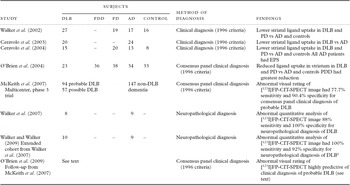
Most evidence for the utility of dopaminergic imaging in the diagnosis of DLB comes from studies of the presynaptic dopamine transporter using [123I]FP-CIT-SPECT (Table 3).
Summary of studies of [123I]FP-CIT-SPECT imaging in dementia with Lewy bodies (DLB) and Parkinson's disease dementia (PDD)

EPS = extrapyramidal symptoms; PD = Parkinson's disease; AD = Alzheimer's disease.
*Quantitative analysis of [123I]FP-CIT-SPECT images for 2009 study was based on striatal binding in posterior putamen on most severely affected side. 2007 analysis used bilateral measures.
Following a number of small studies investigating the utility of dopamine transporter imaging in DLB, a large European multicenter phase 3 trial was completed. Over 300 subjects with DLB or non-DLB dementia underwent [123I]FP-CIT-SPECT imaging with follow-up at one year. Sensitivity and specificity for visual rating of [123I]FP-CIT-SPECT scanning was initially assessed against baseline consensus clinical diagnosis (McKeith et al., Reference McKeith2007). Sensitivity of an abnormal [123I]FP-CIT-SPECT scan for detection of probable DLB was 77.7%, whilst specificity was 90.4%. This cohort was followed up for one year and data were available for 235 subjects, including repeat consensus clinical diagnosis at this time point (O'Brien et al., Reference O'Brien2009). Specificity and sensitivity for probable DLB versus non-DLB dementia were essentially unchanged. Clinical diagnosis of possible DLB was unstable: of the 44 subjects with a clinical diagnosis of possible DLB at baseline, 19 were reclassified as probable DLB at follow-up. [123I]FP-CIT-SPECT imaging was abnormal in 11 of these 19 subjects. Imaging results were normal in all seven patients reclassified as having non-DLB dementia at follow-up. Overall, an abnormal [123I]FP-CIT-SPECT scan at baseline was highly predictive of a diagnosis of DLB at one year. Consensus clinical diagnosis was used as the gold standard in these studies (McKeith et al., Reference McKeith2007; O'Brien et al., Reference O'Brien2009), and is likely to have been more rigorous than in general clinical diagnostic practice. Despite this approach, the study is limited by the lack of neuropathological confirmation of diagnosis.
In a separate study, a cohort of 45 patients with clinically diagnosed DLB (27 patients), AD (17) and corticobasal degeneration (CBD; 1) was followed up prospectively (Walker et al., Reference Walker2002). Autopsy data are currently published for 23 of this cohort. Ten met neuropathological diagnostic criteria for DLB, nine for AD and four for other dementia subtypes (Walker et al., Reference Walker2007; Walker and Walker, Reference Walker and Walker2009). Sensitivity for clinical diagnosis compared to autopsy diagnosis in this study was 80%, whilst specificity was 46%. Sensitivity for [123I]FP-CIT-SPECT imaging based on a visual reading was 80%, whilst specificity was 85%. When semi-quantitative analysis was employed for analysis of [123I]FP-CIT uptake, sensitivity was 100% and specificity 92%. There were no false-positive scan results in this study. One false-negative scan result was found in a patient with a vascular lesion; this type of misinterpretation could be avoided in clinical practice by correlation with MRI data.
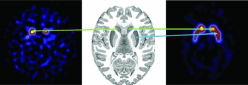
[123I]FP-CIT-SPECT imaging has therefore been shown to have greater diagnostic accuracy than clinical diagnosis when compared to the gold standard of neuropathological diagnosis. Examples of images from a patient with DLB and a healthy control subject are shown in Figure 2.
[123I]FP-CIT-SPECT images from a patient with neuropathologically confirmed DLB (left) and a healthy control subject (right). In the DLB patient there is reduced uptake of [123I]FP-CIT in both the caudate (green arrow) and putamen (blue arrow). In the control subject, there is good uptake throughout the bilateral caudate and putamen.

Other dopaminergic studies
The dopaminergic degeneration that is present in DLB and PDD can be imaged using ligands other than [123I]FP-CIT-SPECT. However, because of cost, availability and practical considerations, they have not been evaluated in large phase 3 studies for the purpose of DLB diagnosis. Dopamine turnover in DLB has been imaged using [18F]DOPA-PET and shown to be significantly reduced in both caudate and putamen compared to healthy controls and patients with AD (Hu et al., Reference Hu2000). There was no difference between control and AD groups.
Imaging of the vesicular monoamine transporter T2 has consistently shown reduction of striatal binding values for [11C]DTBZ using PET. This method of imaging has the advantage that early images can be used to assess perfusion, but the disadvantage is that it requires continuous infusion of the ligand (Gilman et al., Reference Gilman2004; Koeppe et al., Reference Koeppe, Gilman, Junck, Wernette and Frey2008).
A study of post-synaptic D2 receptors demonstrated that the D2 receptor caudate/putamen ratio imaged with [123I]IBZM-SPECT was significantly lower in DLB than in AD on a group comparison. However, the overlap between the two groups meant that discrimination between the two diagnostic groups at an individual patient level was not possible (Walker et al., Reference Walker, Costa, Janssen, Walker, Livingstone and Katona1997). Another study of six patients with DLB showed reduced striatal uptake of [123I]IBZM, but no difference in putamen/caudate uptake ratio when compared to patients with essential tremor as a control group (Plotkin et al., Reference Plotkin2005). At present, there is no clear clinical role for D2 receptor imaging in DLB.
Although [123I]FP-CIT-SPECT is widely used to image the presynaptic dopamine receptor, in some research centers [123I]βCIT-SPECT is used for this purpose. Results are similar to those with [123I]FP-CIT-SPECT imaging, but it is less practical for outpatient use because the ligand must be injected the day before the scan is performed.
Alzheimer's disease
AD is the most common type of dementia and is characterized by early deficits in episodic memory followed by more extensive cognitive and functional deterioration, including disturbance of language and executive function, apraxia, agnosia and changes in personality and behavior. The characteristic pathological features of AD are amyloid (neuritic) plaques and neurofibrillary tangles, predominantly in medial temporal lobe structures and the cerebral cortex. There is also loss of neurons, proliferation of astrocytes and loss of synapses.
In contrast to DLB, the dopaminergic system in AD, including synthesis, storage and dopamine transporter number, is essentially well preserved. The clinical relevance of dopaminergic imaging for AD relates to the fact that cases of suspected or possible DLB with negative (i.e. normal) [123I]FP-CIT-SPECT are likely to have AD. In AD the majority of studies have demonstrated no differences in dopaminergic measures compared to controls, although a few have reported slight reductions in AD. For example, reduced striatal [123I]IBZM has been reported in a group of 13 AD patients compared to controls (Pizzolato et al., Reference Pizzolato1996). A [11C]FLB 457-PET study of hippocampal D2 receptors demonstrated reduced binding in AD subjects compared to controls (by 12–34%), which correlated with deficits in memory and language (Kemppainen et al., Reference Kemppainen2003). It is not clear if these findings represent contamination of AD samples by patients with DLB, as none has included neuropathological diagnosis. As yet, there is no clinical application for these findings.
“Parkinson plus” syndromes
There are a number of “Parkinson plus” syndromes which are associated with cognitive decline and dementia, either at presentation or later in the course of the disease. These disorders form part of the differential diagnosis of dementia. The most common of these disorders are progressive supranuclear palsy (PSP), CBD and multiple system atrophy (MSA). Generally, the parkinsonism is more rapidly progressive than that seen in idiopathic PD and shows a relatively poor response to levodopa. The additional features are key to the clinical diagnosis.
PSP is a tauopathy characterized pathologically by degenerative change in the basal ganglia, frontal cortex, brainstem and spinal cord. Cognitive decline and personality change often occur early in the course of the disease. Postural instability with recurrent falls is an early feature. The other characteristic feature is supranuclear ophthalmoparesis, particularly for down gaze.
In CBD, degeneration of the nigrostriatal motor system is accompanied by pathological changes in several subcortical structures and the fronto-parietal cortex. Like PSP, CBD is a tauopathy. The movement disorder is often complex with an asymmetrical akinetic–rigid parkinsonian syndrome that may be accompanied by dystonia, myoclonus, and cortical motor and sensory disorders, including apraxia and “alien limb” phenomena. Progressive cognitive decline usually occurs with advanced disease but may occur early on.
In MSA there is accumulation of α-synuclein in association with nigrostriatal dopaminergic neuronal degeneration and atrophy of the globus pallidus and olivopontocerebellar structures. MSA usually starts either with parkinsonism or with cerebellar ataxia. Autonomic failure affecting blood pressure and bladder control ensues, along with signs of upper motor neuron degeneration. Although cognitive decline may occur as the disease progresses, frank dementia is uncommon.
Imaging of dopamine storage using [11C]DTBZ-PET in PSP and MSA has consistently shown reduced uptake on group comparisons between small numbers of patients compared to healthy controls, with no differences between idiopathic PD and MSA or PSP groups. However, individual patients with “Parkinson plus” syndromes may show normal striatal [11C]DTBZ binding (Raffel et al., Reference Raffel2006).
Results from imaging of dopamine synthesis using [11F]DOPA-PET in “Parkinson plus” syndromes also consistently show reduced binding in PSP, CBD and MSA compared to control groups (Burn et al., Reference Burn, Sawle and Brooks1994; Laureys et al., Reference Laureys1999).
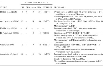
There have been many studies of the dopamine transporter in “Parkinson plus” syndromes, using both [123I]FP-CIT-SPECT and [123I]βCIT-SPECT. [123I]FP-CIT-SPECT studies are summarized in Table 4. Reduced striatal uptake of both ligands occurs in PSP, CBD and MSA in comparison with control groups, although uptake may be normal in a minority of patients. Some studies have demonstrated differences between idiopathic PD and “Parkinson plus” syndromes (e.g. based on overall uptake measures, ratios or asymmetry), but no reliable measure has yet been developed for widespread clinical application of these findings.
Summary of [123I]FP-CIT-SPECT studies in “Parkinson plus” syndromes

PSP = progressive supranuclear palsy; CBD = corticobasal degeneration; MSA = multisystem atrophy; IPD = idiopathic Parkinson's disease; PS = parkinsonian syndrome; HC = healthy controls; ET = essential tremor.
*Three patients underwent [123I]βCIT-SPECT imaging rather than [123I]FP-CIT-SPECT.
The results of studies of post-synaptic D2 receptors in PSP, CBD and MSA are less consistent and at present have little clinical utility. The inconsistency of the results of these studies mirrors the changes that occur with illness progression, e.g. upregulation of D2 receptors in the early disease stages and subsequent decompensation.
One of the challenges of interpreting dopaminergic imaging studies in “Parkinson plus” syndromes is that the majority are based on small samples of clinically diagnosed patients. These disorders are essentially clinical syndromes with heterogeneous clinical presentations and underlying neuropathology. Rates of clinical misdiagnosis are high, and there is a paucity of studies based on autopsy diagnosis.
Frontotemporal dementia
Frontotemporal dementia (FTD) is characterized by frontal and anterior temporal lobar atrophy. In the behavioral variant of FTD, there is a profound change in personality and social behavior, which is associated with wide-ranging deficits in executive and language function (for a recent review see Piguet et al., Reference Piguet, Hornberger, Mioshi and Hodges2011). Early in the disease, presentation is frequently atypical or unusual and there is a high rate of misdiagnosis, e.g. as DLB, AD, or psychiatric disorder. Other less common subtypes of FTD are progressive non-fluent aphasia and semantic dementia.
There is an overlap between FTD and other disorders associated with parkinsonism, including PSP and CBD, which all share abnormal tau pathology. Extrapyramidal symptoms are relatively common early in FTD, but few published data are available and the high rate of neuroleptic prescribing in this group clouds the issue (Pasquier et al., Reference Pasquier, Lebert, Lavenu and Guillaume1999).
Autopsy findings in FTD show bilateral frontotemporal atrophy with neuronal loss, microvacuolation and a variable degree of astrocyte gliosis. Most cases are either tau or ubiquitin positive and there is a mild degree of neuronal loss in the substantia nigra with associated atrophy in the basal ganglia.
Dopaminergic imaging studies have shown abnormalities in the presynaptic dopamine transporter in FTD. A [123I]FP-CIT-SPECT study of seven carefully characterized FTD patients with minimal extrapyramidal symptoms showed 30% reduced uptake in the striatum in FTD compared to control groups (Sedaghat et al., Reference Sedaghat2007). A second study of the dopamine transporter using [11C]CFT-PET showed a reduced ligand uptake in the caudate (14%) and putamen (18%) (Rinne et al., Reference Rinne, Laine, Kaasinen, Norvasuo-Heila, Nagren and Helenius2002). In both studies, the reduced ligand uptake correlated with measures of motor features of parkinsonism.
To summarize, reduced uptake of [123I]FP-CIT has been demonstrated at a group level in FTD, but as yet no data have been published regarding individual cases. It is likely, therefore, that some patients with FTD will have an abnormal [123I]FP-CIT-SPECT scan result, which may be relevant in cases where the differential diagnosis includes both DLB and FTD.
Clinical applications of dopaminergic imaging
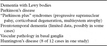
Dopaminergic imaging has been shown to be of help to the clinician in the differential diagnosis of dementia syndromes associated with dopaminergic degeneration. At present, the only imaging method with established clinical utility is [123I]FP-CIT-SPECT. [123I]FP-CIT-SPECT imaging has been validated in a phase 3 clinical trial and in a neuropathological study for differentiation between probable DLB and AD. However, it is important to bear in mind that [123I]FP-CIT-SPECT imaging is abnormal in a number of other dementia syndromes associated with parkinsonism (Table 5).
Dementia syndromes associated with parkinsonism for which [123I]FP-CIT-SPECT imaging is abnormal

Dopamine transporter imaging has been incorporated into several guidelines for the assessment and diagnosis of dementia. Current DLB consensus criteria include abnormal dopamine transporter imaging using PET or SPECT as a suggestive feature of DLB (McKeith et al., Reference McKeith2005). In the UK, the National Institute for Health and Clinical Excellence guidelines recommend the use of [123I]FP-CIT in those with suspected DLB but in whom the diagnosis is in doubt (NICE, 2006). Guidelines from the European Federation of the Neurological Societies (EFNS) on the diagnosis and management of AD and other disorders associated with dementia state that dopamine transporter imaging is useful to differentiate AD and DLB (Hort et al., Reference Hort2010).
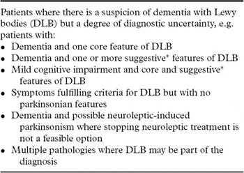
In the majority of cases of dementia, careful clinical assessment leads to reasonable certainty of diagnosis, and advanced neuroimaging techniques are not required. [123I]FP-CIT-SPECT imaging is an adjunct to clinical assessment and is not a diagnostic test. Use of this tool has resource implications and is therefore most appropriately used in cases where diagnosis is uncertain or complicated and the scan result is likely to provide clarification. The main indications for [123I]FP-CIT-SPECT imaging are listed in Table 6.
Clinical indications for FP-CIT-SPECT imaging

*excluding low dopamine transporter uptake in basal ganglia demonstrated by SPECT or PET imaging.
Conclusions and further research
Over the past two decades, dopaminergic imaging has improved diagnostic accuracy in dementia, particularly in distinguishing between DLB and AD. Use of [123I]FP-CIT-SPECT imaging has been incorporated into national and international clinical guidelines, and is widely available in clinical practice.
Studies that will be of clinical relevance in the future include clinico-pathological imaging studies in DLB and other parkinsonian dementia syndromes, to further clarify the diagnostic accuracy of dopaminergic imaging. Studies in prodromal dementia states, e.g. mild cognitive impairment, will provide information about the early stages of dopaminergic degeneration and in the future may inform the direction of disease-modifying treatments.
[123I]FP-CIT-SPECT imaging is the most widely used form of dopaminergic imaging in clinical practice. With the development of more sophisticated ligands, targeting dopaminergic and other neurotransmitter systems, it is likely that neuroimaging will play an increasingly important role in the facilitation of early accurate diagnosis in dementia, in addition to monitoring disease progression and response to treatment.
Conflict of interest
Z. Walker has received consultancy and speaker fees and research support from GE Healthcare, consultancy fees from Bayer Healthcare, and research support from Lundbeck.
Description of authors' roles
Both authors contributed equally to the planning, research, writing and editing of the paper.