Introduction
Polarized light equipment has been an important part of many assisted reproduction laboratories for many years. The ability to visualize birefringent structures in the oocyte (such as the meiotic spindle (MS) and the zona pellucida) in a non-invasive way, has offered many possibilities to researchers. The MS is a crucial structure during meiosis. Alterations in its structure are directly related to anomalies in chromosome segregation (Sharma et al., Reference Sharma, Azeem and Agarwal2013) that are related to implantation failures and abortions (Wang et al., Reference Wang, Meng, Hackett and Keefe2001a; Hunt and Hassold, Reference Hunt and Hassold2008).
Numerous authors have focused their studies on evaluating different characteristics of the MS (presence/absence, position in respect to polar body, size, structure, refringence) and have tried to relate these characteristics to different parameters such as fertilization (de Almeida Ferreira Braga et al., Reference de Almeida Ferreira Braga, de Cássia Sávio Figueira, Rodrigues, Madaschi, Pasqualotto, Iaconelli and Borges2008; Cohen et al. al., Reference Cohen, Malcov, Schwartz, Mey-Raz, Carmon, Cohen, Lessing, Amit and Azem2004; Konc et al., Reference Konc, Kanyó and Cseh2004; Madaschi et al., Reference Madaschi, de Souza Bonetti, de Almeida Ferreira Braga, Pasqualotto, Iaconelli and Borges2008; Moon et al., Reference Moon, Hyun, Lee, Son, Yoon and Lim2003; Rama Raju et al., Reference Rama Raju, Prakash, Krishna and Madan2007; Shen et al., Reference Shen, Stalf, Mehnert, de Santis, Cino, Tinneberg and Eichenlaub-Ritter2006; Wang et al., Reference Wang, Meng, Hackett, Odenbourg and Keefe2001b), embryo quality and blastocyst formation (Wang et al., Reference Wang, Meng, Hackett and Keefe2001a; Moon et al., Reference Moon, Hyun, Lee, Son, Yoon and Lim2003; Cohen et al., Reference Cohen, Malcov, Schwartz, Mey-Raz, Carmon, Cohen, Lessing, Amit and Azem2004; Chamayou et al., Reference Chamayou, Ragolia, Alecci, Storaci, Maglia, Russo and Guglielmino2006; Fang et al., Reference Fang, Tang, Li, Peng, Zhou, Zhuang and Leong2007; Rama Raju et al., Reference Rama Raju, Prakash, Krishna and Madan2007; Madaschi et al., Reference Madaschi, de Souza Bonetti, de Almeida Ferreira Braga, Pasqualotto, Iaconelli and Borges2008) and implantation rate (Chamayou et al., Reference Chamayou, Ragolia, Alecci, Storaci, Maglia, Russo and Guglielmino2006; Shen et al., Reference Shen, Stalf, Mehnert, de Santis, Cino, Tinneberg and Eichenlaub-Ritter2006; Madaschi et al., Reference Madaschi, de Souza Bonetti, de Almeida Ferreira Braga, Pasqualotto, Iaconelli and Borges2008; Kilani et al., Reference Kilani, Cooke, Kan and Chapman2009; Heindryckx et al., Reference Heindryckx, De Gheselle, Lierman, Gerris and De Sutter2011; Kilani et al., Reference Kilani, Cooke, Tilia and Chapman2011; Magli et al., Reference Magli, Capoti, Resta, Stanghellini, Ferraretti and Gianaroli2011; Molinari et al., Reference Molinari, Evangelista, Racca, Cagnazzo and Revelli2012; Korkmaz et al., Reference Korkmaz, Sakinci, Bayoglu Tekin and Ercan2014; García-Oro et al., Reference García-Oro, Rey, Rodríguez, Durán, Devesa and Valverde2017) among others. Published results, however, have been very heterogeneous.
A meta-analysis conducted by Petersen and colleagues (Petersen et al., Reference Petersen, Oliveira, Mauri, Massaro, Baruffi, Pontes and Franco2009), concluded that presence of the MS is related to better fertilization rates, embryo quality and blastocyst formation. Most studies have focused on variables (such as fertilization, embryo quality, or blastocyst formation) that do not necessarily predict a better reproductive outcome. Despite the positive results related to MS, this meta-analysis failed to show any relationship for two of the most important aspects of reproductive success: implantation and live birth rates. The objective of this review was to investigate a possible relationship between MS characteristics and embryo implantation and live birth rates.
Materials and methods
A systematic literature search was performed using the PubMed database, the Cochrane Library and Science Direct with the following keywords: ‘meiotic spindle’, ‘polarized light’ and ‘retardance’. The publications retrieved were analyzed and only those in which embryonic implantation rate was evaluated were selected. From 56 starting studies, we initially included 10 papers in this review (Chamayou et al., Reference Chamayou, Ragolia, Alecci, Storaci, Maglia, Russo and Guglielmino2006; Shen et al., Reference Shen, Stalf, Mehnert, de Santis, Cino, Tinneberg and Eichenlaub-Ritter2006; Madaschi et al., Reference Madaschi, de Souza Bonetti, de Almeida Ferreira Braga, Pasqualotto, Iaconelli and Borges2008; Kilani et al., Reference Kilani, Cooke, Kan and Chapman2009; Heindryckx et al., Reference Heindryckx, De Gheselle, Lierman, Gerris and De Sutter2011; Kilani et al., Reference Kilani, Cooke, Tilia and Chapman2011; Magli et al., Reference Magli, Capoti, Resta, Stanghellini, Ferraretti and Gianaroli2011; Molinari et al., Reference Molinari, Evangelista, Racca, Cagnazzo and Revelli2012; Korkmaz et al., Reference Korkmaz, Sakinci, Bayoglu Tekin and Ercan2014; García-Oro et al., Reference García-Oro, Rey, Rodríguez, Durán, Devesa and Valverde2017).
Only the relevant results for implantation rates in embryos that were well differentiated according to MS presence or absence were finally evaluated. Studies that included live birth data in their results were also reviewed.
Statistical analysis was performed with the relevant embryo implantation and live birth data obtained from the literature. We compared implantation and live birth rates using the chi-squared test. Differences were deemed to be statistically significant at P ≤ 0.01. The studies initially are described briefly here:
Shen and colleagues (Shen et al., Reference Shen, Stalf, Mehnert, de Santis, Cino, Tinneberg and Eichenlaub-Ritter2006) conducted their study in two different clinics. In the Giessen cohort, conception cycles (n = 42) were compared with non-conception cycles (n = 61). The average refringence of the transferred embryos was evaluated in each transference. The authors did not find any differences between average MS retardance in the transferred embryos in conception cycles (1.65 nm) versus non-conception cycles (1.67 nm). In the Milan cohort, 59 embryos transferred from 28 patients were analyzed. The mean retardance of the embryos in conception cycles was 2.76 nm, significantly higher than that observed in non-conception cycles (1.78 nm).
Chamayou and colleagues (Chamayou et al., Reference Chamayou, Ragolia, Alecci, Storaci, Maglia, Russo and Guglielmino2006) evaluated the presence or absence of MS in oocytes from 227 transferred embryos. The author compared the implantation percentage in groups of embryo transfers: embryos with MS (n = 130) versus embryos without MS (n = 97). The results did not show significant differences.
Madaschi and colleagues (Madaschi et al., Reference Madaschi, de Souza Bonetti, de Almeida Ferreira Braga, Pasqualotto, Iaconelli and Borges2008) compared the implantation rate in groups of embryo transfers, embryos with MS (63 patients) versus embryos without MS (22 patients). Results showed a higher implantation rate in the first group. The authors also evaluated the transferences performed with both kinds of embryos, but these were not included in this study.
Kilani and colleagues (Kilani et al., Reference Kilani, Cooke, Kan and Chapman2009) compared 22 oocytes from 18 conception cycles versus 30 oocytes from 19 non-conception cycles. The MS was present in all oocytes. Twenty-two oocytes in the first group had normal MS (according to the criteria defined previously by the author), while only 33% of the oocytes in the second group had a normal MS. The average retardance of the MS in the first group was significantly higher than the average retardance observed in the second group (3 nm vs 2.5 nm).
Magli and colleagues (Magli et al., Reference Magli, Capoti, Resta, Stanghellini, Ferraretti and Gianaroli2011) compared 55 embryo MS transferences versus 10 embryos without MS transferences. In the first group embryonic implantation was observed, while in the second group there were no pregnancies. The author also evaluated the transferences performed with both kind of embryos, but these were not included in this study.
Heindrycks and colleagues (Heindryckx et al., Reference Heindryckx, De Gheselle, Lierman, Gerris and De Sutter2011) evaluated both the MS presence and retardance in 121 oocytes. The author compared the results of conception cycles versus non-conception cycles. In both groups, the percentage of embryos with MS (82.4% vs 81.8%) and the mean retardation of MS (1.48 nm vs 1.51 nm) observed were similar.
Kilani and colleagues (Kilani et al., Reference Kilani, Cooke, Tilia and Chapman2011) conducted a prospective study. The authors evaluated 808 oocytes from 100 patients. Fifty-two blastocysts were transferred in non-conception cycles from oocytes with MS that did not meet the criteria of normality defined by the author (n = 506), whereas for oocytes with normal MS (n = 205), 68 blastocysts were transferred, obtaining high implantation rates. In the 37 clinical pregnancies achieved, there were 30 live births (28 singletons and two sets of monozygotic twins) and seven miscarriages.
Molinari and colleagues (Molinari et al., Reference Molinari, Evangelista, Racca, Cagnazzo and Revelli2012) compared the mean retardance of the MS embryos transferred in 34 conception cycles versus 52 non-conception cycles. The results were not significantly different (1.7 nm vs 1.6 nm).
Korkmaz and colleagues (Korkmaz et al., Reference Korkmaz, Sakinci, Bayoglu Tekin and Ercan2014) performed 53 single-embryo transfers (20 with gestation and 33 without gestation) and analyzed these retrospectively. A MS was observed in all embryos transferred. The average retardance of embryos transferred in each group was similar (1.98 nm vs 2.02 nm).
In a retrospective study published by our group (García-Oro et al., Reference García-Oro, Rey, Rodríguez, Durán, Devesa and Valverde2017), the presence and retardance of MS of 195 embryos transferred was analyzed individually. A higher implantation percentage in embryos with visible MS (32.9%) versus embryos without MS or in telophase (8.8% and 0%) was observed. In addition, we found a higher implantation rate in embryos with high MS retardance values (>1.41 nm), specifically when the retardance value was greater than 1.61 nm, with implantation percentages of 53.8%. This study has been completed recently, although the remaining data have not yet been published. In the 55 pregnancies achieved, there were 39 live births (36 in the MS group), there have been 12 miscarriages (all in the MS group) and there were five gestations of which the outcome is not known (all in the MS group).
Results
Some authors compared the average retardance of embryos transferred in conception versus non-conception cycles (Shen et al., Reference Shen, Stalf, Mehnert, de Santis, Cino, Tinneberg and Eichenlaub-Ritter2006; Kilani et al., Reference Kilani, Cooke, Kan and Chapman2009; Heindryckx et al., Reference Heindryckx, De Gheselle, Lierman, Gerris and De Sutter2011; Molinari et al., Reference Molinari, Evangelista, Racca, Cagnazzo and Revelli2012; Korkmaz et al., Reference Korkmaz, Sakinci, Bayoglu Tekin and Ercan2014). Except for Kilani and Shen (in the part of the study carried out in Milan), none of the authors found differences in the mean retardance of the MS for embryos transferred in each group. When the average retardances of embryos transferred in conception and non-conception cycles were compared, the retardances range observed in each group was similar (for embryos in conception cycle, 1.48−2.76 nm, and for embryos in non-conception cycles, 1.5−2.5 nm). The methodology used by these authors to relate the MS with the implantation rate was not the most appropriate. The average retardances of all embryos transferred in each cycle had little predictive value for establishing a relationship between MS characteristics and embryo implantation rates. For this reason, we believe that these five publications should not be considered for the present review.
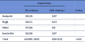
Other authors compared the implantation rate of cycles in which only embryos with visible MS were transferred versus cycles in which only embryos without visible MS were transferred (Chamayou et al., Reference Chamayou, Ragolia, Alecci, Storaci, Maglia, Russo and Guglielmino2006; Madaschi et al., Reference Madaschi, de Souza Bonetti, de Almeida Ferreira Braga, Pasqualotto, Iaconelli and Borges2008; Magli et al., Reference Magli, Capoti, Resta, Stanghellini, Ferraretti and Gianaroli2011). Chamayou was the only one of three authors who does not find significant differences in implantation rates between both groups. The range of implantation percentages of transferences performed in MS embryos was 21.6−24.6%. The range of implantation percentage of transferences performed in embryos without visible MS was 0−19.6%.
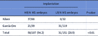
Conversely, Kilani et al. (Reference Kilani, Cooke, Tilia and Chapman2011) compared the implantation rate of cycles in which only embryos with normal MS have been transferred versus cycles in which only embryos with abnormal MS have been transferred. In the first group, the implantation rate was 54%, while in the second group there is no implantation.
In the last published study (García-Oro et al., Reference García-Oro, Rey, Rodríguez, Durán, Devesa and Valverde2017), the implantation rates of each embryo were compared individually, according to the presence and retardance values of the MS. Embryos with visible MS had an implantation percentage of 32.9% and embryos with retardance values >1.61 nm had an implantation percentage of 53.8%.
We finally selected for the present review only four publications, those in which transferences were performed in both MS embryos and non-MS embryos independently. We excluded the Chamayou’s paper from the results because the MS detection rate was very low and probably reflected some issues (discussed later).
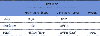
The results of the selected papers were divided in two groups, the first results showed the implantation rates in embryos with or without MS (Table 1) and the second results showed the implantation rates in embryos with MS (high retardance values or normal MS defined by Kilani’s criteria versus low retardance values or abnormal MS defined by Kilani’s criteria; Table 2). In addition, in the second group, we also showed data of live birth rates, the main reproductive outcome (Table 3). In the García-Oro’s study (unpublished data) there were five gestations of which we do not know the reproductive outcome. We have decided not to include these in the live birth rate data to avoid possible bias in the results, therefore the numbers of embryos transferred in Tables 2 and 3 do not match.
Implantation rates in MS embryos versus NMS embryos

MS, meiotic spindle presence; NMS, meiotic spindle absence.
Implantation rates in HR/N MS embryos versus LR/AB MS embryos

HR, high retardance values (>1.61); N, Kilani’s normality criteria; LR, low retardance values (<1.61); AB, embryos excluded from normality Kilani’s criteria.
Live birth rates in HR/N MS embryos versus LR/AB MS embryos

HR, high retardance values (>1.61); N, Kilani’s normality criteria; LR, low retardance values (<1.61); AB, embryos excluded from normality Kilani’s criteria.
Discussion
Several authors have tried to relate implantation rates to the presence and retardance of MS. In the three publications in which the implantation rates of transferred embryos with and without visible MS were compared, the results were very similar. Chamayou et al. (Reference Chamayou, Ragolia, Alecci, Storaci, Maglia, Russo and Guglielmino2006) reported an implantation percentage of 24.6% in embryo transfers with a MS, whereas Madaschi et al. (Reference Madaschi, de Souza Bonetti, de Almeida Ferreira Braga, Pasqualotto, Iaconelli and Borges2008) reported 23% and Magli et al. (Reference Magli, Capoti, Resta, Stanghellini, Ferraretti and Gianaroli2011) 21.6%. The implantation rates obtained by the different authors do not show differences. For implantation rates of embryo transfers without a MS, Chamayou et al. reported an implantation percentage of 19.6%, Madaschi et al. 8.7% and Magli et al. 0%. Analyzing the Chamayou study in depth, the percentage of oocytes for which the MS was seen was very small (43.5% oocytes) compared with the MS detection rate published by most authors (Cohen et al., Reference Cohen, Malcov, Schwartz, Mey-Raz, Carmon, Cohen, Lessing, Amit and Azem2004; Konc et al., Reference Konc, Kanyó and Cseh2004; de Santis et al., Reference de Santis, Cino, Rabellotti, Calzi, Persico, Borini and Coticchio2005; Rama Raju et al., Reference Rama Raju, Prakash, Krishna and Madan2007; Heindryckx et al., Reference Heindryckx, De Gheselle, Lierman, Gerris and De Sutter2011; Kilani et al., Reference Kilani, Cooke, Tilia and Chapman2011; Korkmaz et al., Reference Korkmaz, Sakinci, Bayoglu Tekin and Ercan2014). The MS is a dynamic structure (Montag and van der Ven, Reference Montag and van der Ven2008) and evaluation of its presence could affect detection (Cohen et al., Reference Cohen, Malcov, Schwartz, Mey-Raz, Carmon, Cohen, Lessing, Amit and Azem2004). Similarly, the MS is a highly sensitive structure and certain culture conditions (like pH and temperature) (Montag and van der Ven, Reference Montag and van der Ven2008) can affect it. Both situations could alter the reduced percentage of oocytes observed with MS. Worse reproductive results in the oocytes without MS should be observed but, surprisingly, the implantation rates reported were similar. Another possible explanation for this result could be related to the MS visualization technique used, which requires an experienced evaluator and exhaustive manipulations with successive rotations of the oocyte in different directions before absence of MS is confirmed. Therefore, due to possible overestimation of the results, this publication was not considered.
Kilani’s study (Kilani et al., Reference Kilani, Cooke, Tilia and Chapman2011) was very well designed for our objectives. It was the only prospective study evaluated and a single blastocyst transfer was performed. The only limitation was the MS parameter on which the results were based. as the authors defined qualitative criteria for normality of MS, which could be subjective. Despite this, in the group of embryos with MS that meet these criteria, the implantation rate was very high.
The study published recently by our group (García-Oro et al., Reference García-Oro, Rey, Rodríguez, Durán, Devesa and Valverde2017) alone described the individual and quantitative evaluation of the MS of each of the transferred embryo. This methodological approach is probably one of the best used, although also with some limitations. The implantation rate observed in the values of retardation >1.61 nm (53.8%) was equal to that observed by Kilani and colleagues for embryos with normal MS. Retardance is a measure of the structural order, in this case the level of organization of the microtubules that forms the MS. A high retardance value can be interpreted as a high degree of macromolecular organization and structural density, which would be an indicator of well aligned or organized structures. It is reasonable to assume that the high values of retardance in this study would be similar to that of normal MS observed by Kilani and colleagues and probably that the definition of normality was related to the degree of structural organization, which reflects retardance values.
These results showed a clear relationship between MS characteristics and the embryo implantation rate. In embryos without visible MS the implantation percentage was 7.7%, in those embryos with abnormal MS or retardance values ≤1.61 it was 20.5% and in those embryos with normal MS or retardance values >1.61 it was 54.2%. The implantation rates observed in each group were significantly different.
Only two studies related the characteristics of the MS with the live birth rate. Despite this, the data published by Kilani et al. (Reference Kilani, Cooke, Tilia and Chapman2011) and presented by our group showed significant results in favour of certain characteristics or retardance values for MS and its relationship with live birth rates.
In conclusion, the data evaluated in the present review suggested that the presence of the MS in oocytes is positively associated with embryo implantation. High retardance values, as well as the strict criteria for MS structural normality, were significantly related to higher embryo implantation and live birth rates.
The main limitation of this revision was the risky comparison of implantation data obtained from studies with different methodological approaches. To find the true predictive value for MS in embryo implantation and live birth rates, a broader and prospective study with a single blastocyst transfer would be necessary. Moreover, each MS would be evaluated individually and quantitatively, therefore avoiding possible biases due to the subjectivity of the qualitative parameters.
Financial support
This research received no specific grant from any funding agency, commercial or not-for-profit sectors.
Statement of interest
None
Ethical standards
Not applicable.