INTRODUCTION
Parasitic nematodes belonging to the genus Thelazia (Spirurida, Thelaziidae), commonly known as eyeworms, live in the orbital cavities and surrounding tissues of mammals, including horses, cattle and humans, and are transmitted by invertebrate vectors. Of the 16 eyeworm species that have been described, Thelazia gulosa Railliet and Henry 1910, Thelazia rhodesii Desmarest 1828 and Thelazia lacrymalis Gurlt 1831, are frequently recorded in cattle and horses, and are of major concern to veterinarians all over the world (reviewed by Otranto and Traversa, 2005).
A different species, Thelazia callipaeda Railliet and Henry 1910, is found in dogs, cats, rabbits, foxes and humans (Anderson, 2000). The adult nematodes live under the eyelid and nictitating membrane, causing injuries with mild to severe symptoms (e.g. lacrimation, epiphora, conjunctivitis, keratitis, and even corneal ulcers). Thelazia callipaeda is commonly known as ‘oriental eyeworm’, since it has been reported in humans and dogs in Russia and the former Soviet republics, and Asia (reviewed by Otranto et al. 2003 a). In Europe, this nematode was reported initially in dogs in northern (Rossi and Bertaglia, 1989) and southern Italy, in dogs, cats and foxes (Otranto et al. 2003 a) and subsequently in France (Chermette, Guillot and Bussiéras, 2004) and Germany (Hermosilla, Herrmann and Bauer, 2004).
Over the past couple of years, several studies have been carried out reporting the epidemiology and dissemination of thelaziosis (Otranto et al. 2003 a; Chermette et al. 2004; Hermosilla et al. 2004), the biological life-cycle of T. callipaeda in the definitive host (Otranto et al. 2004), the molecular features of some gene markers (Otranto and Traversa, 2004) and chemotherapy of thelaziosis (Rossi and Peruccio, 1989; Lia et al. 2004; Bianciardi and Otranto, 2005).
The life-cycle of T. callipaeda in naturally infected dogs has been studied and seasonality in the reproductive activity of female nematodes over a 2-year period reported (Otranto et al. 2004). In particular, the development of mature nematodes and the presence of 1st-stage larvae (L1) and/or of 3rd infective stages (L3) were linked to the presence/absence of the vector/s (Otranto et al. 2004). One of the most interesting aspects of the life-cycle of the parasite that still remains unclear is/are the insect/s that act as intermediate host. It has been suggested that more than one species of Diptera is involved in the transmission of T. callipaeda. For example, Amiota okadai Okada, 1956 was found to be infected by T. callipaeda in China (Wang et al. 2002 a; Wang, Wang and Chen, 2002 b) and Phortica variegata Fallén, 1823 under experimental conditions in the USSR (Kozlov, 1963). Other dipterans, such as Musca domestica Linnaeus, have also been reported on rare occasions to be naturally infected with T. callipaeda (Shi et al. 1988). However, a recent study in which this species was infected and allowed to feed directly on the eyes of an infected dog or by feeding the fly with L1 of T. callipaeda, demonstrated that M. domestica was unlikely to act as a vector for T. callipaeda (Otranto et al. 2005). Despite the spread of dog thelaziosis across Europe in recent years, our lack of knowledge of the natural vector for this parasite hinders our understanding of its biology, epidemiology, ecology, control and, most importantly, zoonotic risk.
Among the target genes [i.e. ribosomal DNA (rDNA) and mitochondrial DNA (mtDNA)] most commonly used to study the systematic, epidemiology, biology and diagnosis of parasitic nematodes (Gasser, 1999; Gasser and Newton, 2000), the ribosomal Internal Transcribed Spacer 1 (ITS1) sequence has been characterized for T. callipaeda, T. gulosa, T. rhodesii, T. skrjabini and T. lacrymalis (Otranto and Traversa, 2004). The Thelazia spp. ITS1 sequences provide information on the molecular identification of these nematodes in their definitive and intermediate hosts, irrespective of their developmental stage, as shown recently for the vectors of Thelazia spp. in cattle (Otranto et al. 2003 b).
The aim of the present work was to (1) identify the species of fly that may act as vector/s of T. callipaeda in southern Italy and China; and (2) describe the larval development of T. callipaeda in the body of flies maintained at different temperatures in the laboratory.
MATERIALS AND METHODS
Study area
Two distinct trials were carried out in the Basilicata region of southern Italy (site A) from September to December 2004 and in Wuhe County, Ahnui province, south eastern China (site B) from August to April 1990. Site A was in the municipality of Oliveto Lucano (Potenza province, Italy, altitude about 700 m above sea level (a.s.l.) – latitude 39° and 41° North, longitude 15° and 16° East). Site B was in Wuhe County (Ahnui province, China, altitude about 23 m a.s.l. latitude 30° and 35° North, longitude 115° and 120° East). Both collection sites were characterized by an extremely humid climate and thick undergrowth, and a high prevalence of thelaziosis in dogs (Wang et al. 2002 a; Otranto et al. 2003 a).
Collection and identification procedures
Flies were collected using a white cloth bag containing sliced fruit (about 2 kilos of apples and pears) fermented at 25°C for 48 h. The bag was then strung to the north side of a tree about 60 cm from the undergrowth. Flies were captured twice daily by netting them around the bag for about 2 h each time, in the early morning and evening (temperature, 20–25°C; relative humidity (R.H.), 55–75).
Once captured, the flies were transferred from the net into a clear plastic bag. Initial screening was based on their morphological features, such as 3 dark bands on the legs, a white ring around the eyes and speed of movement (Phortica flies ‘run slowly’ compared to other drosophilids). All flies from sites A and B were then identified morphologically to species level using identification keys proposed by Bächli et al. (2005). Briefly, Phortica was characterized by having a scutum with grey spots around, wing hyaline with 2 interruptions along the costal vein, legs with brown coax and yellow tibia with 3 dark rings.
Breeding of Phortica species
After preliminary identification, the flies were transferred into a cage and the cages then taken to the laboratories of the parasitological unit of the Faculty of Veterinary Medicine, University of Bari, Italy (site A) and the Department of Microbiology and Parasitology, Anhui Medical University, Hefei, China (site B). Phortica spp. were bred in 70×30×30 cm cages comprising a wooden frame covered by netting (diameter 1 mm mesh openings) with one side formed by a long closed corridor for easy access to the inside. Flies were kept at 25°C, with about 65 R.H., with artificial light for natural sun supplementation. Flies were fed with fruit (apples and bananas) cut into 2 pieces and placed on the roof of the cage as food and as a pabulum for fly oviposition. In the laboratory, fruit on top of the cage containing Phortica flies was covered to avoid contamination by the sister group species belonging to Drosophilidae family, the common ‘fruit fly’.
Infection procedures
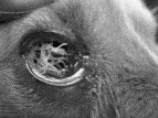
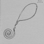
In both sites, several specimens of nematode were collected using a cotton swab from the eyes of a naturally infected dog (Fig. 1) and transferred to the eye of a rabbit. The latter served as a source of T. callipaeda for subsequent fly infection. To infect Phortica flies experimentally, the lachrymal secretions and mucus on the fornix of the rabbit's eyes were checked daily for infective L1 stages characterized by a ‘balloon’ (residue of the egg-shell) at one of the extremities and by a typical rolled shape (Fig. 2). As soon as infective L1 were retrieved in the lachrymal secretions, adult females of T. callipaeda (i.e. 8 in site A and 10 in site B) were collected from the rabbits by ocular flushing with saline solution (NaCl 0·9). First-stage larvae were then collected by slitting the uterus of mature females and expressing the contents into a drop of saline solution under a stereomicroscope.

Fig. 1. Eye of a dog with massive Thelazia callipaeda infection (site A).

Fig. 2. Light micrograph of mature 1st-stage larva outside the nematode presenting a shell membrane. ×200.
After collection, L1 were transferred to a concave slide with few drops of water and fruit juice (water-saline-fruit juice 1[ratio ]1[ratio ]2) fermented by standing for 12 h at 37°C. About 14 h before experimentally infecting the Phortica flies, their cage was covered with a dark wool cloth to increase the temperature and make the flies thirsty. The cage was also lined with black plastic except for one window (7×2 cm) designed to attract the flies to the light; the slide with the L1 in the above medium was placed in that window for about 7 h. Hungry and thirsty flies were attracted by the light and by the smell of the fermented fruit juice and fed on the concave slides.
A total of 245 P. variegata (site A) and 602 A. okadai (site B) were infected in 3 and 4 trials respectively.
Infection of dogs and rabbits
To demonstrate that Phortica may act as a vector for T. callipaeda, 2 L3 collected from infected flies were deposited onto the cornea of 1 dog with the permission of the owner (who was a co-author of the present paper) (site A). The dog was bred in a Thelazia-free area (i.e. the province of Bari). At site B, 50 and 18 nematodes were deposited onto the cornea of the left eye of 3 rabbits and 3 dogs, respectively. Infected animals were isolated for day 39 post-infection (p.i.). From day 30 to day 39 p.i., animals in sites A and B were checked for the presence of adult worms and L1 by ocular flushing with saline solution.
Examination procedures
At Site A, to recover T. callipaeda L3 from the proboscis of flies, Phortica were examined by dissection and visual inspection on day +14 (group 1a), +21 (group 2a) and +53 (group 3a) p.i. from September to December 2004 (Table 1). All dead flies were sexed and stored in 70 ethanol in individual vials for T. callipaeda molecular detection (see below).

In Site B, all flies (both alive and dead) from the 3 groups (1b, 2b and 3b) were dissected daily until day 24 p.i.
In addition, out of the 123 flies in group 4b, 93 were dissected until the day on which the first L3 was found in the proboscis (i.e. +50 days p.i. see Results section) and the remaining 30 flies were maintained through the winter (temperature: min. 5 max. 15°C, average temperature from 8 to 10°C) and dissected after 180 days p.i. (Table 2). Based on the available flies, an average of between 1–4 and 10–20 flies were dissected daily in the 4 groups.

In both sites, flies were examined firstly by stretching the proboscis of Phortica to detect infective L3 and, progressively, dissecting the head, thorax and abdomen to detect the presence/absence of other developmental stages of eyeworms. The number and location of larvae in the fly body were reported and sketched.
DNA isolation, PCR procedures, sequencing and analyses
Flies from site A that died before the dissection date were subject to molecular processing by PCR as previously described (Otranto et al. 2003 b; Otranto and Traversa, 2004). Briefly, genomic DNA was extracted from each individual fly sample using a commercial kit (QIAamp Tissue Kit, Qiagen GmbH, Hilden, Germany).
A partial ITS1 sequence, sizing about 500 bp, was amplified using a conserved primer, namely rDNA-A (5′AGGTGAACCTGCGGAAGGA3′) (Bachellerie and Qu, 1993) and a primer, G2 (5′GACACCAACAGTGAACACCG3′) designed (using the criteria of Sharrocks, 1994) on the ITS1 to previous sequences of T. callipaeda (Accession number: AY207464). Genomic DNA (4 μl) was added to the PCR reaction mix (46 μl) containing 2·5 mm MgCl2, 10 mm Tris-HCl, pH 8·3 and 50 mm KCl, 250 μm of each dNTP, 50 pmol of each primer and 1·25 U of Ampli Taq Gold (Applied Biosystems). PCR was performed in an Applied Biosystems 2700 thermal cycler using the following cycling protocol: 94°C for 12 min (polymerase activation), followed by 30 cycles of 94°C for 30 sec (denaturation); 58°C for 45 sec (annealing); 72°C for 45 sec (extension), followed by 7 min at 72°C (final extension). Negative and positive control reactions were also carried out by substituting the fly DNA template with DNA from non-infected P. variegata and T. callipaeda, respectively. PCR amplifications were also conducted on T. callipaeda DNA spiked with P. variegata DNA to investigate the presence/absence of PCR inhibitors. Amplicons were resolved in 1·8 w/v agarose gels (Ambion), stained with ethidium bromide (10 mg/ml) and then photographed using the Gel Doc 2000-Gel Documentation System (Bio-Rad).
Amplicons were then purified in Ultrafree-DA columns (Amicon, Millipore) and sequenced directly in a ABI-PRISM 377, using the Taq DyeDeoxyTerminator Cycle Sequencing Kit (Applied Biosystems). Sequences were determined in both directions (using the same primers individually as for the PCR) and the electro-pherograms verified by eye. Sequences were aligned using the ClustalX program (Thompson et al. 1997). The alignments were verified by eye and compared with the sequence available for T. callipaeda (Accession no. AY207464; Otranto and Traversa, 2004) to establish the starting and ending region.
RESULTS
Detection of infective larval stages
The 245 flies from sites A and 602 from site B were identified as P. variegata and A. okadai Okada, 1956 respectively. Phortica flies laid eggs on the fruit 6 days after being captured in the field and the L1 appeared 2 days later. The L3 pupated after 13 days and the first adult fly of the new generation emerged 3 weeks later.
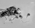
In site A, 3 out of the 104 (2·9) P. variegata dissected were found to be infected by T. callipaeda L3 in the proboscis at days +14 (group 1a), +21 (group 2a) and +53 (group 3a) p.i. respectively (Table 1). In particular, 2 L3 were found on the infected fly from group 1a (Fig. 3), 1 from the fly of group 2a and 1 from the fly of group 3a.

Fig. 3. Third-stage larvae of Thelazia callipaeda in the proboscis of Phortica variegata on day+14 p.i. (site A).
Of the 141 Phortica flies from site A, processed by PCR, 26 (18·4) produced amplicons detectable on agarose gel. The timing in which individuals of P. variegata resulted positive at PCR is reported in Table 1. As expected, amplicons of the positive samples and reference control were approximately 500 bp in length. Conversely, no amplicons were produced from any of the non-infected P. variegata (negative control). Sequences from 26 positive PCR products showed 99 similarity with sequences of the corresponding species available in GenBank (AY207464). In particular, 5 mutations falling within the range of intra-specific differences (Otranto and Traversa, 2004), all represented by transversion (T/G), occurred at positions 386, 394, 399, 404 and 446 bp.
At site B, 106 (17·6) out of 602 A. okadai dissected were found to be infected with T. callipaeda larvae (different stages). In particular, 62 L3 (between 1 and 8) were found in the proboscis of 34 (5·6) flies. The shortest time in which L3 developed and were retrieved was at day +14 (group 2b), +17 (group 1b), at day +19 (group 3b) and at day +50 (group 4b) p.i. (Table 2). Of the 30 flies overwintered (group 4b), 6 L3 were detected at day +180 p.i. in 3 flies (10).
Larval development in Phortica
The larval development described below is based on data collected from the dissection of 602 infected flies. On day +1 p.i. 14 L1 (i.e. newborn larvae) were found in the crop of eight Phortica. By days +2 and +3 p.i., 5 flies presented with 8 L1 in the celomatic cavity (no L1 were found in the crop). On day +4 p.i. L1 were found in the testis of Phortica males (Fig. 4A) and in the body cavity of females (Fig. 4B). A capsule appeared around the L1 which assumed the appearance of a ball on the surface of the testis (Fig. 4A). The cyst, containing L1 in females, was linked to the internal body cavity by a peduncle (Fig. 4B). At this stage, the L1 was curved like the letter ‘C’ with the posterior part sharper than the anterior part and a vesicle visible at the anus. By days +5 and +6 p.i. a capsule appeared around the L1. The larvae in capsules were still ‘C’-like, thick and short, and the posterior part was sharp and thin. The intestine of the larvae was under-developed, with 2 enlargements at the end of the tract (Fig. 4C). At this stage the larvae had a ‘sausage-like’ shape (second stage, L2).

Fig. 4. (A) Drawing of the parasitic ‘cyst’ formed on the surface of the Phortica testis (day +4 p.i.). (B) Drawing of the parasitic ‘cyst’ linked to the internal body cavity of a female by a peduncle (day +4 p.i.). (C) Drawing of the ‘sausage-like’ 2nd-stage larva (L2) with the intestine semi-developed (2 enlargements at end of the tract) (day +5 and +6 p.i.). (D) Drawing of the ‘sausage-like’ 2nd-stage larva (L2) (day +9 p.i.). (E) Drawing of the 3rd-stage larvae (L3) of pre-infective stage in development, showing a well-developed digestive tract (days +11 and +12 p.i.). (F) Drawing of the 3rd-stage larvae (L3) showing a well-developed digestive tract the and genital stem cells.
On days +9 and +10 p.i., the larvae shed the first capsule and the preliminary alimentary tract appeared (Fig. 4D). On days +11 and +12 p.i., the larvae (1·4+/−0·93 mm) were thread-shaped: the alimentary tract was almost complete and ganglion visible. The cuticular striation was evident on the body surface (Fig. 4E) and became more evident over the next few days. The larvae in the capsules measured about 2·24+/−0·093 mm at that time.
Between days +10 and +14 p.i., anatomical development of the L3 was completed and the genitalia in the abdomen were visible. The L3 in capsules started displaying movement, finally leaving their capsules for the haemocoel of Phortica, through the thorax and head to the proboscis. By days +14 to +17 p.i., L3 were present in the head and proboscis of Phortica, although some L1 and L2 were also found in the testis (males) and in the capsules of haemocoel (females). Mature L3 measured 2·5−3·2+/−0·91−0·98 mm (Fig. 4F).
Twenty-two of 30 Phortica originally infected in October survived to the following May, from which a total of four infective L3 were harvested from 3 flies (proboscis and head). At site A, only 1 of the 2 L3, recovered from P. variegata flies (Fig. 3) and deposited onto the cornea of the eye of a dog, developed to day +30 p.i. At site B, 39 days p.i. 16 worms (4 pre-adult stage and 12 adults) were found in 3 dogs' left eye and eight worms (6 pre-adult stage and 2 adults) in 3 rabbits' left eye of the 50 and 18 L3 recovered from P. okadai and deposited onto the cornea of the eyes of 3 dogs and 3 rabbits respectively. Newborn L1 were found in 1 dog's left eye +35 days p.i.
DISCUSSION
One major issue concerning thelaziosis associated with T. callipaeda is the identification of the intermediate host, not only the species of insect acting as a vector but also the timing of larval development in the flies. This work provides evidence that P. variegata and P. okadai, collected in the field in sites A and B and experimentally infected with L1 of T. callipaeda, may act as a vector for this nematode in southern Europe and China. The vectors of canine thelaziosis in Europe and China are similar from an ecological point of view since they both occur in rural environments characterized by a humid climate with thick undergrowth at latitudes in the range 39° to 46° North. This latitude is shared by other Far Eastern and Asian countries in which canine and human thelaziosis has been previously reported (between 10° and 45° North for India and Japan, respectively) (reviewed by Otranto et al. 2003 a). Nevertheless, the altitude at which P. okadai and P. variegata live is very different (i.e. 23 m a.s.l. and 700 m a.s.l respectively) and further entomological investigation into the biology and ecology of these drosophilids is needed. In fact, the taxonomy of the Amiota and Phortica genera (Family: Drosophilidae and Sub-Family: Steganinae) is a widely debated and still an unresolved issue; only recently were the two above taxa recognized as distinct genera (Màca, 2003). For this reason, previous reports of A. okadai as a vector of T. callipaeda (Wang et al. 2002 a,b) should instead refer to Phortica okadai. Genera Amiota and Phortica comprise the few species of drosophilid flies with zoophilic feeding habits, in that they feed on fruit like a common ‘fruit fly’, as well as on the lachrymal secretions of animals and humans. Within this particular taxon, both Amiota and Phortica flies were present in sites A and B (Bächli et al. 2005).
Although this study ought properly to be considered as a laboratory infection trial to detect insect/s acting as the intermediate host of T. callipaeda, it started from a field approach working on species of flies (Phortica) which were present in areas with a history of dog (sites A and B) and human (site B) thelaziosis. The proportion of Phortica flies found to harbour T. callipaeda was similar in groups A and B (i.e. 18·4 and 17·6 respectively), even though 2 different methodologies were employed (i.e. PCR amplification and fly dissection respectively). The fact that the flies in group A were dissected only on days +14, +21 and +53 p.i. to retrieve L3 infective stages in the proboscis may explain the low percentage of flies positive to L3 (2·9). Conversely, in group B, the percentage of flies positive for L3 was higher (5·6) than group A. This difference may be due to either (1) the large number of flies infected in group B (602) and to different susceptibility levels of P. okadai compared to P. variegata; or, more likely, (2) to the fact that at site B flies were dissected every day to retrieve all larval stages, whereas in group A flies were dissected on only 3 specific dates p.i. to retrieve infective L3. Moreover, dead flies were processed by PCR which would also have reduced the number of L3-infected flies observed. This explanation is also supported by the high percentage of dead flies from group A that were positive for T. callipaeda at PCR (18·4) after +14 p.i., when L3 infective larvae presumably developed inside the insect body (see below).
The PCR-based approach for the study of nematodes in the vector/s has both advantages and disadvantages: molecular detection in the vector was an effective tool for retrieving T. callipaeda in Phortica, as previously demonstrated for other species of Thelazia (Otranto et al. 2003 b), and was also the only method available to detect T. callipaeda larvae in dead flies (flies become dehydrated in a matter of hours). However, PCR detection of T. callipaeda is not adequate for identifying the developmental stage of larvae or describing the morphology of immature stages. Hence, the development of infective L3 (collected from experimentally infected Phortica) into adult parasites in dogs (sites A and B) and rabbits (site B) demonstrated in the definitive host provided the evidence needed to verify the role of these flies as a vector of T. callipaeda.
On the basis of the developmental stages recovered by daily dissection of infected Phortica flies, it is possible to reconstruct the following developmental larval cycle. The L1 (newborn larvae), present on day +1 p.i. in the crop of Phortica flies, migrate (days +2 and +3 p.i.) through the celomatic cavity into the testis of males or into the body cavity of females (day +4 p.i.). At this stage a capsule appears around the L1 and it assumes the shape of a ball on the surface of the testis while in females the cyst is typically linked to the internal body cavity by a peduncle. In the cyst, L1 is bent like the letter ‘C’ and grows in size (days +5 and +6 p.i.). At this stage a capsule appears around the L1, the posterior part is sharp and thin and the intestine is semi-developed. Then the larvae shed the first capsule (days +9 and +10 p.i.) and on days +11 and +12 p.i. the larvae has a thread-like shape: the alimentary tract is almost complete and the ganglion visible. From day +10 to +14 p.i. every anatomical part of the L3 is complete and movement is detected. Finally, L3 leave their capsule for the haemocoel of Phortica, reaching the proboscis through the thorax and the head. From day +14 to +17 p.i., the first L3 appears in the head and proboscis of Phortica; meanwhile some sausage-like larvae (L2) and pre-infective larvae appear in the testis (males) and the capsules of the haemocoel (females). Based on the results, T. callipaeda L1 are infective to flies when they are in the lachrymal secretions outside the shell membrane and with the shell residue forming a balloon at the posterior extremity. This finding contrasts with the results reported by Kozlov (1963), who concluded that Thelazia L1 are contained in the eggs when they are ingested by the vector.
In the present study, different larval developmental times were found, with the shortest time to moult into L3 ranging between +14 and +21 days p.i. (mean value +17 days p.i.) depending on temperature (see below). Conversely, Kozlov (1963) demonstrated that the second moult into L3 occurred on day +19 to +22 p.i. (mean value +21 days p.i.), without considering the effect of endo- or exogenous factors on larval development time (see below). Since the development from L3 to the mature nematode takes about 30 days, Kozlov stated that only 2 generations of Thelazia can parasitize the definitive host during summer and fall, since the activity of P. variegata lasts from June to September (Kozlov, 1963). Based on our results, about 5 generations of Thelazia may be completed between April and October at the latitude of sites A and B. In fact Phortica flies are active from April to October (Bächli, personal communication). These findings are confirmed by the detection in the eyes of naturally infected dogs, of L4 in March 2002 and April, July and October 2003 (Otranto et al. 2004), which indicates that flies acting as the intermediate hosts of T. callipaeda are present from very early spring to early autumn (specifically, the third moult of T. callipaeda from L3 to L4 takes place 7–9 days after the flies transmit L3 to dogs; Kozlov, 1963).
The simultaneous finding (day +14 to +17 p.i.) of L3 in the head and proboscis of Phortica together with some sausage-like (L2) and pre-infective larvae, suggests that the development of T. callipaeda larvae in the intermediate host is not synchronized and that different developmental stages may occur at the same time. This is probably due to the fact that the timing of larval development reported here may vary from 2 to 4 days for each developmental stage in the fly, and influenced by environmental temperature. In fact, the higher the temperature, the faster the first L3 appears (e.g. in site B, the first L3 appeared at +14 days p.i. – max temp. 29·7°C; and at +50 days p.i. – max temp. 15·3°C). In addition, an important phenomenon which confirms the above finding is the overwintering of the larval T. callipaeda in Phortica.
Overall, the development of larvae in the Phortica flies is consistent with the biology of T. callipaeda eyeworms reported in naturally infected definitive hosts (Otranto et al. 2004). In fact, infective L1 were detected in the lachrymal secretions of dogs in spring, summer and early fall (Otranto et al. 2004), precisely when Phortica flies are present. This means that L1 may then be ingested by flies feeding on the animals’ lachrymal secretions. The finding of L3 in Phortica flies (infected on October) after 180 days p.i. (i.e. April) demonstrates that T. callipaeda may overwinter in the vector. This is probably related to the hibernation phenomenon of the vectors as already demonstrated in face flies Musca autumnalis (Chirico, 1994). The same author observed that fly hibernation was characterized by both the presence of hypertrophied fat bodies (HFB) and arrested ovarian development. In this study, only non-gonoactive female flies with HFB were infected by Thelazia spp. The overwintering in Phortica flies is also confirmed by the presence of L3 in the eyes of naturally infected dogs in early spring (Otranto et al. 2004) as soon as the vector is re-activated after hibernation and transmits T. callipaeda to dogs. Meanwhile, in the definitive host, female nematodes begin to release L1 at the start of spring when Phortica begin flying again. In the definitive host, female nematodes (which survive at least 9 months with overlapping generations) possess L1 in uteri throughout the year. In particular, the occurrence of blastomerized eggs and larvae in uteri close to the vulvar opening during winter may be explained by the need to release larvae in the following spring and summer season as soon as the vector appears (Otranto et al. 2004). Conversely, the absence of blastomerized eggs in the uteri of female nematodes from May to November (2002–2003) may be due to the fact that the mature females strive to release L1 when the vector is present, as confirmed by the finding of L1 in June-July both in uteri and in the animals’ lachrymal secretions (Otranto et al. 2004).
The phenomenon of overwintering, described here for the first time for T. callipaeda, is probably one of the most interesting behavioural strategies of T. callipaeda to survive the winter in an area in which both the host and the vectors are present. The overwintering of T. callipaeda may increase its infective potential thus resulting in an interesting cross-infection strategy which allows the infection of susceptible animals when the activity of Phortica is resumed. It is clear that the overwintering of T. callipaeda may follow two different modalities. The first is ‘endogenous-overwintering’ which occurs in female nematodes in the infected definitive host; it may be caused by a low metabolic rate of the females in the host's eyes at low temperatures. The second is ‘induced-overwintering’ caused by changes to fly metabolism leading to fly hibernation. Other phenomena, such as the presence of HFB and arrested ovarian development, may also concur (Chirico, 1994).
Phortica is indeed an interesting example of how drosophilids can develop zoophilic habits and a wonderful model of co-evolution between both the nematode (i.e. T. callipaeda) and the host on which it feeds (i.e. dogs and humans). It is conceivable that one of the factors driving Phortica spp. towards adopting zoophilic behaviour is infection by T. callipaeda, which makes it more awkward for Phortica to feed on fruit, whereas animal lachrymal secretions are a more easily available food. Phortica may originally have ‘met’ T. callipaeda during an ‘eye secretion meal’ and it may have evolved conveniently as its vector.
The results of this study contribute an interesting new ‘piece of the jigsaw puzzle’ and a valuable step towards understanding how this ‘oriental’ infection found its way into Europe, and modelling its possible spread to habitats and environments favourable to Phortica flies. They are also important for preventing and controlling the spread of this disease to animals and humans (mainly children and the elderly) living in socio-economically disadvantaged regions of the Far East, where public health warning standards are very low and may precipitate in the event of natural calamities.
The authors wish to acknowledge Mark T. Fox (Department of Pathology and Infectious Diseases, The Royal Veterinary College, University of London, UK) for his helpful comments and critical suggestions. The authors wish to express their thanks to Gianluca Galli (Bayer, Milano, Italy), Norbert Mencke (Bayer HealthCare AG, Animal Health Division, Germany) and Giovanni Girone (Chancellor of the University of Bari) for funding this project. Thanks to Gerhard Bächli (Zoological Museum, Zurich, Switzerland) for his invaluable help and assistance in the identification of Phortica flies. Experimental infections were carried out according to the animal welfare rules and regulations of each in the respective countries (Italy and China).