Introduction
Oocytes and zygotes rely on precise mechanisms to ensure accurate chromosomal segregation during meiosis and mitosis to maintain their euploid state. Errors in the exact execution of these mechanisms cause abnormal segregation of chromatids, resulting in the generation of ‘aneuploid’ daughter blastomeres. Aneuploid embryos reflect both gains/losses of entire chromosomes; and aneuploidy is associated with critical abnormalities such as implantation failure, spontaneous abortion, and conditions such as Down’s syndrome (DS), Turner’s syndrome (TS), and Klinefelter’s syndrome (KS) (Orr et al., Reference Orr, Godek and Compton2015). In routine in vitro fertilization (IVF), aneuploidy, structural variants, monogenic disorders, and mosaicism can be detected with next-generation sequencing through a single or several cells biopsied from embryos. However, these chromosomal disorders are difficult to detect without the use of invasive methods.
Extended culture is a potential self-selection process used to identify competent embryos that are expected to continue to develop into blastocysts. Embryos transferred at the blastocyst stage have a higher implantation rate compared with those transferred on day 3 (Gardner et al., Reference Gardner, Lane, Stevens, Schlenker and Schoolcraft2000, Reference Gardner and Lane2003, Reference Gardner, Surrey, Minjarez, Leitz, Stevens and Schoolcraft2004; Milki et al., Reference Milki, Hinckley, Fisch, Dasig and Behr2000; Alpha Scientists in Reproductive Medicine and ESHRE Special Interest Group of Embryology, 2011; Rocafort et al., Reference Rocafort, Enciso, Leza, Sarasa and Aizpurua2018). However, this practice involves prolonged in vitro culture and may increase the chance of altered gene expression and epigenetic inheritance (Niemitz et al., Reference Niemitz and Feinberg2004; Horsthemke et al., Reference Horsthemke and Ludwig2005; Manipalviratn et al., Reference Manipalviratn, DeCherney and Segars2009).
The introduction of time-lapse (TL) microscopy in IVF has enabled consecutive observations of embryonic development, and has generated morphokinetic variables that constitute a novel non-invasive method to continuously monitor and assess the dynamic processes of early embryonic development (Rubio et al., Reference Rubio, Galán, Larreategui, Ayerdi, Bellver, Herrero and Meseguer2014). Such use of morphokinetic variables as measured by TL for improved embryo selection has been reported previously (Wong et al., Reference Wong, Loewke, Bossert, Behr, De Jonge, Baer and Reijo Pera2010). TL studies currently focus on translating manual scoring into computerized scoring under the Istanbul consensus on embryo assessment. However, more effective morphology assessment is still needed (Gruhn et al., Reference Gruhn, Zielinska, Shukla, Blanshard, Capalbo, Cimadomo, Nikiforov, Chan, Newnham, Vogel, Scarica, Krapchev, Taylor, Kristensen, Cheng, Ernst, Bjørn, Colmorn, Blayney, Elder, Liss, Hartshorne, Grøndahl, Rienzi, Ubaldi, McCoy, Lukaszuk, Andersen, Schuh and Hoffmann2019; Tran et al., Reference Tran, Cooke, Illingworth and Gardner2019).
Blastomere movement occurs after the first cell division in human embryos, and has been suggested to be a prognostic parameter for the prediction of pregnancy outcome following cleavage-stage embryo transfer (Ohata et al., Reference Ohata, Ezoe, Miki, Morita, Tsuchiya, Kaneko, Okimura, Uchiyama, Yabuuchi, Kobayashi, Montag and Kato2019). Embryos manifesting chaotic division exhibit a lower quality on day 3 and a reduction in rates of subsequent blastocyst formation (Athayde et al., Reference Athayde Wirka, Chen, Conaghan, Ivani, Gvakharia, Behr, Suraj, Tan and Shen2014; Yang et al., Reference Yang, Shi, Gong, Zhang, Lu, Tan, Leng, Hao, He, Gu, Lu and Lin2015). Disordered cleavage causes a deterioration in embryonic quality, but its underlying mechanism(s) of action remains unclear; although we assumed that disordered cleavage of early-stage embryos possessed an abnormal chromosomal status or a disordered genome that impairs subsequent development. We aimed to assess the relationship between embryonic cleavage model in first mitosis and the chromosomal status of human embryos, and to discuss the potential biological and clinical implications for the cleavage model as a single parameter that can be used to assess embryonic quality in the present study.
Materials and methods
Donating couples and clinical treatment
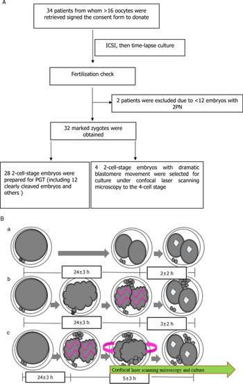
Between April 2019 and June 2020, 34 infertile couples who underwent their first IVF cycle at the Reproductive Center were recruited to donate normal 2-cell-stage embryos for this study. The infertility factors of the recruited couples were all male factors, including teratozoospermia, asthenozoospermia, oligozoospermia, and azoospermia. Abnormal karyotypes from either the men or women in this study were excluded. The female partner’s pituitary downregulation and controlled ovarian stimulation protocol were achieved as described previously by Ye et al. (Reference Ye, Huang, Zeng and Pei2009). When at least three follicles reached a mean diameter of 18 mm, 10,000 IU of hCG (Ovidrel, Merck Serono, Italy) was administered to induce final follicular and oocyte maturations. Oocyte retrieval was performed by transvaginal ultrasound-guided follicle aspiration 36 h after hCG administration. All retrieved oocytes undergoing intracytoplasmic sperm injection (ICSI) were collected with informed consent from the couples who agreed to donate normal zygotes or 2-cell-stage embryos for research use. After ICSI, all fertilized oocytes were cultured in TL incubators (Embryoscope, VitroLife AB, Sweden) to record fertilization to the 2-cell stage (27–28 h after ICSI; Fig. 1A).
Flow chart of study and cleavage model at first cleavage. (A) Flow chart of the present study. Twenty-eight 2-cell embryos were prepared for PGT and four entangled embryos in the first mitotic division were stained with Hoechst and cultured in a confocal laser scanning microscope. (Ba) Clear cleavage. The relatively static distribution of blastomeres is a characteristic that fundamentally distinguishes clearly cleaving embryos from entangled-cleavage embryos in the first mitosis. (Bb, Bc) Entangled cleavage. Entangled cleavage includes two models: embryos with wobbling blastomeres in a moderate motion (Bb), and embryos with blastomeres that turn around in a dramatic fashion (Bc). The time point of relatively static distribution of blastomeres is visible nuclei appearance in both blastomeres of entangled cleaved embryos. But in clear cleaved embryos the profiles of the blastomeres are static elliptical just after mitosis, then the visible nuclei appear peacefully after 2 ± 2 h.

Sources of 2-cell-stage embryos (or zygotes)
The 32 embryos under study were selected from 32 patients with a favourable prognosis, who each produced more than 12 normally fertilized oocytes with two pronuclei (2PN) after a fertilization check on day 1. The 32 donated zygotes were cultured in TL incubators for use in recording the first cleavage model (CM); and then two blastomeres underwent genomic testing 32 h after fertilization (28 in 32 embryos). One zygote was donated at maximum for each patient after the first round of cleavage checking. Four embryos with chaotic cleavage in the first mitosis and that were graded as unviable were taken out of the TL incubators (32 h after fertilization) and then cultured in the confocal laser scanning microscope to record their subsequent blastomere motions and chromosomal distribution until the four-cell stage. None of the 32 embryos was used for embryo transfer or preservation (Fig. 1A).
The CM was previously described by Ohata et al. (Reference Ohata, Ezoe, Miki, Morita, Tsuchiya, Kaneko, Okimura, Uchiyama, Yabuuchi, Kobayashi, Montag and Kato2019) with some mild modifications. First, the blastomeres’ movements were not instant events but began at the appearance of the cellular furrow and then lasted for 2–8 h, except for clear cleavage embryos. Second, the embryo fragments occurred in rare clear cleavage groups, but they showed higher frequently in entangled-cleavage embryos. Finally, the marker of blastomere shift in status from mobile to static was the appearance of visible nuclei in both blastomeres (Fig. 1B). The relatively static distribution of blastomeres was the characteristic that fundamentally distinguished the clearly cleaving embryos from entangled-cleavage embryos in the first mitosis. Entangled cleavage included two models: embryos with wobbling blastomeres in a moderate motion and embryos with blastomeres that turned around in a dramatic motion (Fig. 1B).
Confocal laser scanning microscopy
Four 2-cell embryos showing chaotic division were extracted from TL and stained with 5-μM Hoechst stain (Thermo Fisher, USA) to visualize their DNA. Images were then captured at 30 min intervals for 24 h using a confocal laser scanning microscope (Leica TCS SP8; Mannheim, Germany) equipped with a Plan Apochromat ×20 magnification objective, as well as an observation dish and incubator system (Tokai Hit Co., Japan), with medium at 37°C and under 5% CO2 in compressed air (Fig. 1A, B).
Biopsy of 2-cell-stage embryos
Twenty-eight 2-cell embryos were extracted from TL (at 32 h) for genomic testing. Single-blastomere isolation for donated 2-cell embryo biopsy was performed according to previously established methods (Yan L et al., Reference Yan, Yang, Guo, Yang, Wu, Li, Liu, Lian, Zheng, Yan, Huang, Li, Wu, Wen, Lao, Li, Qiao and Tang2013).
Preimplantation genetic testing (PGT)
Following biopsy, the cells were placed into 0.2-ml thinly walled tubes and sealed and frozen by placing them in a freezer at −20°C prior to genetic screening. Single-cell, whole-genome amplification (WGA) with multiple annealing and looping-based amplification cycles (MALBAC) were used.
We performed WGA on cleavage-stage blastomeres using MALBAC following the manufacturer’s protocol (Catalogue No. YK001B; Yikon Genomics).
Cells were lysed by heating (20 min at 50°C and 10 min at 80°C) in 5 μl lysis buffer. Then, 30 μl of freshly prepared preamplification mix was added to each tube and the mixture was incubated at 94°C for 3 min. Next, DNA was amplified using eight cycles of 40 s at 20°C, 40 s at 30°C, 30 s at 40°C, 30 s at 50°C, 30 s at 60°C, 4 min at 70°C, 20 s at 95°C, and 10 s at 58°C, and was immediately placed on ice. Next, 30 μl of the amplification reaction mix were added to each tube and the mixture incubated at 94°C for 30 s; followed by 17 cycles of 20 s at 94°C, 30 s at 58°C, and 3 min at 72°C.
Low-coverage (0.3×), genome-sequenced MALBAC products were purified using a DNA purification kit to construct a DNA library.
PGT-outcome grouping
Normally, ‘PGT normal’ embryos are euploid, without genomic disorders of deletions and/or duplications (including microdeletions and/or microduplications), that is 46,XN. Deletion and/or duplication in chromosomal embryos indicates that the embryos possess small (>10 Mb) or submicroscopic, genomic deletions and duplications (1 kb to 10 Mb), as well as mosaic forms, for example 46,XN, dup(16)(p13.3p13.13) (5.7 Mb) and 46,XN, dup(16)(q23.3q24.2) (5.5 Mb) (mos, 50%).
‘Euploid’ embryos are defined as double (2n) chromosomal sets in embryos regardless of the deletion or addition of chromosomes in the present trial.
Statistics
Statistical data are presented as means ± 1 standard deviation (SD). A chi-squared test or Fisher’s exact-probability test was used when appropriate to compare parameters. A P-value < 0.05 was considered to be statistically significant; Stata12.0 (Statacorp, USA) was used for data analysis.
Results
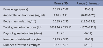
The baseline demographic features describing the 32 patients are presented in Table 1. Mean female age was 26.43 ± 2.67 years, the mean number of oocytes retrieved was 18.25 ± 3.25, and the mean number of transferable embryos was 6.42 ± 2.57. All available transferable embryos were vitrified to prevent potential ovarian hyperstimulation syndrome.
Characteristics and outcomes of assisted reproductive technology treatment of patients (N = 32) who donated zygotes

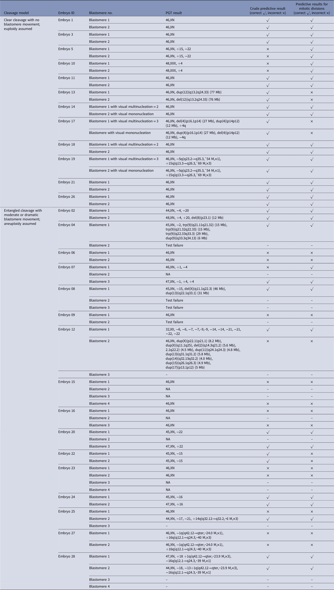
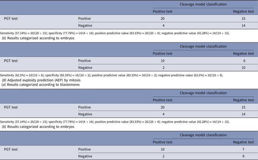
Twenty-eight donated zygotes were used to test chromosomal segmentation in 2-cell-stage embryos 1 day after ICSI (Table 2). To predict euploidy when blastomeres were the objects of observation, we analyzed the raw data (Table 3A-a), we observed a sensitivity of 57.14%, a specificity of 77.78%, a positive predictive value (PPV) of 83.33%, and a negative predictive value (NPV) of 42.28%. When we analyzed the embryos, the sensitivity was 62.5%, specificity was 83.33%, PPV was 83.33%, and NPV was 62.5% (Table 3A-b). When discriminating the results between meiosis and mitosis, sensitivity was 57.14% and 58.82%, specificity was 77.78% and 81.81%, PPV was 83.33% and 83.33%, and NPV was 42.28% and 56.25% when predictions were made for blastomeres and embryos, respectively (Table 3B-a,b).
Cleavage model and PGT results during embryonic first mitosis a

a Crude predictive results indicate euploidy or aneuploidy regardless of deletions or duplications. Predictive results for mitotic divisions mean complementary chromosomal errors including both aneuploidy and deletions and duplications according to PGT test. Here, although embryo ID 5 was correct in chromosomal number, there were errors in chromosomal type, so the prediction was incorrect if held to strict criteria.
NA, not available.
Sensitivity and specificity of using the cleavage model to predict euploidy in 2-cell embryos (raw data adjusted for mitosis)
(A) Crude euploidy prediction (CEP)
(a) Results categorized according to blastomeres

Sensitivity (58.82%) = 10/(10 + 7); specificity (81.81%) = 9/(9 + 2); positive predictive value (83.33%) = 10/(10 + 2); negative predictive value (56.25%) = 9/(7 + 9).
Aneuploidy and deletions/duplications occurring during meiosis and mitosis
When we biopsied the 2-cell-stage embryos and performed PGT (Table 2), we detected aberrant chromosomes in embryo 5 (46,XN, +15, −22 in both blastomeres), embryo 10 (48,XXX, +4 in both blastomeres), embryo 19, embryo 22, and embryo 27. These embryos showed aberrant chromosomal segmentation that might have occurred during meiosis, which was difficult to detect in subsequent mitotic divisions.
In detecting aneuploidy caused by mitosis, which show the complementary chromosomal errors in each blastomere using CM, the predicted results were consistent with the PGT test in some embryos (embryos 2, 4, 7, 8, 12, 20, 24, and 28), although it was low in NPV. In addition, we unambiguously detected chromosomal deletions/duplications, but not aneuploidy, caused by mitosis during the first cellular division using PGT in embryo 13 [46,XN, dup(12)(q13.2q24.33) (77 Mb); 46,XN, del(12)(q13.2q24.33) (76 Mb)] and embryo 17 [46,XN, del(4)(p16.1p14) (27 Mb), dup(4)(p14p12) (12 Mb), +4q; 46,XN, dup(4)(p16.1p14) (27 Mb), del(4)(p14p12) (12 Mb), −4q]. It was difficult using the CM to detect small errors (chromosomal deletions/duplications) at the first mitotic stage in embryo development (embryos 13 and 17). Mosaic status might be formed from different karyotypes in blastomeres of 2-cell embryos after their consequent division.
Chaotic chromosomal segregation in entangled-cleavage embryos
When analyzing confocal laser images using the entangled CM in viable embryos stained with Hoechst, in the entangled CM embryos, fragments with micronuclei were observed. The fragments were increasing coincidence with blastomere motion increased from moderate to dramatic (fragmentation rates in CM with no or slight motion, moderate motion and dramatic motion were 5.25% ± 5.06%, 9.02% ± 9.05%, and 22.79% ± 9.05%, respectively; P < 0.05). Chromosomes in the zygote were split into smaller blastomeres in occasional (sometimes recognizable) fragments. In subsequent cytoplasmic movements, the smaller blastomeres with micronuclei and an anuclear blastomere appeared to fuse together (the torsion on the blastomeres and chromosomes devolved; Fig. 3).
Multinucleation in blastomeres and embryos with more than two daughter cells at the 2-cell stage
Multinucleation in blastomeres (more than one nucleus per blastomere) has previously been considered abnormal, however in our study four embryos (embryos 14, 17, 18, and 19) with visual multinucleation (two and three) engendered correct genomic results (embryos 14 and 18) or incorrect results: embryo 17 [46,XN, del(4)(p16.1p14) (27 Mb), dup(4)(p14p12) (12 Mb), +4q; 46,XN, dup(4)(p16.1p14) (27 Mb), del(4)(p14p12) (12 Mb), −4q], and embryo 19 [46,XN, −5q(q23.2→q35.3, ∼54 M,×1), +15q(q13.3→q26.3, ∼69 M,×3) ×2]. Additionally, embryos might have had more than two cells after zygotes were separated (embryos 15, 16, 20, 23, 25, and 28) in the entangled CM; normally, two blastomeres contain all the genomic material.
Discussion
This study, which was conducted using early-stage human embryos revealed a correlation between CM and PGT outcomes from a resource trial of normal human embryos; and our results shed light on the importance of the early stages of human embryonic development, and provided a parameter for predicting which embryos were euploid. Non-invasive and rapid TL imaging analyzed using the CM before the 4-blastomere stage could significantly improve assessment of embryonic morphology (Guerif et al., Reference Guerif, Le Gouge, Giraudeau, Poindron, Bidault, Gasnier and Royere2007; Chavez et al., Reference Chavez, Loewke, Han, Moussavi, Colls, Munne, Behr and Reijo Pera2012).
PGT outcomes were grouped based on the original causes of chromosomal normalities or abnormalities: PGT normal (including euploidy and no deletions and/or duplications); euploid; or deletions and/or duplications of embryonic chromosomes and their corresponding mosaic forms. Aneuploidy is defined as a chromosomal number that deviates from a multiple of the haploid set (Griffin et al., Reference Griffin and Ogur2018). The resulting classification could help us understand the probable errors reflected in the CM (Fig. 1B). Embryos from the clear cleavage group typically manifest a high possibility of normal PGT outcomes and euploidy rates. However, we could not establish the exact causes of the CM and chromosomal deletion and duplication errors because live staining to distinguish small-fragment DNA errors in human embryos in first mitosis is difficult to perform microscopically. The rate of chromosomal segmentation errors might be higher in entangled CM during mitosis due to blastomere torsion and chromosomal devolution. The mechanisms of blastomere torsion and chromosomal devolution in the entangled CM are unknown, but the outcomes show us that there might be a problem with inner developmental potential when a zygote is cleaved in this particular way.
As a multinuclear embryo might be chromosomally normal at a particular point in time, our data provided direct evidence relative to previous clinical trials (Aguilar et al., Reference Aguilar, Rubio, Muñoz, Pellicer and Meseguer2016; Desai et al., Reference Desai, Goldberg, Austin and Falcone2018). From our observations, the chromosomal abnormalities occurring during mitosis may be higher when compared with previous studies (Lamb et al., Reference Lamb, Freeman, Savage-Austin, Pettay, Taft, Hersey, Gu, Shen, Saker, May, Avramopoulos, Petersen, Hallberg, Mikkelsen, Hassold and Sherman1996; Kuliev et al., Reference Kuliev, Zlatopolsky, Kirillova, Spivakova and Cieslak Janzen2011), where the corresponding errors in two blastomeres could be caused by meiotic chromosomal segmentation, and the complementary errors in two blastomeres could be caused by first mitotic chromosomal segmentation. These complementary errors in two blastomeres might create mosaic embryos in eight cells or during the blastocyst stage.
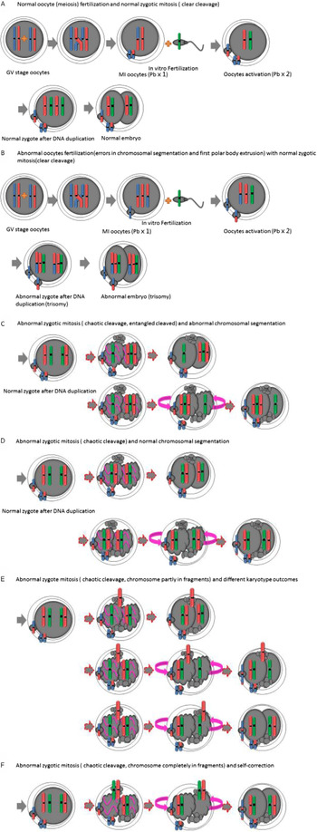
Normal embryos depend upon exquisite mechanisms to maintain their chromosomal euploidy (Fig. 2A). Meiotic errors could not be detected using the CM, as this was an upstream event and not associated with subsequent cell division (Fig. 2B). Entangled-cleavage embryos might be abnormal or show a normal chromosomal distribution (Fig. 2C, D). Embryos with a high proportion of fragments present another possibility for chromosomal distribution, that is chromosome entry into the smaller blastomeres or fragments that are in subsequent motion, and fused or not fused with normally sized anuclear blastomeres (Fig. 2E, F). Based upon our hypothesis, the CM could detect mosaic forms when two blastomeres presented with complementary chromosomal errors. However, due to our use of biopsy and PGT, we have no data available regarding mosaicism. The other potential meiotic and mitotic errors in early human embryos might be associated with the phenomenon of anaphase lagging (Fragouli, Reference Fragouli, Alfarawati, Spath, Jaroudi, Sarasa, Enciso and Wells2013). Lagging chromosomes, which fail to migrate to one of the spindle poles along with the other chromosomes, tended to be left behind, forming separate micronuclei during the interphase before ultimately being lost altogether. Micronuclei are often observed in human embryos at the cleavage stage, providing further evidence for an appreciable frequency of anaphase lag during early embryogenesis (Coonen et al., Reference Coonen, Derhaag, Dumoulin, van Wissen, Bras, Janssen, Evers and Geraedts2004). However, the exactly reasons must be established in further research.
A diagram of the association between the cleavage model and chromosomal segmentation.  indicates the tetraploid configuration before synapsis.
 or
 indicates sister chromatids post-homologous recombination of chromosomes after first meiosis.
 indicates the chromatids of sperm.
 indicates the embryonic chromosome. (A) Chromosomally normal embryo. (B) Trisomic embryo due to chromosomal aberrations during the first meiosis. (C) Chromosomally abnormal embryo (one blastomere is a monosome, and the other is trisomic; this embryo will develop into a mosaic embryo). This error might be the same as that for chromosomal deletions and/or duplications). (D) Chromosomally normal embryo showing entangled cleavage. (E, F) Embryos with a high proportion of fragments manifest another possibility for chromosomal distribution; that is chromosomal entry into smaller blastomeres or fragments that are in subsequent motion, and either fuse or do not fuse with normally sized anuclear blastomeres.

Torsion devolved on the blastomeres and chromosomes. In cytoplasmic movements, the smaller blastomeres with micronuclei and an anuclear blastomere appeared to fuse together (22–23.5 h), the last two images are at inconsistent time points due to a pause of the confocal laser scanning microscope for image extracting and analysis.

From the results of this study, the exact direction of causation between cleavage movement and aneuploidy is difficult to establish, although blastomere movement seems to be a consequence of aneuploidy because the internal force emerging of blastomere movement might be caused by aneuploidy. Relative mechanism studies should be performed to elucidate the relationship between CM and aneuploidy.
This approach had several limitations. Using biopsies and PGT of blastocyst-stage embryos, we observed that predictive outcomes might be slightly inconsistent with respect to 2-cell-stage embryos. The potential reasons for this might be the high false-negative rate for mosaic blastocysts with PGT (Harton et al., Reference Harton, Cinnioglu and Fiorentino2017) and self-repair in morula stage (Coticchio et al., Reference Coticchio, Lagalla, Sturmey, Pennetta and Borini2019), and the deficiency of data on subsequent cell divisions in embryonic development. The location of the biopsy needle might affect the results, examples of when the blastomeres or trophectoderm divide from abnormal blastomeres of 2-cell-stage embryos were missing during biopsy, and the PGT results seemed normal. Therefore, much investigatory work still needs to be performed prior to clinical application of our model.
In conclusion, we observed high specificity and PPV, but low sensitivity and NPV, using the CM in the prediction of euploidy that implied that CM could be considered as a screening method for embryo selection. Due to the relative scarcity of human zygotes, it is virtually impossible to achieve a large sample size for zygotic evaluation, and therefore additional observational studies using the CM to select transferable embryos are needed before clinical practice.
Acknowledgements
We thank Yuan Q. Sun for help with revising the design of the present experiments.
Financial support
This research was supported by the Chongqing Science and Technology Bureau (No. cstc2017shms-zdyfX 0020), and by a generous anonymous donor.
Conflicts of interest
No conflicts of interest exist in the submission of this manuscript, and the manuscript was approved for publication by all authors.
Ethical standards
The local ethics committee approved this investigation on 5 April 2019 (reference number 2019-602). All zygotes used to examine the relationship between the CM and chromosomal segmentation errors were donated for research purposes after informed consent was obtained. The zygotes or 2-cell embryos were approved for research by the Chongqing Obstetrics and Gynaecology Hospital review board, and no confidential health information could be associated with the zygotes.