
I. INTRODUCTION
In the last few decades, it was shown that the cyclic response of materials is governed by physically based microstructural processes. As described, e.g., in the early review Reference Mughrabi1 and works of Mughrabi et al., Reference Mughrabi and Christ2–Reference Mughrabi4 understanding of these elementary mechanisms is necessary for the development of new advanced materials with enhanced fatigue and creep properties. Highly alloyed austenitic stainless steels belong to such a group of advanced materials, which have a very wide spectrum of applications in different branches of the industry. They are used for the design of equipments working both at room and low temperatures as well as high temperatures. Reference Marshall5,Reference Maziasz, Busby and Konings6 In the field of coal-fired power generation, there is an increased demand to improve the efficiency of power plants and thereby reduce carbon emissions which leads to the need for new heat-resistant materials with improved high-temperature strength, enhanced creep properties, and high-temperature corrosion resistance at proposed increased high operating temperatures at 700 °C. Reference Chai, Forsberg and Di Gianfrancesco7
One of the promising cost-effective candidate materials developed for use in future advanced ultra-supercritical coal-fired boilers is the austenitic stainless steel Sandvik Sanicro 25. This steel is based on the Fe–Ni–Cr alloy system with a high content of alloying elements: W, N, Co, and Nb added (presumably for solid solution hardening), as well as a high content of Nb, N, Cr, and Cu (presumably for precipitation strengthening). The 22.5 wt% Cr content results in good steam oxidation and hot corrosion resistance. High Ni content of approximately 25.0 wt% and relatively high amounts of nitrogen contribute to the stabilization of the FCC structure.
Most of the austenitic steels commercially available today are capable of operational service temperatures up to 650 °C. Highly alloyed steel Sanicro 25 exhibits excellent high-temperature strength, creep properties, and corrosion resistance at temperatures up to 700 °C. To understand material characteristics and mechanisms leading to such performance, there is a serious need for the investigation of the interrelated effects of composition, microstructure, deformation mechanisms, and overall material response under static and cyclic loading conditions.
Recently, cyclic plastic response and low cycle fatigue behavior Reference Polák, Petráš, Heczko, Kuběna, Kruml and Chai8,Reference Polák, Petráš, Heczko, Kruml and Chai9 as well as surface profile evolution and fatigue crack initiation at the room temperature Reference Polák, Mazánová, Kuběna, Heczko and Man10,Reference Polák, Mazánová, Heczko, Kuběna and Man11 of this material were reported. Creep properties Reference Chai, Forsberg and Di Gianfrancesco7,Reference Chai, Boström, Olaison and Forsberg12 and corresponding microstructural changes Reference Zurek, Yang, Lin, Huttel, Singheiser and Quadakkers13 after thousands of hours of steam exposure were also studied. However, detailed investigation of mechanisms governing high-temperature cyclic behavior is still missing. Polák and co-workers Reference Polák, Petráš, Heczko, Kuběna, Kruml and Chai8,Reference Polák, Petráš, Heczko, Kruml and Chai9,Reference Heczko, Polák and Kruml14,Reference Petráš, Škorík and Polák15 have reported exceptional cyclic hardening, during low-cycle fatigue at 700 °C, leading to a remarkably high saturated cyclic stress. They have hypothesized that fine scale precipitates and their interaction with mobile dislocations may play a significant role in cyclic strengthening. The purpose of the present work is to reveal further details of overall microstructural changes and corresponding mechanisms leading to the striking and exceptionally high-temperature cyclic hardening of Sanicro 25.
II. EXPERIMENTAL DETAILS
A. Material
The experimental material, Sanicro 25 austenitic stainless steel of grade UNS S31035, was supplied by Sandvik (Stockholm, Sweden) in the form of cylindrical rods of 150 mm in diameter. The nominal composition was (in wt%) 0.1 C, 22.5 Cr, 25.0 Ni, 0.5 Mn, 3.6 W, 1.5 Co, 3.0 Cu, 0.5 Nb, 0.23 N, 0.2 Si, and balance Fe. Cylindrical specimens for high-temperature cyclic straining were machined with the axis parallel to the rod. The gage length was 15 mm and the diameter of the specimens was 6 mm. After the production of the crude shape of specimens, they were annealed at 1200 °C for one hour and cooled in air. This state of material is noted as “initial” in the remainder of the text.
B. Mechanical tests
Mechanical tests were performed using a computer-controlled electrohydraulic MTS system equipped with hydraulic grips, split resistance furnace, and high-temperature axial extensometer with a 12 mm base. Fully-reversed, strain-controlled cyclic loading with a constant strain rate of 2 × 10⁻³ s−1 was applied at 700 °C. The saturated amplitude of plastic strain was evaluated. Further information about the testing conditions can be found elsewhere. Reference Polák, Petráš, Heczko, Kuběna, Kruml and Chai8,Reference Polák, Petráš, Heczko, Kruml and Chai9,Reference Heczko, Polák and Kruml14
C. Transmission electron microscopy
The microstructure was investigated by means of transmission electron microscopy (TEM). Thin plates were cut perpendicular to the loading axis from the gage length of the bulk specimens by an electric-discharge machine. These were mechanically ground to produce thin foils of thickness of 0.08 mm, which were then punched out to produce disks having a diameter of 3 mm. The discs were then electrolytically polished using a double jet device TenuPol2 (Struers GmbH, Willich, Germany). The electrolyte was composed of 95% of acetic acid and 5% of perchloric acid. The polishing conditions were 80–90 V, 0.2 mA, and at a temperature of 13–16 °C. Further details about the oriented TEM foil preparation can be found in Ref. Reference Heczko, Polák and Kruml14.
The spatial arrangement of dislocations in grains was determined using the technique of oriented foils. Conventional transmission electron microscopy (CTEM) was performed using a Phillips CM12 TEM (FEI, Eindhoven, the Netherlands) working at 120 kV and a JEOL JEM-2100F scanning transmission electron microscope (STEM; JEOL Ltd., Tokyo, Japan) operating at 200 kV. For atomic resolution observations, the grains of interest were tilted to perform high-angle annular dark-field (HAADF) zone axis imaging using a probe-corrected FEI Titan3 80–300 kV STEM (FEI, Eindhoven, the Netherlands). High spatial resolution energy dispersive X-ray spectroscopy (EDS) scans were performed at 300 kV using an image-corrected FEI Titan3 60–300 kV with a Super-X EDS detector and quantified using the Bruker Esprit software (Bruker Nano GmbH, Berlin, Germany). The detection system uses four silicon drift detectors that are located radially around the objective pole piece and a specimen stage for improved collection performance. The presence of carbon and nitrogen was verified by electron energy loss spectroscopy (EELS).
III. RESULTS
A. Initial microstructure
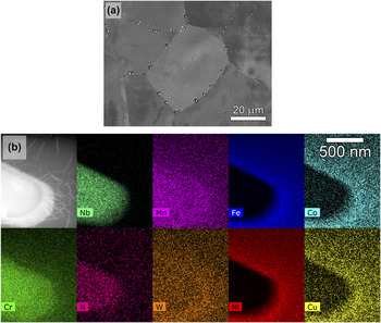
A representative view of the overall microstructure in the initial state is shown in Fig. 1(a). The average grain size, determined by the intercept method, was 60 μm; however, large grains of sizes up to 200 μm were also found. At the grain boundaries as well as inside the grains, large Nb-rich primary particles with sizes varying from 100 to 400 nm were observed. Reported previously, Reference Zurek, Yang, Lin, Huttel, Singheiser and Quadakkers13,Reference Heczko, Polák and Kruml14 these are nitrides designated as Z-phase with a composition of (Cr,Nb)N. Particles of the Z-phase have been observed frequently in Nb stabilized austenitic steels containing a high level of nitrogen. Reference Zurek, Yang, Lin, Huttel, Singheiser and Quadakkers13,Reference Sourmail16
Initial state of the material. (a) SEM micrograph (backscattered electrons mode) of electrolytically polished surface of the Sanicro 25 after annealing for 1 h at 1200 °C and cooling in air. Z-phase precipitates are located at the grain boundaries and also inside the grains. (b) STEM HAADF image of Z-phase particle (bright contrast) embedded in the austenite matrix. EDS showing enrichment of Cr, Nb, N as well as Mn and W in the precipitate. On the contrary, Fe, Ni, Co, and Cu are depleted.

The crystal structure of the Z-phase for the stoichiometry CrNbN has been proposed as distorted body-centered tetragonal (space group P4/nmm) with lattice parameters a = 3.04 Å and c = 7.39 Å. Reference Sourmail16,Reference Danielsen, Hald, Grumsen and Somers17 In austenitic steels, the empirical formula is approximately (Cr0.8Fe0.2)(Nb0.9Mo0.1)N Reference Danielsen, Hald, Grumsen and Somers17 ; however, the composition varies with the composition of the steel, and small concentrations of several substitutional elements (Mn,W) can be dissolved in the Z-phase as illustrated for the present case in Fig. 1(b).
The Z-phase is stable at high temperatures, and after annealing at 1200 °C, it is the predominant precipitate since its dissolution temperature is between 1300 and 1450 °C, in accord with the present results. Except for a small amount of nanometer-sized M23C6 chromium carbides, the Z-phase was the only precipitate observed in the initial state. Reference Zurek, Yang, Lin, Huttel, Singheiser and Quadakkers13,Reference Heczko, Polák and Kruml14
B. Cyclic stress–strain response
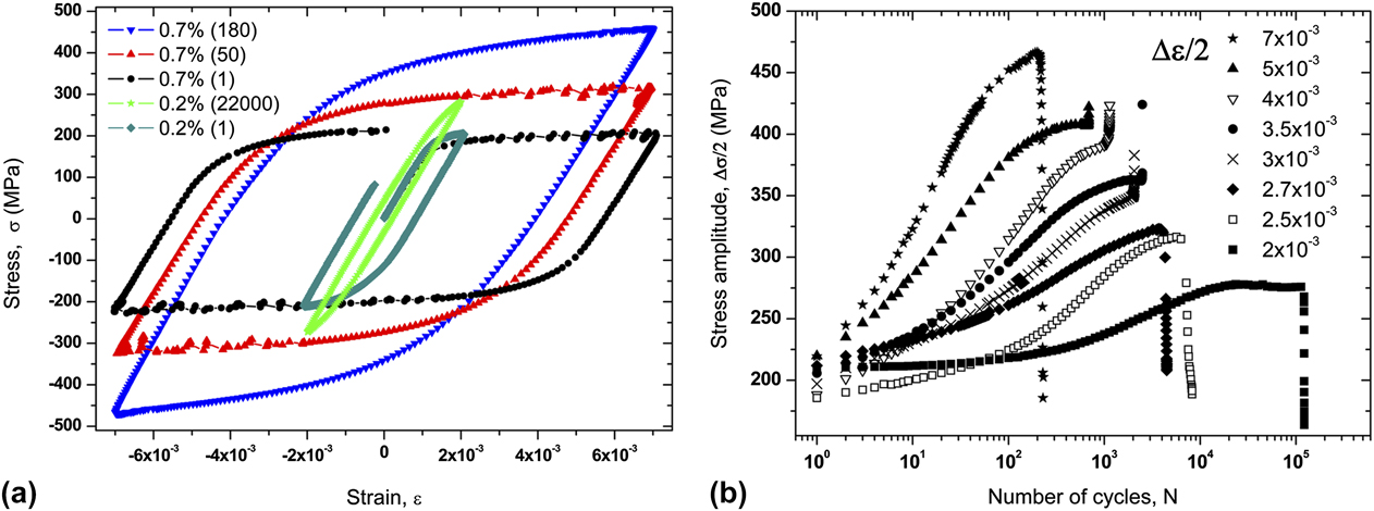
Detailed investigation of the overall stress–strain response under constant total strain amplitude loading was reported earlier. Reference Polák, Petráš, Heczko, Kuběna, Kruml and Chai8–Reference Polák, Mazánová, Heczko, Kuběna and Man11 In the present study, attention is placed on the cyclic stress–strain response at 700 °C. Evolution of the hysteresis loop shape in Fig. 2(a) for total strain amplitudes (Δε/2) 0.2 and 0.7% illustrates the significant increase of cyclic stress as a function of fatigue cycles. Cyclic hardening/softening curves of specimens cyclically loaded over a wide interval of total strain amplitudes are plotted in Fig. 2(b) as a stress amplitude Δσ/2 versus number of cycles N. The initial increase of the stress amplitude with each cycle indicates a notable work-hardening effect. This is observed for all strain amplitudes. The higher the applied strain amplitude, the more significant and rapid the cyclic hardening. After the inflection point, the hardening rate rapidly decreases and a tendency to reach saturation of the cyclic stress is observed for all strain amplitudes. The possible substructural origins of this behavior are now presented.
(a) Evolution of the hysteresis loop shape with cycling at total strain amplitudes 0.2 and 0.7%. (b) Cyclic hardening curves in constant total strain amplitude loading at temperature of 700 °C. Stress amplitude versus number of cycles is plotted.

C. Dislocation structure evolution
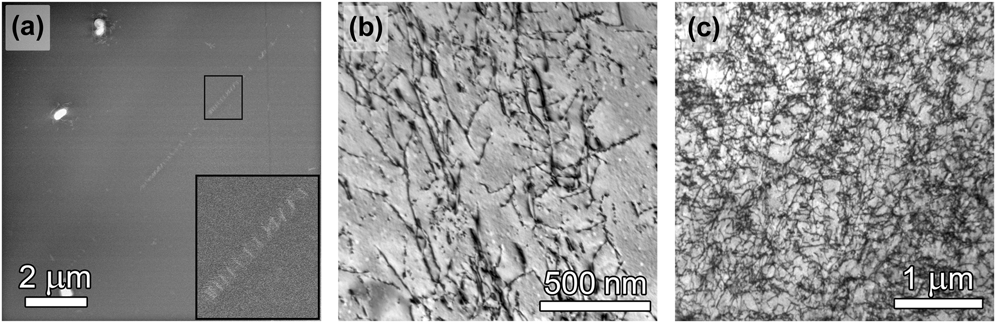
The stacking fault energy in austenitic stainless steels is quite low [lower than 0.020 Jm−2 (Refs. Reference Obrtlík, Kruml and Polák18–Reference Lu, Hultman, Holström, Antonsson, Grehk, Li, Vitos and Golpayegani20)] and as a consequence the dissociation distance between Shockley partial dislocations is large. Therefore, in the initial state of the material, the dislocations tend to be present in planar arrays and pile-ups of individual dislocations and stacking faults, Reference Heczko, Polák and Kruml14 as is shown in the low angle annular dark-field (LAADF) STEM diffraction contrast image (STEM-DCI) in Fig. 3(a). Annealing at 1200 °C for 1 h leads to significantly decreased dislocation density. Most of dislocations observed are individual pile-ups or tangles close to the incoherent Z-phase precipitates or grain boundaries.
(a) STEM LAADF micrograph of representative dislocation structure observed in initial state. As a result of annealing, the dislocation density is very low with individual dislocations and pile-ups observed (inset); CTEM BF image showing increased homogeneous high dislocation density distribution in samples cycled at 700 °C with (b) Δε/2 = 0.27%, and (c) Δε/2 = 0.50%.

With cyclic loading, the dislocation structure evolution strongly depends on the imposed plastic strain amplitude, temperature, and interstitial content. In closely related studies, Reference Polák, Mazánová, Kuběna, Heczko and Man10,Reference Polák, Mazánová, Heczko, Kuběna and Man11,Reference Heczko, Polák and Kruml14 strong planarity of the dislocation slip and plastic strain localization was reported for the room-temperature fatigue of Sanicro 25. At high temperatures, the dislocation structures differ significantly. All grains are filled with a homogeneously distributed and high density of dislocations. Typical and representative arrangements are shown in Fig. 3. When compared to the initial state, cycling with total strain amplitudes of both Δε/2 = 0.27% and Δε/2 = 0.50% leads to a notable increase of the dislocation density. Evidence of cross-slip and climb of dislocations supports the hypothesis about thermally activated effects Reference Caillard and Martin21 contributing to the production of homogeneous, high dislocation density. Cyclic slip localization is suppressed. In contrast to conventional austenitic steels such as 316L, Reference Obrtlík, Kruml and Polák18,Reference Gerland, Alain, Ait Saadi and Mendez22 no cellular structures were found indicating that localized dislocation rearrangement, usually responsible for cyclic softening at high temperatures, Reference Mughrabi4,Reference Pham, Solenthaler, Janssens and Holdsworth23 is not present in Sanicro 25.
D. Precipitation of strengthening nanoparticles
Cyclic straining at 700 °C leads to substantial microstructural changes. The large number of alloying elements with high concentration in Sanicro 25 results in a significant enhancement of diffusion effects and further precipitation of secondary phases. In steels with a high level of nitrogen that are stabilized by niobium, high mutual affinity of these two elements results in a fast Z-phase precipitation. Reference Zurek, Yang, Lin, Huttel, Singheiser and Quadakkers13,Reference Sourmail16,Reference Danielsen, Hald, Grumsen and Somers17 The presence of carbon is known to lead to rapid nucleation of numerous M23C6 chromium-rich carbides inside the grains; however, this occurs mostly at grain boundaries where they cause significant depletion of chromium content in the matrix, especially at low-angle boundaries. Reference Heczko, Polák and Kruml14 As reported earlier, Reference Sourmail16 carbides can be found already after very short aging times (less than one hour at 750 °C) even in stabilized steels.
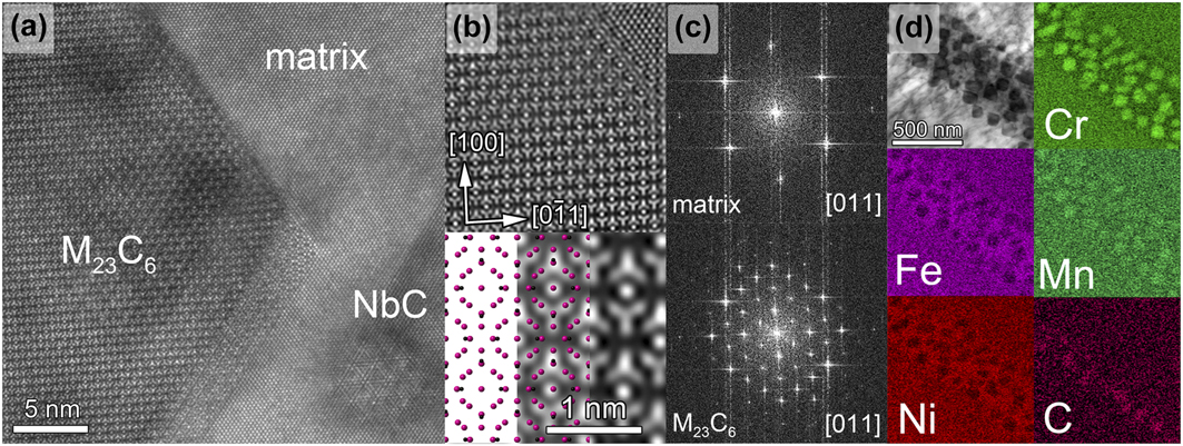
An example of semicoherent, cuboidal M23C6 chromium-rich carbides nucleated within an austenite grain is shown in the atomic resolution STEM HAADF micrograph in Fig. 4(a). The structure is viewed along the [011] zone axis. An image of the interface demonstrates a cube-on-cube orientation of the carbide with the matrix in Fig. 4(b). Fast Fourier transformation (FFT) filtering (frequencies corresponding to the noise and the transmitted beam spot were excluded) of the image was used to emphasize the interface. In detail, the experimentally obtained atomic structure is compared to a Cr23C6 model Reference Bowman, Arnold, Storms and Nereson24 created in the Crystal Maker software. In the model, carbon atoms are colored in black and chromium atoms in pink [Fig. 4(b)]. FFT patterns [Fig. 4(c)] determined from the matrix and M23C6 areas confirm a cube-on-cube orientation. In the reciprocal space, the shorter distance to the carbide diffraction spots (by a factor of 3) relative to that of the FCC matrix corresponds well to the reported lattice parameter of M23C6, which is three times larger than the lattice parameter of the austenitic FCC matrix. Reference Sourmail16 Representative EDS analysis of an array of M23C6 carbides precipitated at a low angle grain boundary (approx. 4°) is shown in Fig. 4(d), indicating that these carbides can also nucleate heterogeneously on such boundaries.
Specimen cycled with Δε/2 = 0.27% (aged 23 h at 700 °C). (a) STEM HAADF micrograph taken close to the [011] zone axis showing M23C6 chromium carbide nucleated in austenitic matrix. In the bottom right is an incoherent NbC nanoparticle. (b) FFT filtered image showing M23C6/matrix interface. In detail, experimentally obtained atomic structure of carbide is compared with the Crystal Maker software model. Black are carbon atoms, pink are chromium atoms. (c) Comparison of FFT taken from the matrix and M23C6 area. (d) STEM EDS mapping of M23C6 precipitates aggregated at the low angle (approx. 4°) grain boundary, which are rich in Cr, C, and Mn.

In addition to the phases reported in the present study, Zurek et al. Reference Zurek, Yang, Lin, Huttel, Singheiser and Quadakkers13 observed also Cr2N, μ-phase of the type Fe7W6, and a small amount of Laves (W-rich) phase. However, these phases were detected after the aging of Sanicro 25 for several thousands of hours. Fatigued samples in this work were aged only up to 153 h, (the lowest strain amplitude Δε/2 = 0.2%) and none of the latter phases were detected. Time needed for their nucleation and coarsening is probably longer than reached during cyclic loading.
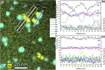
Study of the deformation microstructure Reference Heczko, Polák and Kruml14 revealed possible interactions of dislocations with nanometer-sized microstructural objects. Therefore, high spatial resolution EDS mapping was performed on a specimen cyclically loaded with the total strain amplitude Δε/2 = 2.7 × 10⁻³ (4482 cycles to fracture, i.e., 23 h at 700 °C). As shown clearly in Fig. 5, two different populations of nanometer-sized particles embedded in the matrix are revealed—particles rich in copper and particles rich in niobium. They are discussed in following sections.
(a) High spatial resolution STEM EDS map showing the distribution of Cu-rich and Nb-rich nanoparticles. (b) Line scan across two Cu-rich nanoparticles illustrating Cu atoms enrichment and depletion of main austenite matrix elements, Fe, Ni, and Cr. (c) Line scan across Nb-rich nanoprecipitate showing strong Nb EDS peak.

1. Cu-rich nanoparticles
After cyclic loading, the spherically shaped copper-rich particles are numerous, spatially dense, and homogeneously distributed in the matrix. They were previously observed in several austenitic stainless steels. Reference Tohyama, Minami, Viswanathan and Nutting25,Reference Jiang and Zhu26 In the present study, a combination of both atomic resolution Super-X EDS mapping and STEM-HAADF imaging (using low camera length 73 mm, with inner-angle cut-off 55 mrad and outer angle cut-off 370 mrad, to enhance Z-contrast) was used for characterization of Cu-rich particles.
First, the initial state of the material was inspected by an EDS analysis. Raw data were extracted from the original spectral maps and quantified using the Bruker Esprit software and the Cliff–Lorimer k-factor fit. No Cu-rich phase has been detected. Quantified maps showed only a homogeneous, Cu-enriched solid solution with an average of approximately 3.4 at.%. This result is fully in agreement with the work reported by Chi et al., Reference Chi, Yu, Dong, Liu, Cheng, Liu and Xie27 who used 3D atom probe tomography (APT) for compositional analysis of the Cu-phase precipitation. After solid solution treatment, all Cu atoms dissolved in the austenitic matrix and formed a supersaturated solid solution. It is also noted that a similar distribution and density of particles were also found in samples extracted from the grip section of the same fatigue specimen, indicating that the Cu-rich particles are resulting from a thermally induced decomposition of the austenite matrix.
In Fig. 5(b), an EDS line scan across two Cu-rich particles located close together is shown. The composition (in at.%) is plotted as a function of the distance. It is evident, that after 23 h of exposure at 700 °C and superimposed cycling, spherical regions are formed. They are enriched in copper while depleted in the other elements such as Fe, Cr, and Ni. Further EDS analysis in the matrix revealed about 1.8 at.% of Cu, i.e., notable depletion when compared with the initial state. This again corresponds closely to the APT results of Chi et al. Reference Chi, Yu, Dong, Liu, Cheng, Liu and Xie27 Increase of temperature to 700 °C and an apparently high chemical driving force in the supersaturated solid solution induces a rapid diffusion of copper atoms into Cu-rich regions. As the aging time continues, the base matrix elements (Fe, Cr, Ni) diffuse away, and the Cu-rich nanoprecipitates are formed.
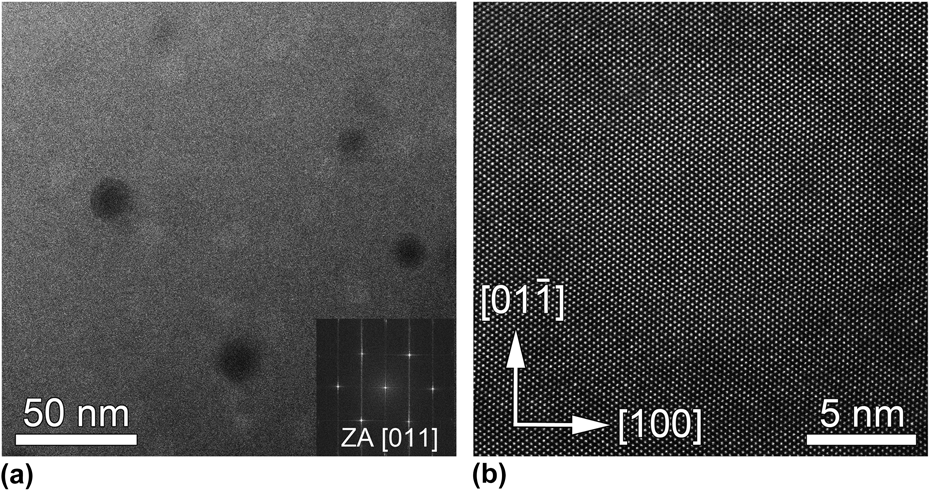
Probe-corrected STEM HAADF imaging was used to obtain crystal structure characteristics. As shown in Fig. 6(a), two types of round objects are observed—with darker and with a brighter contrast relative to the matrix. Further analysis has shown that the dark contrast objects are cone-shaped holes which were previously occupied by Cu-phase particles located close to the surface but were electrochemically attacked and removed during the sample preparation. The bright contrast objects of a similar size scale are the Cu-rich nanoparticles remaining embedded in the matrix. In HAADF Z-contrast imaging, the intensity of atomic columns directly reflects their mean square atomic number (Z). Since Cu has a higher atomic number (29) than Cr (24), Fe (26), and Ni (28), Cu-rich regions have a slightly brighter contrast than the matrix. By counting only cone-shaped holes and dividing that number by two (accounting for holes at the top and at the bottom of the TEM foil), the surface areal density of particles was determined to 3.2 × 10¹⁴ 1/m2. Incorporated EDS mapping and measurement of foil thickness by EELS also enabled determination of the particle volume density, which was about (1.1 ± 0.4) × 1022 1/m3. The average size of particles after 23 h of aging at 700 °C was 7.4 nm, median was 7.0 nm, minimum and maximum were 4.6 and 15.0 nm, respectively.
[011] zone axis STEM HAADF Z-contrast image showing (a) comparison of Cu-rich precipitates (bright contrast) and cone-shaped holes in the matrix where precipitates were electropolished out (dark contrast). (b) Atomic resolution image of coherent Cu-rich nanoparticle embedded in the matrix. Region enriched in copper atoms is brighter than surrounding austenite matrix.

It was reported earlier
Reference Jiang and Zhu26,Reference Chi, Yu, Dong, Liu, Cheng, Liu and Xie27
that in conventional steels Cu-rich nanoparticles are semicoherent with the matrix, since misfit dislocations or ledges were found at the interface. However, in the present work, no discontinuities in atomic column spacing across the precipitate/matrix interface were observed. Pure copper has the same cubic
 $Fm\bar 3m$
(space group 225) crystallographic structure as the austenitic matrix with a lattice parameter a
Cu = 3.615 Å (JCPDS 04-0836). We have used neutron powder diffraction on the instrument MEREDIT@NPI for precise measurement of the Sanicro 25 austenitic matrix lattice parameter in the initial state prior to high-temperature exposure. It was evaluated to be a
γ = 3.60692(8) Å, which is slightly larger when compared to conventional steels.
Reference Jiang and Zhu26,Reference Chi, Yu, Dong, Liu, Cheng, Liu and Xie27
Interestingly, in highly alloyed Sanicro 25 steel, pure copper would have a small positive lattice misfit of only δ = 0.22%, which is apparently sufficiently small to enable perfect coherency between particles and the matrix that has been observed in the present study.
2. Nb-rich nanoparticles
Nb-rich nanoparticles were found after cyclic loading with Δε/2 = 2.7 × 10⁻³ at 700 °C [23 h of aging, see Fig. 5(a)]. They are smaller than the Cu-rich precipitates with an average size of about 5.1 nm. The median was also 5.1 nm, and the minimum and maximum diameters were 3.8 and 6.7 nm, respectively. Contrary to the Cu-rich nanoparticles, their distribution is notably inhomogeneous. They are predominantly found in aggregates of a few particles gathered together and are very rarely observed as isolated separate objects. Furthermore, in many cases they are arranged along dislocation lines, as will be discussed further.
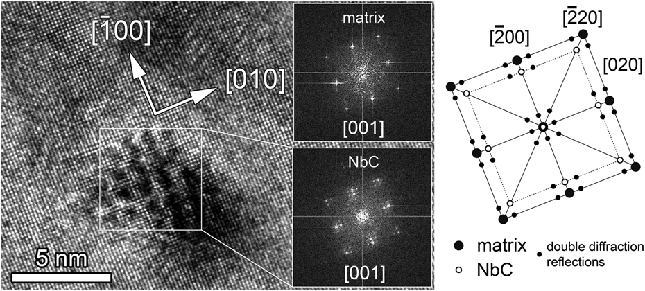
Areas with strong Nb content detected by EDS [as shown e.g., in the line scan in Fig. 5(c)] were analyzed using atomic resolution HR-TEM and STEM. In HR-TEM, for both [001] and [011] zone axes distinctive Moiré fringes were found at assumed nanoparticle locations.
Moiré fringes are parallel lines formed by the interference of diffracting crystal lattice planes from two overlapping crystals, which generally might have different spacing and orientation. Reference Poddar, Cizek, Beladi and Hodgson28,Reference Williams and Carter29 Shown in Fig. 7 is a [001] zone axis HR-TEM phase contrast image showing the Moiré fringes and the corresponding FFT obtained from the particle region. Along with the intense FFT spots from the austenite matrix, weaker satellite reflections are also observed belonging to a second lattice, accompanied by double diffraction reflections. The patterns indicate that the precipitates have an FCC crystal structure oriented in a cube-on-cube relationship with the austenite matrix.
HR-TEM image of the NbC particle viewed along the [001] zone axis. Comparison of FFT diffraction patterns from NbC precipitate and the matrix. The image is complemented by a schematic drawing showing the difference between the spacing of spots corresponding to both crystal lattices.

As described by the kinematical theory, in case of semicoherent or incoherent misfitting precipitates, a waveform in the lattice spacing could potentially generate both positive- and negative-displacement satellite reflections in the diffraction pattern with an offset based upon the planar periodicity. Reference Williams and Carter29,Reference Carroll and Carroll30 The FFTs of both HR-TEM (Fig. 7) and STEM HAADF images of the precipitate demonstrate that these satellites result from the presence of nanoparticles, as a similar strict periodicity exists in the particle/matrix interface due to the positive lattice misfit.
Based on the conclusion that these are in fact Moiré fringes, the lattice plane misfit can be measured by use of the following equation Reference Carroll and Carroll30–Reference Edington32 for the period of modulation, λm:
 $${\lambda _{\rm{m}}} = {{ha} \over {{h^2} + {k^2} + {l^2}}}{G \over {\Delta G}}\quad ,$$
where a is the lattice parameter, G is the distance of the (hkl) spot from the (000) spot, and ΔG is the distance of a satellite spot from the main (hkl) spot. Solving for λm for measured reciprocal values from a {002}-type of reflections reveals a waveform periodicity of ∼1.22. Thus, the lattice parameter of the precipitate is 1.22 times larger than the lattice parameter of the matrix, indicating a positive misfit of about 22%. Using both the lattice parameter of the austenitic matrix and this matrix/precipitate lattice misfit, the precipitate lattice constant is estimated to be about 4.41 Å.
Thus, the prominent features of these precipitates—(a) Nb-rich, (b) observation of Moiré fringes, and (c) corresponding FFT diffraction patterns indicating the FCC structure with a cube-on-cube orientation and a positive lattice misfit—are consistent with the results published earlier by Sourmail Reference Sourmail16 and Haddrill et al. Reference Haddrill, Youngerand and Baker33 In the Fe–Ni–Cr-based steels with a high carbon and nitrogen content, they report a strong tendency for nucleation of NbX (X = C, N) precipitates since Nb has a high affinity for both C and N. Possible phases have a NaCl FCC structure with lattice parameters of NbN and NbC a = 4.4 Å and a = 4.47 Å, respectively. Reference Sourmail16 To identify which type of nanoparticles is present in Sanicro 25, the EELS analysis was used for the detection of light elements. While carbon was verified in the regions of the Nb-rich nanoprecipitates, no nitrogen was found. Nucleation of the NbN phase is presumably less probable since the coarse Z-phase (CrNbN) primary precipitates probably consume most of the available N. Therefore, the experimental evidence suggests the presence of NbC nanoparticles.
Several authors Reference Poddar, Cizek, Beladi and Hodgson28,Reference Carroll and Carroll30,Reference Zhang, Hu, Tu, Schmauder and Wu34,Reference Solenthaler, Ramesh, Uggowitzer and Spolenak35 describe NbC nanoparticles as coherent or semicoherent. Nevertheless, if both the lattice misfit and particle size are taken in account, coherency with the matrix is arguable. No misfit dislocations at the particle–matrix interface, which might be able to accommodate significant lattice misfit, were observed in this work.
In a closely related study presently in progress, a STEM HAADF image simulation based on the model of an incoherent NbC nanoparticle with austenite matrix was performed to validate the structure of nanoprecipitates and the observed Moiré-like contrast. Modeling and simulation has shown excellent agreement with experimental results.
The evolution of strain-induced NbC precipitates in a model austenitic steel compressed at high temperatures was investigated by Poddar et al. Reference Poddar, Cizek, Beladi and Hodgson28 Depending on the length of hold times interrupting high temperature uniaxial straining, different stages of particle nucleation were observed. Longer hold times led to well-defined, facetted precipitates about 10 nanometers in size, while after short interruptions of less than 3 s substoichiometric, cluster-like nanoparticles about 5 nm of diameter were observed.
To explore how the precipitation of NbC nanoparticles is affected by high-temperature cyclic loading compared with static (thermal exposure only), the grip part of the specimen was studied and results were compared to observations from the gage section. A comparison of typical substructure features is shown in Fig. 8. In both cases, dislocations and stacking faults are covered by nucleated NbC precipitates. In Fig. 8(a) and in detail in 8b, nanoparticles approximately 6 nm in size are densely configured along a dislocation. At high magnification, a distinctive Moiré-like pattern is observed. The same arrangements of NbC precipitates were found also in the grip part of specimen. However, in the latter case, they are much larger with diameters up to about 20 nm, and they are detected exclusively on lattice defects as illustrated in Fig. 8(c). A pile-up of 1/2〈101〉 edge dislocations is covered by a high density of NbC particles. Relative to the grip section results, another important difference is that, aggregates of small NbC nanoparticles are found in the cyclically strained gage section, which are not decorating dislocations. However, considering their clustered arrangement, it is hypothesized that these small groups of nanoparticles remain after dislocations detached from them under cyclic loading conditions. Their dispersion is inhomogeneous when compared to Cu-rich precipitates. However, they can act as obstacles for subsequent dislocation movements. A typical example of pinning interaction with a moving dislocation is shown in Fig. 8(d). The STEM HAADF image with enhanced strain contrast (long camera length of 230 mm used) of dislocation lines illustrates pinning of a 1/2
 $\left[ {01\bar 1} \right]$
mobile edge dislocation by two NbC precipitates of sizes about 6 nm.
STEM HAADF image showing (a) NbC particles nucleated on the dislocation viewed along [001] zone axis; (b) detail of Moiré-like pattern observed in locations where NbC particles are present; (c) bright field image of NbC nanoprecipitates nucleated at edge dislocations in grip part of the specimen; (d) [011] zone axis STEM HAADF image with enhanced strain contrast (a large camera length 230 mm was used) showing two NbC particles (marked by black arrows) pinning 1/2
 $\left[ {01\bar 1} \right]$
edge dislocation. A distinctive bowing of the dislocation line between the particles is noted. White bars drawn in the image show traces of selected crystallographic planes projected into the image plane. White arrows are then traces of crystallographic directions projected into the image plane.

Because of large positive misfit, it is further surmised that precipitation of NbC particles in the defect-free austenite matrix is extremely difficult. However, nucleation clearly does occur at lattice defects, where part of the strain energy can be released by replacement of the dislocation line length and strain field with the precipitate nucleus. Reference Sourmail16,Reference Poddar, Cizek, Beladi and Hodgson28 This also helps to overcome the nucleation barrier and enables the existence of nanoprecipitates as incoherent particles in the austenite matrix.
IV. DISCUSSION
The initial condition of the Sanicro 25 alloy, i.e., the chemical composition and microstructural state, plays a fundamental role in the evolution of the deformation microstructure and corresponding mechanical response during cyclic loading at high temperatures. The present work has shown that a high number and content of alloying elements in Sanicro 25 leads to a unusual cyclic behavior, not observed in the case of conventional stainless steels. Reference Obrtlík, Kruml and Polák18,Reference Gerland, Alain, Ait Saadi and Mendez22 Fatigue at 700 °C facilitates diffusion and precipitation effects resulting in the nucleation of two distinctive populations of nanoparticles embedded in the matrix—particles rich in copper and nanosized NbC carbides.
To discuss how significant the influence of these nanoparticles on the extraordinary cyclic hardening is at high temperatures, it is necessary to outline all possible strengthening mechanisms that may be acting. In each cycle, the overall yield stress is determined by several components. First, grain boundary strengthening should be considered. The average grain size of the Sanicro 25 is 60 μm. Since this grain size is relatively large, then, according to the Hall–Petch equation, grain effects can be considered as negligibly small. As was discussed previously, Sanicro 25 presents a higher content of W, Co, N, and Nb. Therefore, solid solution strengthening might be more pronounced. Some conventional austenitic steels exhibit a solute segregation to dislocations causing pinning of their movement. These effects are mostly observed around temperatures close to 400 °C. Reference Gerland, Alain, Ait Saadi and Mendez22,Reference Pham and Holdsworth36 However, Sanicro 25 was tested at 700 °C, and no evidence of solute atmosphere formation via an EDS analysis and/or locking effects as exhibited by dynamic strain aging has been found so far. The third component to be considered is the stress corresponding to the lattice friction. This contribution usually plays an important role at room temperatures but should be negligible at high temperatures.
In the present case, the mutual interaction of two remaining strengthening mechanisms, forest dislocation hardening and precipitation strengthening, appear to play a fundamental role in the high-temperature cyclic loading. The dislocation density is substantially increased in cycling with all strain amplitudes. Reference Heczko, Polák and Kruml14 Both the cross-slip and climb of dislocations are thermally enhanced, with dislocations gradually filling the whole volume of the grain. As the dislocation density rises with each cycle, movement and mutual dislocation–dislocation interactions become more complex and important. Based on the Taylor relation, dislocation forest hardening then can contribute significantly to an increase of the cyclic stress.
The mode of NbC carbide precipitation is profoundly affected by plastic deformation. The application of cyclic strain during testing introduces the necessary density and configuration of matrix dislocations. The increased, homogeneous distribution of dislocations within the grains then causes a significant acceleration in the nucleation, growth, and coarsening of precipitates, possibly aided by the accelerated pipe diffusion of solute elements through dislocation cores. Reference Poddar, Cizek, Beladi and Hodgson28 Despite the continuous cyclic tension-compression loading, cluster-like, incoherent NbC nanoparticles rapidly nucleate on some dislocations since the time needed for their precipitation is short. Dislocations are then prevented from further movement as a result of extensive pinning by the dispersion of NbC particles nucleated on them [see Figs. 8(a) and 8(b)].
As was shown in Fig. 8(d), not all of the existing particles are decorating dislocations, indicating that it is possible for dislocations to break away from the NbC precipitates. Unpinned dislocations will leave behind a nonuniform distribution of nanoparticles demarking the former positions of the dislocation lines. Once mobilized, the dislocations will nonetheless experience a dense array of preexisting precipitates that retards their motion and makes likely the subsequent arrest and additional heterogeneous nucleation of additional NbC particles along the dislocation lines. Provided a sufficient source of free Nb and C in the solid solution, it is possible that this process could repeat numerous times, on an individual dislocation basis, and lead to continually increasing density of NbC obstacles in the microstructure as a function cyclic loading.
Based on the present post-mortem STEM-DCI observations, these particles are smaller than precipitates found gathered along completely pinned dislocation lines (e.g., in the sample after only thermal annealing). This further suggests that once detached from dislocations, those particles do not coarsen significantly. However, they can act as obstacles for further dislocation movement [see Fig. 8(d)]. Being incoherent and in aggregated configurations, they are difficult to be overcome by dislocations even when climb bypass is active. Indeed, these particles appear to present attractive interactions with dislocations [e.g., Fig. 8(d)], analogous to the potent high-temperature strengthening that can be achieved by incoherent particles in oxide dispersion strengthened alloys. Reference Rösler and Arzt37
A third synergistic effect contributing to the dynamic strengthening is the precipitation of fully coherent Cu-rich nanoparticles uniformly distributed in the matrix with an extremely high number density. As they are coherent with the matrix, gliding dislocations can readily cut them. It is interesting to note that pure copper is softer than austenitic matrix, with a shear modulus approximately half that of conventional austenitic steels. As a consequence, it might be expected that they can provide attractive sites (in terms of elastic strain energy) for shearing dislocations. A more quantitative description of this interaction is presently in progress.
The permanent increase in stress levels with the number of cycles in fatigue of Sanicro 25 differs from the cyclic response observed for standard austenitic stainless steels. In these solid-solution-hardened alloys, a so called “plateau” cyclic stress level behavior occurs and is connected with the development of the dislocation cell structure as observed in conventional 304 and 316 steels at 650 °C. Reference Carroll and Carroll30,Reference Kesternich38,Reference Challenger and Moteff39 As a consequence of thermally enhanced cross-slip and climb, increased dislocation–dislocation interactions lead to localized reordering and formation of tangles and later dislocation walls. With further cycling, walls evolve into well-developed low-energy configurations called dislocation cells. Reference Mughrabi4,Reference Gerland, Alain, Ait Saadi and Mendez22,Reference Pham, Solenthaler, Janssens and Holdsworth23,Reference Carroll and Carroll30,Reference Bressers and Steen40 In Sanicro 25, a distinctly different dislocation structure develops, with a remarkably homogeneous high-density distribution of dislocations. This general substructure characteristic suggests that dislocations are fundamentally controlled more by their mobility than by interdislocation forces (leading eventually to dislocation cell development). The intensive precipitation of the coherent, numerous, and spatially dense Cu-rich phase along with incoherent NbC nanoparticles identified in this work is expected to lead to significant retardation of the dislocation movement. The pinning effects and associated obstacles to the dislocation motion prevent normal recovery processes and the conventional development of low-energy cellular structures. As a consequence, this alloy exhibits extensive cyclic hardening, even at elevated temperatures.
V. CONCLUSIONS
The study of the origin of the extraordinary hardening of the highly alloyed austenitic stainless steel Sanicro 25 during cyclic loading at 700 °C by use of advanced scanning transmission electron microscopy leads to the following conclusions:
-
(1) Cu-rich nanoparticles precipitate homogeneously with high number density in the matrix as a result of elevated temperature. They have an FCC structure and are coherent with the austenitic matrix with a positive lattice misfit of only 0.22%.
-
(2) Elevated temperature along with the high dislocation density facilitates the rapid precipitation of NbC nanoparticles on dislocations and stacking faults. Larger particles are found along dislocation lines, smaller precipitates in aggregated groups from which dislocations have apparently detached as a result of cyclic stress. These incoherent NbC precipitates in the matrix can act as effective obstacles for the dislocation movement at a high temperature.
-
(3) The nanoparticles act as obstacles to the dislocation motion and prevent reordering of dislocations. Low-energy cellular structures are not formed, and the homogeneous high dislocation density structure is characterized by a high amount of dislocation–dislocation interactions. As a consequence, this material exhibits extraordinary cyclic hardening leading to the remarkably high saturated cyclic stress at 700 °C.
ACKNOWLEDGMENTS
The present work was conducted in the frame of IPMinfra supported through the project No. LM2015069 and the project CEITEC 2020 No. LQ1601 of MEYS. The support by the project RVO: 68081723 and grant 13-23652S of the Grant Agency of the Czech Republic is gratefully acknowledged. MJM acknowledges the support of the National Science Foundation under contract #DMR-60050072. BDE acknowledges support from the Center for Emergent Materials: an NSF MRSEC under award number DMR-1420451. The support of the Fulbright Fellowship grant awarded to Milan Heczko by The J. William Fulbright Commission in the Czech Republic is gratefully acknowledged.