Stem cells are defined by their ability to give rise to new stem cells (self-renewal) and to differentiate into one or more mature specialised cells of a tissue. There are two types of stem cells: embryonic stem cells and adult stem cells. In vivo, embryonic stem cells constitute the inner cell mass of an early-stage embryo and undergo complete differentiation into the three germ layers within a few days; when isolated from early embryos they possess extended proliferative capacity and the potential to differentiate into all tissues derived from the three germ layers (Refs Reference Thomson1, Reference Reubinoff2). Adult stem cells have been isolated from various tissues – including bone marrow (Ref. Reference Weissman3), skin (Ref. Reference Watt4), gut (Refs Reference Whitehead5, Reference Potten6), muscle (Ref. Reference Wagers and Conboy7), brain (Ref. Reference Gage8), liver (Ref. Reference Alison9), breast (Ref. Reference Clarke10) and pancreas (Ref. Reference Lechner and Habener11) – but their capacity for expansion and multilineage differentiation is thought to be more limited. Adult stem cells are thought to function in maintenance and repair of tissues throughout the lifespan of the individual. Indeed, some adult tissues require extensive proliferation and tissue replacement to survive: in physiological conditions the entire intestinal lining fully replaces itself weekly and the bone marrow produces 108 cells daily.
Adult stem cells and self-renewal
The process by which stem cells give rise to terminally differentiated cells occurs through a variety of committed progenitor cells (or transient amplifying cells), often overlapping in their differentiation capacity. During commitment, stem cells can undergo extensive proliferation and sequential differentiation, accompanied by a decrease in self-renewal capability to produce mature cells. The primary function of this transit population is to increase the number of mature cells produced by each stem cell division.
Self-renewal is defined by a cell division where at least one of the daughter cells retains the identical developmental potential as the mother cell. In principle, this property allows an identical stem cell pool to be maintained throughout the life of the individual, and thus provision of differentiated cells to repair the losses occurring in tissues with time (and hence maintenance of tissue function). Maintenance of the stem cell pool is achieved only if stem cell self-renewal occurs without a change in the genome during DNA replication. This guarantees preservation of the ability to generate the same types and numbers of differentiated progenitors. For decades it has been a common belief that the ability of stem cells to self-renew was, if not indefinite, very extended and that the stem cell pool was maintained for long periods of time. This was based on early experiments where haematopoietic stem cells (HSCs) were shown to be able to serially repopulate up to five mice (Ref. Reference Siminovitch, Till and McCulloch12), suggesting that the few transplanted HSCs were able to undergo extensive self-renewal. In support of this was the concept of asymmetric cell division and the immortal strand formulated by John Cairns in 1975. This proposed that during division adult stem cells would be able to distinguish and segregate the parental chromatids from the newly synthesised ones and retain the parental strands; in this way stem cell DNA would not bear errors that may occur during DNA replication (Refs 13, 14 and references therein).
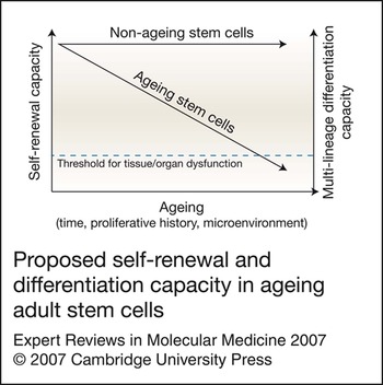
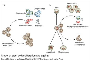
However, despite the extensive self-renewal property of adult stem cells, more recently the notion of stem cell ageing has emerged. Data suggest that with age the process of self-renewal and differentiation are affected, resulting in the generation of differentiated progeny but in fewer numbers or in the generation of progeny skewed towards one particular lineage (Refs Reference Wright15, Reference Rossi16) (Figs 1 and 2). Asymmetric cell division has been observed in a very limited number of cell types and the experimental evidence is weak (Ref. Reference Lansdorp14). By contrast, more convincing evidence have been recently presented suggesting that there is an accumulation of changes in the DNA of stem cells with time, which may affect their functionality (Ref. Reference Rossi17). If DNA changes reach a certain threshold, stem cells may undergo apoptosis or senescence, with increased cellular losses and decreased stem cell numbers. Alternatively, stem cells with DNA changes may escape senescence and have an increased risk of undergoing clonal change (Fig. 2).
Proposed self-renewal and differentiation capacity in ageing adult stem cells. If stem cells age, self-renewal capacity and/or potential for multilineage differentiation may be reduced over time and/or replication cycles. A threshold may then be reached that is inconsistent with efficient normal tissue or organ function.

Model of stem cell proliferation and ageing. (a) Young stem cells – depicted here as haematopoietic stem cells – undergo self-renewal, which allows maintenance of the stem cell pool and production of all the differentiated effector cells required. (b) The accumulation of mutations in the DNA with age leads to an increased loss of stem cells and decreased ability to produce new stem cells, with a net decrease in stem cell number. Moreover, stem cells lose their ability to differentiate into all lineages. Increased cellular loss occurs by apoptosis or senescence when cells accumulate mutations in the DNA to a critical level. Such mutated stem cells that escape apoptosis or senescence become at risk of malignant transformation.

Although evidence for some of these processes is well described, others remain more hypothetical. Moreover, it is unclear whether these changes occur in stem cells as part of a physiological process or only in circumstances of extreme stress, and whether it is the result of a programme intrinsic to stem cells or of changes in the microenvironment in which they reside. It is also a matter of debate whether it is in any way related or causal to the ageing of tissues and eventually loss of their function or alternatively may predispose to development of malignancy. In this review, we discuss the literature available in these areas.
Mechanisms of ageing
Ageing is seen as a side effect of mechanisms that are in place to limit carcinogenesis (Refs Reference Campisi18, Reference Sharpless and DePinho19, Reference Pelicci20). Cells are provided with two defence mechanisms to limit cell proliferation and protect themselves from transformation when exposed to inappropriate stimuli: senescence and apoptosis. While the molecular mechanisms that limit cell proliferation by cell loss through apoptosis are well understood, the contribution of senescence to the ageing processes remains unclear. Moreover, the diverse signalling pathways that cause cells to choose between apoptosis and senescence are controversial (reviewed in Ref. Reference Gil and Peters21). This is mainly because of the lack of reliable markers for the identification of senescent cells.
It is still controversial whether senescence occurs in vivo or only in vitro. Recently, evidence of in vivo senescence has been emerging based on multiple parameters. Senescent cells have been identified in ageing human skin (Ref. Reference Dimri22), and human and mouse liver (Refs Reference Paradis23, Reference Lechel24, Reference Wiemann25, Reference Satyanarayana26). Senescent cells were considered as such when they were metabolically viable but irreversibly arrested in the G1 phase of the cell cycle, had undergone morphological changes (enlarged and flattened morphology), and expressed the senescence-associated β-galactosidase, higher levels of the tumour suppressors p21CIP1/WAF1 (encoded by CDKN1A) and p16INK4a (encoded by CDKN2A) and hypophosphorylated retinoblastoma (Rb) protein.
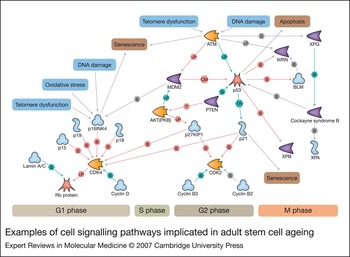
Several pathways control cell proliferation (Fig. 3), including the p16INK4a–Rb pathway, the p19ARF–p53 pathway and the p53–p21CIP1/WAF1 pathway (hereafter named as p16, p19 and p21) (reviewed in Refs Reference Sharpless and DePinho27, Reference Kim and Sharpless28). These pathways can be activated by telomere shortening or dysfunction (Ref. Reference Sharpless and DePinho27) or by a variety of stresses such as DNA damage and oncogenic insults (Ref. Reference Serrano29). More recently, the PTEN–p27Kip1 (hereafter named p27) pathway has also emerged as important in controlling cell proliferation (Ref. Reference Maehama and Dixon30; reviewed in Ref. Reference Di Cristofano and Pandolfi31). It is beyond the scope of this review to fully cover developments in this field, but the most important aspects in relation to stem cells are summarised here.
Examples of cell signalling pathways implicated in adult stem cell ageing. The figure shows possible relationships between cellular processes such as oxidative stress and telomere dysfunction via signalling pathways through to outcomes such as cellular senescence, apoptosis and cell cycle control. Pathway analysis was carried out using using Metacore MapEditor (GeneGo Inc: http://www.genego.com). The signalling pathways include gene products now associated with cellular senescence (p21, p27 and p16), and possible links to genes and proteins implicated in progeroid syndromes [e.g. Werner syndrome (WRN), Cockayne syndrome B; see Table 1] and stem cell ageing (see Table 2) are indicated. Red arrows show inhibitory effects; turquoise arrows show activation; grey arrows indicate unknown effect of interaction. Abbreviations: B, binding event; CM, covalent modification; P, phosphorylation event; TR, transcription regulation. Details on individual genes can be found at the GeneCards website: http://www.genecards.org/index.shtml.

Replicative senescence was first described by Hayflick in 1961 (Ref. Reference Hayflick and Moorhead32). The biological clock underlying the limited potential of somatic cells is the length of telomeres (the repeated DNA sequence at the end of each chromosome). Telomeres provide stability to the chromosomes and protect them against DNA loss associated with cell division (Ref. Reference Blackburn33). Telomeres progressively shorten in all somatic cells at each cell division (Refs Reference Harley, Futcher and Greider34, Reference de Lange35). When telomeres reach a critical short length, cells undergo replicative senescence (Refs Reference Hayflick and Moorhead32, Reference Harley, Futcher and Greider34). Telomere length can be maintained by the enzyme telomerase (Ref. Reference Geserick and Blasco36), but most adult somatic cells lack this enzyme. Stem cells, T cells and germ cells express it at different levels.
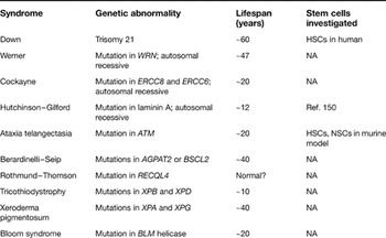
An alternative mechanism to replicative senescence is stress-induced senescence, which is independent of telomere length (Refs Reference Sherr and DePinho37, Reference Drayton and Peters38). It can be induced by telomere dysfunction [which can occur when telomeres undergo a change of state (uncapping) or structure or DNA sequence], or as a result of DNA damage, oncogenic insults, and oxidative damage to cellular constituents leading to accumulation of abnormal proteins, lipid and macromolecule. The importance of these mechanisms in ageing are illustrated by progeroid (premature ageing) syndromes, most of which are due to single gene defects in one of the DNA repair enzymes or structural proteins of the nuclear membrane (Table 1).
Progeroid syndromes and the investigation of stem cell ageinga

aFor reviews on progeroid syndromes, see Refs 151 and 152.
Abbreviations: HSC, haematopoietic stem cell; NA, not applicable; NSC, neural stem cell. Information on genes mentioned in the table can be found at http://www.genecards.org/index.shtml.
Replicative senescence occurs mainly via the p21 pathway, and stress-induced senescence via p16 and/or p19 (Refs Reference Kim and Sharpless28, Reference Chin39). Interestingly, both p16 and p19 pathways are controlled by a transcription factor repressor of the Polycomb group gene family called BMI-1, which has been shown to be important in the self-renewal of neural stem cells and HSCs (Refs Reference Molofsky40, Reference Iwama41, Reference Park42). BMI-1 induces cell progression into S phase of the cell cycle by blocking p16 and p19 (Refs Reference Jacobs43, Reference Itahana44). BMI-1 was recently shown to be a direct transcriptional target of MYC and Sonic Hedgehog (SHH), which are also important in stem cell self-renewal. Decreased MYC led to decreased BMI-1, increasing the probability of switching on p16 (Ref. Reference Guney, Wu and Sedivy45). Moreover, SHH was shown to control proliferation through BMI-1-mediated repression of p16 and p19 (Ref. Reference Bruggeman46).
PTEN (phosphatase and tensin homologue) is a phosphatase that negatively regulates phosphoinositide 3-kinase (PI3K) signalling (Ref. Reference Cantley and Neel47; reviewed in Ref. Reference Stiles48). PTEN thus inhibits growth factors signals that act through PI3K. Among the downstream signalling molecules, AKT is the most characterised. This in turns acts on cell cycle regulators p27 and p21 and induction of senescence (Fig. 3). The PTEN–PI3K/AKT signalling pathway also interacts with signalling pathways known to be essential for stem cell self-renewal, including the transforming growth factor (TGF)-β–SMAD pathway and the WNT/β-catenin pathway.
Detecting ageing in stem cells
To define whether stem cells undergo ageing, suitable methods to assess stem cell numbers and function are required. Moreover, as any ageing phenotype develops over time, suitable models that allow monitoring of stem cell numbers and function for a long period of time need to be identified. However, this is difficult to achieve because of technical difficulties in the identification of stem cells and differences in ageing mechanisms in different species, particularly mouse and human. It is important to understand the limitations of the assays and models available to be able of interpret the often contrasting data. To date, the majority of data available on ageing in stem cells are in relation to HSCs. Limited data are available in other stem cell systems, such as epithelial stem cells, neural stem cells, satellite cells and mesenchymal stem cells.
Identification of stem cells
The identification, isolation and enumeration of stem cells are elusive. Identification of stem cells is carried out by immunophenotype, by their ability to retain a dye due to their relative quiescent state or by their location, such as in the case of epithelial stem cells in the bulge and basal lamina or intestinal stem cells in the crypt. However, none of those methods is able to identify stem cells uniquely and to differentiate them from the transient amplifying cells. HSCs are the most extensively characterised type of stem cells (reviewed in Ref. Reference Kondo49).
Several markers have been proposed for HSCs, such as CD34. CD34 is expressed on 0.5–5.0% of human bone marrow cells, is found on early haematopoietic progenitor cells but not on their mature counterparts, and has been used to provide cells that achieve clinical engraftment following transplantation (Refs Reference Civin50, Reference Brugger51). Other surface markers have been used in the laboratory in association with CD34 to identify more-primitive populations of HSCs, such as CD34+CD38low cells (Refs Reference Bhatia52, Reference Glimm53). However, even with the most rigorous combination of markers only a small percentage of enriched cells are bona fide stem cells, which replicate infrequently but have the capacity of extended growth and when transplanted are responsible for long-term engraftment (Refs Reference Kondo49, Reference Lajtha54, Reference Barrandon and Green55, Reference Morrison56). The majority of the cells are progenitor (or transient amplifying) cells, which replicate frequently but cycle only a limited number of times before undergoing terminal differentiation, and on transplantation provide at best only short-term engraftment (Refs Reference Lajtha54, Reference Barrandon and Green55, Reference Morrison56, Reference Lavker57).
Other stem cells types are even less characterised in terms of markers. Although knowledge of epithelial stem cells and satellite cells has advanced at a fast pace in the past five years, prospective isolation is still in its infancy and is based on a combination of markers such as presence of the marker CD34, ability to retain bromodeoxyuridine (5-bromo-2-deoxyuridine; BrdU) over time, integrin expression levels for epithelial stem cells (Ref. Reference Tumbar58), and properties of size and granularity for satellite cells (Ref. Reference Wagers and Conboy7). This results at best only in enrichment for multipotent epithelial or satellite cells. Neural stem cells and mesenchymal stem cells are isolated by selective culture conditions. Very little is known about markers to isolate and enrich these stem cell types (Refs Reference Tamaki59, Reference Quirici60, Reference Bensidhoum61, Reference Dennis62). More-detailed studies employing prospective isolation and clonal marking are required to define the precise lineage progression of these stem cell populations.
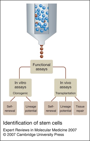
The alternative to enumeration of stem cells is to rely on a range of functional in vitro and in vivo assays (Fig. 4). These have been well developed over the last 20–30 years in an attempt to identify HSCs. In vitro assays assess cells based on their proliferative capacity and retention of multipotent differentiation. In vivo assays assess the ability of stem cells to repopulate tissues with cells of multiple lineages in a host depleted of stem cells (e.g. lethally irradiated in the case of HSCs) before transplantation. Transplantation of limiting dilution numbers of stem cells in a competitive repopulating assay allows a quantitation of the stem-cell-repopulating unit present in a transplanted population. The analysis of the types and numbers of progenitors arising following transplantation inform on the multipotential capacity of the transplanted stem cells.
Identification of stem cells. Cell-surface markers can be used to enrich for rare stem cells (red) within populations. However, functional assays are then required to confirm stem cell properties of self-renewal and lineage commitment (in vitro and in vivo assays). In vitro assays are based on the extent of the clonogenic capacity of the progenitor cells and their multilineage differentiation. In vivo assays are based on transplantation and assessment of tissue reconstitution.

In the past it was thought that some in vitro assays that tested the ability of a putative stem cell population to proliferate for extended periods of time were surrogate assays for the in vivo transplantation assay. However, with the development of gene-marking protocols, views on the most appropriate assays to test for stem cell function have changed, at least for HSCs. Initially, in vitro assays such as long-term culture initiating cell (LTC-IC) (Ref. Reference Szilvassy63) and high proliferative potential (HPP) cell assays (Ref. Reference McNiece64) were postulated to test the ability to generate additional stem and progenitor cells for variable periods of times. However, the generation of gene-marked LTC-IC proved very efficient, whereas transplantation of these same cells in an ablated host resulted in very low (<1%) levels of circulating, gene-marked cells. This suggested that in vitro assays detected progenitor cells or transient amplifying cells responsible at most for short-term engraftment (Refs Reference Bhatia65, Reference Larochelle66), and that a transplantation assay was the only conclusive assay for HSCs to assess their ability to give rise to cells of the lymphoid and myeloerythroid lineages.
Animal models
As the in vitro assays available for HSCs became inadequate, animal models were required. Moreover, as stem cells such as HSCs maintained their function for longer than the average human lifespan, this required models that allowed monitoring for long periods of time to assess any effect of ageing. For murine HSCs in vivo, competitive repopulation assays were performed in mice, and long-term engraftment of HSCs was assessed by secondary and tertiary transplant. However, the assessment of human HSCs was more problematic. Xenogenic hosts, particularly the immunodeficient NOD/SCID murine model (nonobese diabetic mice with severe combined immunodeficiency disease), were proposed (Ref. Reference Bonnet67). Transplantation of HSCs in these mice resulted in terminally differentiated human cells from multiple haematopoietic lineages including B cells, immature progenitor cells, mature erythrocytes and all lineages of myeloid cells, but in low and variable numbers. In these systems multilineage engraftment of human cells could be achieved only for up to 4 months in optimised systems (Refs Reference Larochelle66, Reference Larochelle68, Reference Cashman69), and secondary transplants were very difficult to achieve. Engraftment levels of baboon haematopoietic cells in NOD/SCID mice were different to engraftment levels of the same cells in baboons (Ref. Reference Horn70). In baboons, clones responsible for long-term engraftment and contributing to all lineages for nearly two years appeared only 6–8 weeks post-transplantation (Ref. Reference Schmidt71). This time is usually at the limit of the follow-up period in most studies where NOD/SCID mice have been used. Therefore, although NOD/SCID repopulating cells have been used as a measure of long-term repopulating cells, they probably more closely resemble short-term repopulating cells, leading to the conclusion that the NOD/SCID model may not be very useful for the study of human stem cell ageing.
The discrepancies between in vitro and in vivo assays found with HSCs may not apply to all stem cell types. Recent evidence from epithelial stem cells suggests that in vitro assays may be more predictive of what happens in vivo. Bulge cells form large colonies in vitro that can proliferate for extended periods of time. Holoclones are believed to have properties suggestive of stem cells in that they have a high colony-forming efficiency, and give rise to meroclones and paraclones, which have progressively less colony-forming efficiency and behave like transient amplifying cells (Ref. Reference Barrandon and Green55). Transplantation of single human holoclones gave rise to hair follicle, interfollicular epidermis, sebaceous gland and bulge stem cells in immunodeficient mice (Refs Reference Blanpain72, Reference Claudinot73) and this was long term, as shown by their ability to participate in several hair cycles and to contribute to all epithelial lineages of the hair follicle and sebaceous gland following secondary transplant (Ref. Reference Claudinot73). If these data were to be confirmed it suggests that, in contrast to HSCs, studies on the in vitro ageing of human holoclones may be informative on the changes occurring in bulge stem cells in vivo.
For other stem cell types such a relationship has not been established and transplantation studies are only at early stages (Refs Reference Collins74, Reference Collins75, Reference Ostenfeld76). For example, single myofibres with their associated satellite cells were transplanted into irradiated dystrophic mdx-nude mouse muscle. The single fibres could give rise to at least 100 new myofibres and up to tenfold more Pax-7+ satellite cells, a marker used to identify satellite cells (Ref. Reference Collins74). These cells were able to contribute to muscle repair. However, the follow up of these experiments was short (5–8 weeks), and in a subsequent study no correlation was found between the number of Pax-7+ cells present in a population and its ability to regenerate muscle (Ref. Reference Collins75), suggesting that Pax-7 may not be an appropriate marker for satellite cells and any conclusion on the ageing of satellite cells needs to be taken with caution.
These studies highlight how often data on stem cells have been based on assumptions regarding their identity that more-detailed studies subsequently revealed to be incorrect. Each individual stem cell type should be properly assessed, possibly by more than one method. Regardless of the assay used, one has to bear in mind that both in vitro and in vivo assays have drawbacks. In vitro assays may be predictive of changes in the transient amplifying population rather than the stem cell population. Transplantation assays have the problem that they relate to the future potential of stem and progenitor cells, which implies that the nature of a certain stem cell type can be deduced only retrospectively on the basis of their ability to produce cells of all lineages. Any statement based on transplantation, therefore, does not tell us anything about the behaviour of a single cell but rather about the probabilistic behaviour of a population of cells. It can underestimate frequencies because it detects only those cells that not only have sufficient self-renewal potential and multipotent differentiation but also express the homing molecules necessary to seed to the tissue of interest.
Of mice and men
As a result of the problem related to the assessment of stem cell numbers and lineage potential in humans, most of the data generated on stem cell behaviour and ageing are in murine models. Yet, when considering data generated from mice to assess ageing in stem cells some points need to be kept in mind. There is compelling evidence that mice do not use the same mechanisms as humans to regulate the maximal numbers of cell division (Refs Reference Wright and Shay77, Reference Wright and Shay78, Reference Smogorzewska and de Lange79). Laboratory mice have far longer telomeres (40–60 kb) than humans (10–15 kb), and telomerase can be expressed by differentiated rodent cells such as fibroblasts (Refs Reference Prowse and Greider80, Reference Blasco81). It is likely that human stem cells undergo a far higher proliferative stress than mice. For example, it has been calculated that the number of HSCs at birth in mice and humans is similar and therefore the number of committed and mature blood cells produced by each stem cell changes with the size of the animal (Ref. Reference Abkowitz82). As a human is about 3000 times larger than a mouse and lives about 40 times longer, even considering that the lifespan of some cell types, such as erythrocytes, is shorter in mice, the proliferative stress on each human HSC is clearly higher. Therefore, the role of telomere shortening in mice is likely not to have the same relevance as in humans. In addition, it has been shown that murine cells are much more sensitive to oxidative stress and do not repair DNA damage as efficiently (Ref. Reference Parrinello83). In murine embryo fibroblasts, the p16 and p19 pathways, mostly activated by oxidative stress, seem to be more dominant; in human cells the p21 pathway seems dominant, although the p16 pathway can also play a role (Ref. Reference Itahana, Campisi and Dimri84). Understanding these differences is essential to evaluate evidence of ageing in stem cells and to design the appropriate model to investigate it.
In this respect, the telomerase knockout mice (mTerc −/−) (Ref. Reference Blasco81) may become a very useful model. The first generation of these mice do not show any distinguishing phenotype from the wild-type littermates. However, cells from the fourth-generation mTerc −/− mice start showing significant telomere shortening and genomic instability. Cells from fifth- and sixth-generation mTerc −/− showed p21-dependent growth arrest (Ref. Reference Chin39). These mice also showed reduced lifespan, several signs of premature ageing including decreased HSC repopulation ability (Ref. Reference Samper85), and reduced capacity to respond to stresses such as following haematopoietic ablation (Refs Reference Samper85, Reference Rudolph86, Reference Lee87). It is possible that generating knockout mice on an mTerc −/− background will result in models with ageing features that better resemble what is observed in humans. This is best exemplified in ataxia telangiectasia (A-T) which results from the loss of ataxia telangiectasia mutated (ATM) function and is characterised by accelerated telomere loss, progressive neurological degeneration, premature ageing and increased neoplastic transformation. Several Atm −/− mouse strains have been generated. However, relative to ataxia telangiectasia patients they show only modest neuronal and premature ageing signs (Refs Reference Barlow88, Reference Xu89). When Atm −/− mice were crossed with successive generations of the mTerc −/− mice, the model better recapitulated several aspects of the human disease such as multisystem failure and accelerated ageing (Ref. Reference Wong90). This correlated with levels of telomere dysfunction and affected stem cell reserve. Thus, mouse cell growth arrested by telomere erosion similar to human cells may be able to give a valuable insight into the role of telomere shortening or dysfunction and human stem cell ageing.
Progeroid syndromes
The long lifespan of humans makes the study of human ageing difficult to dissect. However, progeroid syndromes – mostly single gene defects that result in accelerating ageing phenotypes (Table 1) – may provide insight into some of the mechanisms of normal ageing.
For most of these syndromes it is unknown whether stem cells from these patients are affected. However, where this has been investigated, a clear association has been established between stem cell depletion, organ failure, reduced longevity and increased risk of clonal change. Studies on ataxia telangiectasia establish the link between stem cell telomere erosion, depletion of stem cell reserve and progressive multiorgan compromise with neurological degeneration, premature ageing and early death (Ref. Reference Wong90). Down syndrome has emerged recently as an excellent model to study events leading to stem cell ageing. Down syndrome is associated with many of the signs of premature ageing including early abnormalities typical of Alzheimer disease, T-cell deficiency, increased incidence of myelodisplastic-type disease and leukaemia, premature reduction in skin elasticity, increased incidence of osteoporosis and early death (Refs Reference Epstein and Scriver91, Reference Nespoli92, Reference Rumble93, Reference Angelopoulou94, Reference Creutzig95).
By contrast to ataxia telangiectasia, where stem cell ageing is detected once organs have been compromised and the ageing process has ensued, the presence of trisomy 21 in Down syndrome allows the study of events occurring in the early phase of ageing development. Similarly to ataxia telangiectasia, the mean telomere restriction fragment (mTRF) of peripheral blood lymphocytes declines more rapidly in individuals with Down syndrome than in normal individuals, reflecting an accelerating HSC telomere shortening and thus HSC ageing (Ref. Reference Vaziri96). The accelerated telomere shortening in Down syndrome is present already in fetal peripheral blood lymphocytes (Ref. Reference Holmes97). This is associated with stem/progenitor cell deficiency as shown by a reduction in cells possessing the phenotype of haematopoietic progenitor cells and HSCs (detected as CD34+ cells) in fetal blood and bone marrow of Down syndrome children and in the number of LTC-ICs in bone marrow of children affected by Down syndrome (Ref. Reference Holmes97). It is of interest that both ataxia telangiectasia and Down syndrome patients have increased risk of developing malignancies, supporting the hypothesis that telomere dysfunction may overcome tumour suppressive mechanisms and predispose to genomic instability and cellular transformation.
Mechanisms of ageing in stem cells
Triggering signals
There are several indications that mechanisms of ageing apply to stem cells. We have already seen how critical telomere shortening has been shown to affect the self-renewal capacity of stem cells such as HSCs (Refs Reference Samper85, Reference Rudolph86). Epithelial stem cells seem also to be regulated by telomere length and telomerase. In telomerase-deficient mice, telomere shortening affects the function of epithelial stem cells. In particular, mobilisation of epithelial stem cells out of the hair follicle upon mitogen-induced proliferation is more affected when telomeres are critically short. This results in decreased proliferation of the stem cells in the bulge and decreased production of transient amplifying cells, with lack of hair growth (Ref. Reference Flores, Cayuela and Blasco98). Analysis of adult mTERC −/− mice with shortened telomeres showed impaired in vitro expansion of neurospheres (cultured cell aggregates from neural stem and progenitor cells) (Ref. Reference Ferron99).
Very recently, it has been shown that DNA damage can result in HSC exhaustion. Ercc1 knockout mice, which are defective in nucleotide excision repair, have decreased responses to haematopoietic stress and exhaustion of haematopoietic progenitor activity (Ref. Reference Prasher100). Moreover, it has been shown that increased production of reactive oxygen species (ROS) leads to bone marrow failure in Atm −/− mice (Ref. Reference Ito101).
Tumour suppressive pathways
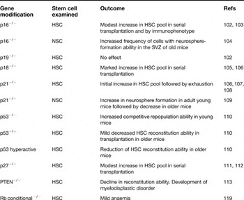
A role for tumour suppressive pathways in self-renewal has clearly been shown for murine HSCs and to a limited extent for neural stem cells (Table 2; Fig. 3). Loss of p16 but not p19 resulted in an increase, although modest, of the HSC pool as shown by serial transplantation (Refs Reference Stepanova and Sorrentino102, Reference Janzen103) and of the neural stem cell pool. An increase with age of the number of cells from the subventricular region that were capable of forming neurospheres in culture was reported, as well as an increase in their multipotent self-renewal ability (Ref. Reference Molofsky104). p18INK4c−/− (Cdkn2c −/−) mice showed a marked increase in the number of HSCs, which retained a repopulation advantage over wild-type cells in secondary transplants (Refs Reference Yuan105, Reference Yu106). Complete deficiency of p21 led to increased HSC proliferation and amplification of the size of the stem cell pool under homeostatic conditions in mice, but eventually led to exhaustion of the HSC pool through serial transplantation (Refs Reference Yu106, Reference Choudhury107, Reference Cheng108). By contrast, increased expression of p21 initiated the process of cellular senescence in response to stress. This was also seen in postnatal neural stem cells. In vitro culture of neural stem cell progenitors from p21−/− mice showed an increase in proliferation of the neural stem cell progenitors at a young age followed by a decline when neural stem cell cultures were derived from mice at 16 months of age, suggesting a similar phenomenon of exhaustion with age (Ref. Reference Kippin109).
Effects of changes in expression of tumour suppressor genes in stem cells

Abbreviations: HSC, haematopoietic stem cell; NSC, neural stem cell; SVZ, forebrain lateral ventricle subventricular zone. For gene information, refer to section ‘Mechanisms of ageing’ and http://www.genecards.org/index.shtml.
Studies on p53−/− have been more difficult to carry out because of the increased incidence of tumours in those mice and death at a young age. However, studies in young p53−/− mice showed an increased capacity of self-renewal in HSCs. Reducing p53 levels (p53−/+ mice) increased HSC proliferation with age compared with wild-type mice, whereas mice with a hyperactive p53 mutation showed a reduction in the number of proliferating HSCs with age (Ref. Reference Dumble110). Deletion of p27 resulted in an increased frequency of serially transplantable HSCs in addition to an expanded progenitor compartment (Refs Reference Walkley111, Reference Cheng112). PTEN inactivation resulted in short-term expansion of HSCs in the bone marrow but long-term decline as a result of HSCs entering the cell cycle (Ref. Reference Zhang113). Interestingly, PTEN mutant mice also developed a myeloproliferative disorder, reinforcing the notion that the factors limiting stem cell self-renewal have a role in malignant transformation. However, in the case of PTEN two independent PTEN-driven mechanisms seemed to drive HSC self-renewal and leukaemogenesis: rapamycin blocked the generation and maintenance of leukaemia-initiating cells but restored normal self-renewal properties of HSCs (Ref. Reference Zhang113).
Self-renewal genes involved in senescence pathways
Genes known to promote stem cell self-renewal have been shown to be involved in senescence pathways. It has been demonstrated that BMI-1 functions in vivo to maintain the pool of neural stem cells and HSCs by inhibiting the senescence programme through p16 (Refs Reference Molofsky40, Reference Iwama41, Reference Park42, Reference Lessard and Sauvageau114). Mice lacking Bmi-1 showed induction of both p16 and p19 in the haematopoietic and neural tissue, and double deletion of p16 and p19 partially rescued the phenotype observed in Bmi-1-deficient mice, suggesting that p16 and p19 are downstream of Bmi-1 (Refs Reference Bruggeman46, Reference Molofsky115). Similarly absence of Hoxb4, a gene of the homeobox family considered important in regulating the size of the HSC pool (Refs Reference Brun116, Reference Bjornsson117), significantly increased levels of p21 in fetal liver haematopoietic progenitors (Ref. Reference Brun116). Moreover, p21 deficiency markedly enhanced the Hoxb4-mediated increase in HSC self-renewal (Ref. Reference Miyake118).
Together, these data establish the relationship between senescence pathways and stem cell self-renewal. However, there is no strong evidence that any of the factors discussed change expression with age and thus play a role in the induction of ageing in stem cells. Most studies have been carried out in animal models with germ-line gene deletions, which resulted in a persistent defect present in all cell types. It is possible that less pronounced and prolonged changes in the expression of any of these factors, as expected with ageing, would not be sufficient to promote any significant change in stem cell function. For example, conditional somatic mutagenesis of Rb, a central player of the senescence pathway and embryonic lethal, did not result in a great defect of the HSCs in primary and secondary transplants but only in a mild anaemia (Ref. Reference Walkley and Orkin119). Thus, a careful analysis of the effect of these factors on stem cell self-renewal and ageing in physiological conditions is required. Moreover, if all cells harbour the defect a question to be addressed is whether some of the effects seen are intrinsic to the stem cell compartment or are related to the microenvironment. Indeed, transplantation of wild-type bone marrow in p27−/− mice resulted in expansion of the lymphoid compartment similar to that seen in the p27−/− animals. (Ref. Reference Walkley111), raising the possibility that the expansion of the HSC population is in part the result of a microenvironment defect (Ref. Reference Chien120).
Evidence of ageing in stem cells
These experimental limitations have shed doubts on the possibility that stem cells undergo a process of ageing. One of the main points of dispute follows the reasoning that if stem cells undergo ageing one would expect a decrease in stem cell number with age and/or a change in the number and type of progenitors produced in physiological conditions, and this has not been consistently shown. In mice, not only has a decrease in HSC numbers with an immunophenotype of the long-term repopulating cell not been observed but an absolute increase with age has been reported by several groups (Refs Reference Rossi16, Reference Sudo121, Reference Pearce122, Reference Morrison123). In other systems, estimates of changes in satellite cell number with age varied depending on the methods used, with some showing a decrease and some an increase in numbers (Refs Reference Gibson and Schultz124, Reference Conboy125, Reference Brack, Bildsoe and Hughes126).
However, such contrasting data have been generated by enumerating stem cells on the basis of their immunophenotype. Estimates of HSC numbers using transplantation assays are more reproducible. HSCs have been shown to undergo a loss of repopulation capacity with reduction in number and quality of progenitors under proliferative stress or with age in transplantation studies. In competitive repopulation assays, after the second or third serial transplant the host HSCs were shown to have a competitive advantage over the serially passaged donor cells (Ref. Reference Ogden and Mickliem127). When comparing HSCs from young and older mice, older animals showed reduced repopulation potential when compared with younger organisms. Moreover, the stem cell developmental programme in old age was restricted and was able to contribute mainly to the myeloid lineage. There was a reduction in the number of common lymphoid progenitors, and in gene expression profiling the transcription factors important for commitment to common lymphoid progenitors were downregulated in favour of those required for common myeloid progenitors commitment (Ref. Reference Rossi16). Similar data were also described with satellite cells (Ref. Reference Brack, Bildsoe and Hughes126). One idea used to reconcile the opposing evidence is that the decrease seen in stem cell activity during transplantation is due to defects in stem cell homing ability coupled with an aged microenvironment and not to a decrease in stem cell numbers. To this end it has been shown that homing efficiency of old stem cells was considerably diminished compared with stem cells of young mice (Refs Reference Morrison123, Reference Liang, Van Zant and Szilvassy128). However, this is not incompatible with the idea of an intrinsic change of the stem cell properties with age. A cell no longer fit for long-term survival could be made less competitive by its decreased homing ability. The possibility that changes in the microenvironment may also be responsible for the decreased engraftment is also not sufficient on its own. Young stem cells transplanted in an old host showed a decrease in B-cell development in the short term (Ref. Reference Rossi16). However, in long-term analysis B-cell development was found to be unaffected by the old microenvironment, suggesting that the defect in the microenvironment is minor (Ref. Reference Rossi16).
The more recent evidence that points to a role of the microenvironment as a major player in the poor regenerative capacity of satellite cells with age needs to be interpreted with caution. In young and old mice sharing a circulatory system, old muscle progenitor cells were reactivated by an unknown factor in the serum of young mice (Ref. Reference Conboy153). Although these data are very thought provoking, they require a more detailed characterisation of satellite cells as stem cells and their lineage progression before any conclusion can be drawn and generalised to all stem cells. Moreover, the microenvironment itself is likely to be derived from a stem cell population that undergoes ageing. For example, the mesenchymal stem cell is the progenitor of osteoblasts, adipocytes and myelosuppportive stromal cells – all essential components of the bone marrow microenvironment to support the survival, proliferation and differentiation of the HSC. Mesenchymal stem cells age both in vitro and in vivo, with a decrease in the number of progenitors and multipotentiality with age of the donor (Ref. Reference Baxter129). It would be interesting to know whether a stem cell population is responsible for the microenvironment in which stem cells reside in other tissues outside the bone marrow.
An alternative explanation for the discrepancy observed in stem cell numbers with age may be found in the changes in gene expression occurring in the long-term repopulating stem cells with age. Analysis of gene expression profiling of this population in mice revealed that of the 317 genes found upregulated in aged stem cells, 16 have been implicated in the genesis of various subtypes of human leukaemias (Ref. Reference Rossi16). Although it is recognised that most of those genes are important for stem cell self-renewal, it is tempting to speculate that increased expression of leukaemia-associated genes may be responsible for the expansion of the long-term HSC population with age and represent a state of preleukaemic transformation, which would be more easily observed in mice because of the weaker protection of tumour suppressor mechanisms. This would lead to the expansion of cells with the phenotype of long-term repopulating stem cells but that have lost the ability to appropriately differentiate and repopulate the haematopoietic system. Indeed, in the same study the number of cells with a phenotype of long-term repopulating cells, from which common myeloid progenitors and common lymphoid progenitors derive, increased with serial transplant, suggesting that this population was transplantable and had some stem cell properties. However, the balance between myeloid and lymphoid cells was shown to be altered following serial transplantation, and haematopoietic stem cell differentiation in the secondary transplant was skewed towards myeloid cells at the expense of lymphoid cells (Ref. Reference Rossi16), suggesting that the long-term repopulating stem cells were undergoing an altered differentiation program.
A careful examination of human stem cell number and quality is therefore required. Nevertheless, although data on the ageing of human stem cells are limited and mostly indirect, they all favour a decrease in stem cell number and quality with age. Most evidence comes from human HSCs. The incidence of anaemia and neutropaenia increases in old age for unknown reasons (Ref. Reference Lipschitz, Mitchell and Thompson130), the amount of active bone marrow in the pelvis sternum and vertebrae decreases from childhood to old age (Ref. Reference Harstock, Smith and Ketter131), the number of CD34+ circulating in blood is reduced (Ref. Reference Bagnara132), and the number and function of CD34+ cells harvested from bone marrow declines with age (Ref. Reference Ho, Wagner and Mahlknecht133). In women heterozygous for X-linked glucose-6-phosphate dehydrogenase, the chimaerism remained balanced until middle age but was heavily skewed towards one active X-linked allele or the other in old age, consistent with a decline in the number of recruitable stem cells (Ref. Reference Gale134). Ho et al. (Ref. Reference Ho, Wagner and Mahlknecht133) reported that over 20 years of experience, age was the main variable and worst prognostic factors for clinical outcome of transplantation. These data all favour the notion of a decrease in stem cell number in humans with age.
This is supported also by the presence of mechanisms important in cellular ageing. Despite the fact that HSCs produce telomerase, telomeres of stem cells have been seen to progressively shorten with age (Refs Reference Morrison135, Reference Lansdorp136, Reference Rufer137, Reference Vaziri138) and following haematopoietic stress, such as in HSC transplantation (Refs Reference Wynn139, Reference Notaro140). This suggests that telomerase is expressed in amounts insufficient to prevent telomere erosion but sufficient to delay it (Ref. Reference Allsopp141). The importance of telomere shortening in HSC ageing and organ failure is well represented in individuals suffering from dyskeratosis congenita: this arises from mutations in one of the components of the telomerase enzyme and results in reduced levels of telomerase activity, shorter telomeres and bone marrow failure as one of the principal cause of death (Ref. Reference Mason, Wilson and Bessler142). These data suggest that although more accurate studies in appropriate models are required, human stem cells can undergo a process of ageing, which may have important outcomes at least in certain pathological conditions.
Outstanding research questions on stem cell ageing and therapeutic intervention
Organismal ageing is characterised by functional decline of tissues and organs with age and this is most obvious in situations of injury or stress. As stem cells are involved in the repair and maintenance of tissues, questions arise as to how important the role of stem cells is in ageing in the decline of tissue function and whether there is potential for intervention.
There is clearly no evidence that stem cells are the master switch of tissue function and have any effect on longevity. This is a very difficult hypothesis to test as ageing is a global process affecting multiple organs. However, it is of interest that in a study of men and women of 60–97 years of age it was observed that telomere length in peripheral blood, which has been shown to reflect telomere length of the HSC pool (Ref. Reference Rufer137), was related to subsequent survival, with longer telomeres associated with a significantly lower risk of death (Ref. Reference Cawthon143). Moreover, in progeroid syndromes where stem cells have been studied a clear association among accelerated telomere shortening, decreased number of stem cells, organ failure and lifespan is present (Refs Reference Wong90, Reference Holmes97). Further studies are required to ascertain the impact of stem cell ageing in physiological conditions as opposed to conditions of stress only.
One of the arguments that place stem cells at the low level of contribution in the development of the ageing phenotype is the reasoning that tissues such as the central nervous system have low turnover and the majority of neurons are not replaced, despite the fact that the central nervous system is severely affected by ageing. Although there may be differences among stem cell types in the requirements for self-renewal, the view that there is no cell replacement in some tissues is outdated. Even stem cells such as neural stem cells, which were thought to be inactive in postnatal life, are now known to contribute to neuron replacement in some brain regions under steady-state conditions throughout life (Ref. Reference Kruger and Morrison144), suggesting that ageing may have a role in all stem cell types. A more careful understanding of tissue turnover and the role that stem cells have in it may reveal that stem cell dysfunction is involved in a wider number of degenerative pathologies than previously thought.
Regardless of the impact that stem cell ageing can have on organ failure and longevity, the ability to intervene and increase even modestly the number of stem cells in the pool may have great impact in areas of chronic diseases or regenerative medicine. Pathologies such as ataxia telangiectasia show stem cell defects and organ failure due to increased production of ROS. Very recently, treatment with antioxidant restored the reconstitution ability of ATM-deficient HSCs and prevented bone marrow failure (Ref. Reference Ito101). Limited proliferation and favoured differentiation has been shown to compromise the ex vivo expansion and restrict the broader use of adult stem cells. mTerc −/− mice with p21−/− showed an increase in the stem cell pool with no further incidence of cancer (Ref. Reference Choudhury107), suggesting that temporary inhibition of p21 expression may extend the self-renewal property of stem cells sufficiently to allow rescue of tissue function in a situation of limited regeneration capacity available or when this tends to be lost such as during in vitro culture of HSCs. Indeed, valproic acid was used to increase the proliferation and self-renewal of HSCs through downregulation of p21 (Refs Reference De Felice145, Reference Bug146). The temporary inhibition of p21 expression allowed preservation of the self-renewal capacity of HSCs during ex vivo culture and gene modification. Therapeutic strategies developed so far are few but they are a proof of principle of the importance of an in-depth understanding of the mechanisms involved in stem cell ageing and the potential for intervention. A better understanding of the molecular basis of stem cell ageing will uncover novel molecules capable of extending tissue survival and repair.
Most of the data available on the molecular players in stem cell ageing are derived from transgenic animals. Considerable more information is needed on the identity of the molecular pathways that play a role in ageing of human stem cells in physiological conditions. However, the uncovering of these pathways will open up opportunities for drug discovery and strategies to identify compounds that can be used to manipulate stem cells for therapeutic or biotechnology purposes. Progress is well under way with proof-of-concept approaches in small-molecule manipulation of both embryonic stem cells and adult tissue stem/progenitor cells (Refs Reference Chen147, Reference Trowbridge148, Reference Sato149). These early studies are pointing the way to a bright future for ageing research and stem cell biology.
Acknowledgements and funding
Research in the authors' laboratories is supported by Cancer Research UK, University of Glasgow and European Community grant LSHC-CT-2004-502943, Strategic Promotion of Ageing Research Capacity, Biotechnology and Biological Sciences Research Council, Leukaemia Research Fund, and the University of Sheffield.