Introduction
Schistosomiasis is one of the most devastating neglected tropical diseases. It is considered a major cause of morbidity and mortality in Africa, South America, the Caribbean, the Middle East and Asia, due to the prevalence of freshwater snails that act as intermediate hosts for the Schistosoma species (Ortu et al., Reference Ortu, Ndayishimiye and Clements2017). Almost 732 million people are vulnerable to infection all over the world (WHO, 2014), and more than 200 million people have already been infected (Colley & Secor, Reference Colley and Secor2014). Schistosoma was first discovered by the German surgeon Theodor Bilharz, who identified Schistosoma haematobium eggs in the urine of an Egyptian farmer (Othman & Soliman, Reference Othman and Soliman2015). Despite the steady decrease in incidence of S. haematobium in Middle and Upper Egypt, infection still persists in most southern governorates, at an average rate of 7.8% of the population (El-Khoby et al., Reference El-Khoby, Galal and Fenwick2000). Schistosoma mansoni is another species that is endemic in the Egyptian Nile River Delta, due to the presence of the snail Biomphalaria alexandrina (Nour, Reference Nour2010). According to Barakat (Reference Barakat2013), the average rate of infection has reached 36.45% among the villagers in the Nile Delta governorates.
Schistosoma mansoni has a complex life cycle in the human body. Infection starts with skin penetration by furcocercous cercariae. Schistosomula pass into the circulatory system and reside in the liver until they become adult worms. On fertilization, every adult copula migrates against the portal blood flow to reach the inferior mesenteric venous plexus around the colon, where females lay their eggs. Only 30% of eggs penetrate the colonic mucosa and exit the human body with the stool. The rest of the eggs remain embedded in the colonic mucosa or are swept back to the liver in the portal blood flow (Chistulo et al., Reference Chistulo, Loverde and Engels2004). Around these eggs, granulomas develop. Granuloma formation is responsible for tissue distortion, irreversible liver fibrosis and portal hypertension (Adisa et al., Reference Adisa, Egbujo, Yahaya and Echejoh2012; Barda et al., Reference Barda, Coulibaly, Hatz and Keiser2017).
Another species, Schistosoma japonicum, is endemic in countries of the Far East. It has become extremely rare in Japan due to strict preventive measures. Nevertheless, Yanagisawa et al. (Reference Yanagisawa, Yuasa and Tanaka2010) reported a mysterious case of S. japonicum infection that presented with infective endocarditis and liver cirrhosis. The patient was an immigrant worker from the Philippines, where S. japonicum is still prevalent. Absence of eggs in the stools initially obscured detection of the infection, and it was only diagnosed using serological tests and imaging techniques. Since repeated blood cultures were negative, schistosomiasis was suspected to precipitate the patient's endocarditis (Yanagisawa et al., Reference Yanagisawa, Yuasa and Tanaka2010).
Hamburger et al. (Reference Hamburger, Turetske, Kapeller and Deresiewicz1991) designed a molecular technique for the detection of S. mansoni DNA in the stool. It was a challenge to overcome the natural polymerase chain reaction (PCR) inhibitors in the stool and avoid false-negative results (Schrader et al., Reference Schrader, Schielke, Ellerbroek and Johne2012). In 2009 Wichmann et al. developed a molecular technique that detects cell-free parasite DNA (CFPD) in the blood, which is similar to the technique used for the detection of solid-tumour cell-free DNA in metastasis.
In the current study, we sought to determine the incidence of S. mansoni infection in Egyptian endocarditis patients from S. mansoni-endemic areas. Different diagnostic techniques were used to identify all the infected cases. We also investigated the presence of S. mansoni CFPD in the blood and cardiac samples of S. mansoni-infected mice.
Materials and methods
The human study
Study area, design and sample size
This part of the study was a baseline cross-sectional survey to investigate the incidence of S. mansoni infection in endocarditis patients. The study was carried out from June 2015 until January 2017, in Tanta University Faculty of Medicine. Tanta University Hospitals are located in the centre of the Nile Delta, Egypt. Villagers inhabiting the surrounding governorates are commonly engaged in farming activities and frequently exposed to furcocercus cercariae in infected fresh water. We aimed at a minimum of 100 participants. Allowing for a drop-out of 45–55%, we enrolled a total of 186 patients who were admitted to Tanta University Cardiology Department Hospital suffering from atypical Duke criteria for infective endocarditis (Pérez-Vázquez et al., Reference Pérez-Vázquez, Fariñas, García-Palomo, Bernal, Revuelta and González-Macías2000).
Inclusion criteria
Patients suffering from the manifestations of infective endocarditis and having one of the following criteria were included: a past history of schistosomiasis; exposure to fresh water or from rural areas endemic with S. mansoni; hepato-splenomegaly or shrunken liver and splenomegaly; negative or only one positive blood culture; more than 4 years old; having a cardiac valve replacement (aortic or mitral regurgitation) or already having a prosthetic valve insert after a history of endocarditis.
Exclusion criteria
Patients suffering from the manifestations of infective endocarditis and having one of the following criteria were excluded: congenital heart anomalies; under 4 years old; autoimmune disease, e.g. systemic lupus erythematosus (SLE); from urban areas and having no history of exposure to infected fresh water in endemic areas; a history of rheumatic endocarditis and no manifestations of schistosomiasis; two positive blood cultures.
Control group
For the control group, 100 cardiac patients with manifestations other than infective endocarditis were chosen. They were age, sex and habitat cross-matched with the endocarditis group.
Collection of samples
Stool, blood and urine samples were collected from endocarditis patients to detect S. mansoni eggs, specific antibody and antigen, respectively. Repeated urine and stool samples were collected on alternate days. Blood samples were also investigated for the presence of CFPD. Stool and blood samples were collected from control group patients for the detection of eggs and specific antibody, respectively.
Detection of S. mansoni eggs
The quantitative Kato–Katz procedure (Katz et al., Reference Katz, Chaves and Pellegrino1972) was performed for two large stool samples collected on alternate days before reporting the patient to be free of infection.
Detection of specific anti-Schistosoma mansoni antibody
The presence of specific anti-Schistosoma antibodies in the blood was investigated by ELI.H.A Schistosoma kit (EliTechGroup, Puteaux, Paris, France, catalog number 66600) according to the manufacturer's instructions. A titre ≥1/160 was considered a significant reaction and presumption of active infection.
Detection of Schistosoma mansoni antigen
A commercially available POC-CCA cassette test was used to detect S. mansoni antigen in urine (Rapid ABC Diagnostics, New Damietta City, Egypt). The results were recorded as negative, trace or positive. If one sample gave a trace, while the other gave a negative, the patient was considered to be antigen negative. In the case of recording a trace in two samples from the same patient, he or she was reported as positive for S. mansoni infection (Coulibaly et al., Reference Coulibaly, N'Goran, Utzinger, Doenhoff and Dawson2013).
The animal study
Twenty-five BALB/c mice infected with S. mansoni for 4 months were purchased from the Theodor Bilharz Institute for parasitic research, Imbaba, Giza, Egypt. Another five apparently healthy BALB/c mice were used as the control group. They were bought from the same institute and cross-matched with infected mice to limit tissue variation. The least number of animals to allow statistical analysis of the data was used, in accordance with the ethical considerations for research on experimental animals.
Collection of samples
Stool samples were examined microscopically on two alternate days. The 20 infected mice were anaesthetized with halothane. Blood samples were withdrawn by cardiac puncture and freshly used for DNA extraction. Afterwards, animals were euthanized and hearts were retrieved. Each heart was thoroughly washed with cold phosphate-buffered saline (PBS), and then divided into two parts. One part was fixed in 10% formaldehyde for histopathological examination, and the other was freshly used for DNA extraction.
Collection of adult worms
The other five infected mice were euthanized by intraperitoneal injection of sodium pentobarbital and heparin, to inhibit intrahepatic blood clotting and enhance movement of adult worms to mesenteric vessels. Adult schistosomes were collected by hepatic perfusion (Tucker et al., Reference Tucker, Karunaratne, Lewis, Freitas and Liang2001). Intestines were also extracted, chopped and washed several times in cold PBS to extract adult worms. After thorough washing, the collected worms were prepared for DNA extraction.
Histopathological examination
Heart samples were fixed in paraffin blocks, sliced into 5 μm-thick sections and prepared for haematoxylin and eosin (H&E) staining. Five cardiac sections from each mouse were blindly examined by the acknowledged pathologist who was unaware of the infection status of the sample. Detected histopathological features in infected and healthy mice were compared afterwards.
CFPD detection by real-time PCR
Real-time PCR was performed to detect S. mansoni free DNA in blood and heart samples. The extracted S. mansoni worms were treated as a fresh tissue sample. DNA was extracted using DNeasy Blood & Tissue Kit (Qiagen, Hilden, Germany) according to the manufacturer's instructions. The extracted schistosomal DNA was quantified using a fluorometric method (Qubit® 2.0, Invitrogen Life Technology, GmbH, Hilden, Germany). A serial dilution of DNA was prepared to generate a standard curve. DNA extraction from fresh blood (10 ml) and cardiac samples was performed.
Real-time PCR was carried out using Applied Biosystem Stepone™ Real-time PCR Systems (Applied Biosystems, Foster City, California, USA) to target the 121 bp tandem repeat sequence of S. mansoni described by Hamburger et al. (Reference Hamburger, Turetske, Kapeller and Deresiewicz1991). The following primer sequences were used: forward (CCACGCTCTCGCAAATAATCT), reverse (CAACCGTTCTATGAAAATCGTTGT) and the non-specific sequence SYBR Green Master Mix. Assessment was done by melting-curve analysis. Each dilution was tested in triplicate. The cycle threshold (CT) mean was plotted against the DNA concentration to construct the standard curve.
Each reaction included 3 μl of DNA, 2.5 pmol primers, 12.5 μl of SYBR Green Master Mix (Applied Biosystems) and 7 μl nuclease-free water, to reach a final volume of 25 μl. The PCR cycling conditions were as follows: initial denaturation for 10 min at 95°C and 40 cycles of 95°C for 15 s, 60°C for 30 s and 72°C for 30 s. Melting-curve analysis was done by increasing the temperature from 60 to 95°C (0.1°C/s) while monitoring the fluorescence. Negative control wells (non-template controls) were included to exclude any false-positive results. Samples were tested in triplicate (Wichmann et al., Reference Wichmann, Panning, Quack, Kramme, Burchard, Grevelding and Drosten2009). Quantification of schistosomal DNA was done by comparing the CT of each sample to that of the standard curve.
Statistical analysis
Values of the measured parameters were expressed as mean ± standard deviation. Chi-square was used to detect the significance between positive and negative samples, while the t-test was used to compare means. Differences were considered significant at values of P < 0.05. Sensitivity, specificity, positive predictive value (PPV), negative predictive value (NPV) and accuracy were measured. Statistical analysis was processed using the Statistical Program of Social Sciences (SPSS) for windows, version 14.0 (SPSS Inc., Chicago, Illinois, USA).
Results
The human study
This study was carried out on 186 patients who were diagnosed with, or suspected to have, infective endocarditis. They were chosen from patients of Tanta University Cardiology Department according to the previously mentioned inclusion and exclusion criteria. Regarding gender, 72% (i.e. 134) were males and 27.95% (i.e. 52) were females. Regarding residency; 148 patients were living in rural areas and 38 had a past history of living in villages during their childhood. One hundred and nineteen patients (63.97%) were 21–62 years old (mean = 40.66 years), and 36.02% were 6–20 years old (mean = 13.75 years).
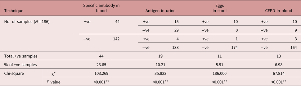
Forty-four patients (i.e. 23.65%) were seropositive for Schistosoma-specific antibody, 19 (10.21%) were positive for S. mansoni antigen, and only 11 cases (5.91%) were positive for eggs in stool. Schistosoma antigen was detected in 43.1% of seropositive patients. The percentage of S. mansoni-infected patients was significantly less than that of non-infected patients in the studied cohort of endocarditis patients. Eggs were detected in 4% of control patients, while the specific antibody was detected in 7% (tables 1 and 2).
Rate of S. mansoni infection in the human study (endocarditis versus non-endocarditis patients).

** Highly significant.
Schistosoma mansoni detection in endocarditis patients.

CFPD = cell-free parasite DNA. ** Highly significant.
The sensitivity, specificity, accuracy, NPV and PPV of the applied diagnostic tests are demonstrated in table 3. The egg detection technique recorded the least sensitivity (69), but the highest specificity (100), accuracy (97.3) and PPV (100). The antigen detection technique showed the highest sensitivity (79), while CFPD detection gave the highest negative predictive value (98).
Sensitivity and specificity of Schistosoma diagnostic techniques in the human study.

PPV, positive predictive value; NPV, negative predictive value; CFPD, cell-free parasite DNA.
The animal study
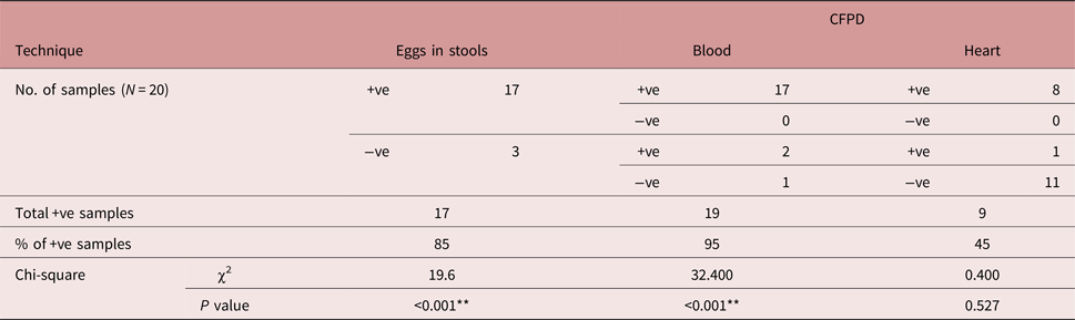
Eighty-five per cent of infected mice were positive for eggs in stools. Free DNA was detected in 95% of blood samples (table 4). Neither eggs nor CFPD positivity were reported in control mice.
Schistosoma mansoni detection in the different samples of infected BALB/c mice.

CFPD, cell-free parasite DNA. **, Highly significant.
The molecular findings
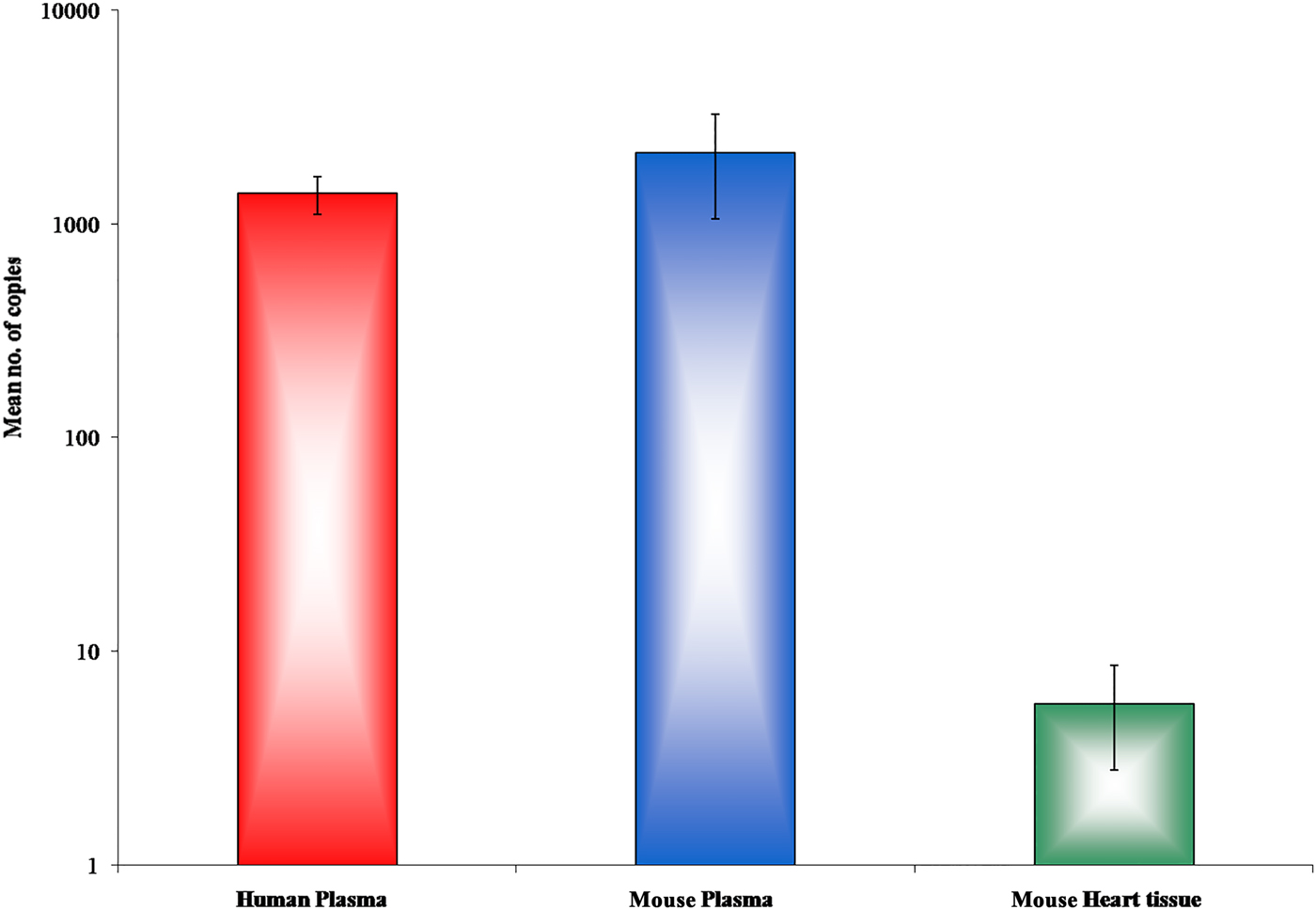
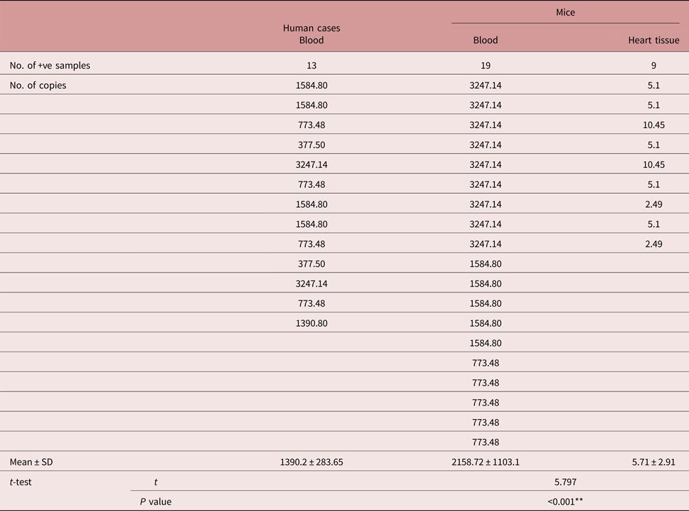
As shown in table 4; free DNA was detected in 95% of blood samples of infected mice and 45% of heart samples. Out of the 44 seropositive endocarditis patients; ten had CFPD in blood (table 2). Another three patients were positive for CFPD but seronegative. Ten endocarditis patients were positive for both S. mansoni eggs and CFPD. Two patients were positive for CFPD, but negative for eggs. The mean numbers of CFPD copies (table 5) are illustrated in fig. 1.
Comparison of S. mansoni CFPD in human and mouse samples by real-time PCR. Mean number of copies ± standard deviation = 1390.2 ± 283.65 in human blood, 2158.72 ± 1103.1 in mouse blood and 5.71 ± 2.91 in mouse heart samples.

Means of S. mansoni CFPD copies detected in the endocarditis patients, and infected BALB/c mice.

CFPD, cell-free parasite DNA; SD, standard deviation. ** Highly significant.
The histopathological findings
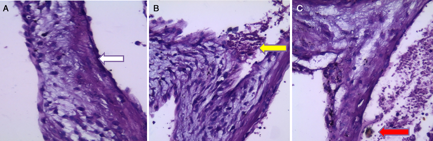
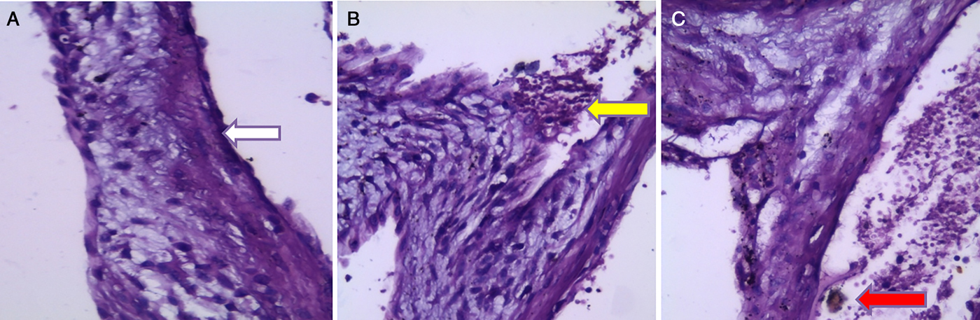
As demonstrated in fig. 2; subendothelial fibroblastic proliferation, collagen strands and inflammatory cellular infiltrate in a myxoid oedematous matrix were observed in valve sections of infected mice. Endothelial damage, subendothelial haemorrhage and brown pigmentation were seen in 30% of infected mice.
Valve sections, stained with H&E (×200), from BALB/c mice infected with S. mansoni, showing (A) an increase in collagen deposition under the endothelial lining (white arrow), (B) inflammatory cellular infiltrate (yellow arrow) and (C) subendothelial haemorrhage with haematin deposition (red arrow).
Discussion
The human heart is not one of the body organs commonly affected by parasitic infections. However, some parasites can disturb the cardiac structure and function either directly or indirectly. According to Hidron et al. (Reference Hidron, Vogenthaler, Santos-Preciado, Rodriguez-Morales, Franco-Paredes and Rassi2010), the parasites most associated with cardiac invasion are American Trypanosoma, Toxoplasma gondii, Trichinella spiralis, Entamoeba histolytica, Taenia solium (cysticercus cellulosae) and Echinococcus granulosus. Manifestations include myocarditis, pericarditis and pancarditis (Papamatheakis et al., Reference Papamatheakis, Mocumbi, Kim and Mandel2014). It is therefore important for cardiologists in parasite-endemic areas to consider parasitic infections in the differential diagnosis of vague cardiac diseases.
Cardiac tissue invasion is an uncommon finding in schistosomiasis. In Reference Victor, Lira, Arruda, Monteiro and Lima1996, Victor et al. reported a case of schistosomal endomyocardial fibrosis in a 14-year-old girl who presented with refractory ascites and progressive atrio-ventricular block. Cardiac granuloma was identified only post mortem, when the autopsy revealed endomyocardial inflammatory infiltrates and fibrosis around schistosomal eggs. Schistosoma is considered to be among the main parasites that can affect the heart indirectly in the advanced stages of infection. Schistosomal liver fibrosis and portal hypertension can lead to the hepatopulmonary syndrome known as cor pulmonale. After eggs have been shunted to the pulmonary circulation via the re-opened portosystemic anastomosis, granulomae and fibrosis start to develop and to deprive the lung of its elasticity and gas-exchange function. The patient starts to suffer from shortness of breath, dyspnoea, hypoxaemia, right ventricular hypertrophy and right-side heart failure (de Cleva et al., Reference de Cleva, Herman, Pugliese, Zilberstein, Saad, Rodrigues and Laudanna2003; Fouad & Yehia, Reference Fouad and Yehia2014). According to Papamatheakis et al. (Reference Papamatheakis, Mocumbi, Kim and Mandel2014), pathogenesis in cor pulmonale may also include arterial embolism (pulmonary arteriopathy due to endothelial damage). Pulmonary artery thrombosis, arrhythmias and sudden cardiac death syndrome have also been reported after schistosomiasis. Unfortunately, schistosomal cardiac involvement carries a grave prognosis (Hidron et al., Reference Hidron, Vogenthaler, Santos-Preciado, Rodriguez-Morales, Franco-Paredes and Rassi2010).
We carried out the current study at Tanta University Hospital in the centre of the Nile Delta, where S. mansoni is endemic. Following the unfamiliar association between schistosomiasis and endocarditis reported in a Filipino case in Japan by Yanagisawa et al. (Reference Yanagisawa, Yuasa and Tanaka2010), we aimed to provide a rational analysis of the phenomenon in the context of Egyptian endocarditis patients. Yanagisawa and his team were the first to report endocarditis as a complication of Schistosoma infection. A 43-year-old patient complained of persistent fever and oedema in the lower limbs. Although he had no history of heart disease, examination revealed a diastolic murmur, cardiomegaly, pulmonary congestion, elevated serum C-reactive protein and pancytopenia with relative eosinophilia. Infective endocarditis was initially excluded by repeatedly negative blood cultures, but transthoracic echocardiography revealed regurgitation and large vegetations on the aortic valve, and ultrasound revealed fibrosis of liver septae. The impaired liver function and negative hepatitis virus markers aroused suspicion of schistosomiasis. Meanwhile, repeated stool samples were negative for eggs, and serological tests reported a moderate elevation of anti-S. japonicum IgG (Wichmann et al., Reference Wichmann, Panning, Quack, Kramme, Burchard, Grevelding and Drosten2009).
In our study, we used various techniques to identify any possible S. mansoni infection (recent or otherwise) in the cohort of endocarditis patients. Seropositivity was reported in 23.65% of the patients, while antigen and eggs were detected in only 10.21% and 5.91%, respectively. According to Rose et al. (Reference Rose, Zimmerman, Hsu, Golby, Saleh, Folkerth, Santagata, Milner and Ramkissoon2014), direct pathological evaluation of the affected tissue is the gold standard for diagnosis. However, human tissue biopsies were not available in the current study, and stool examination gave the lowest number of positive cases of all the techniques used.
Where positive cases are diagnosed only by techniques other than coproscopy, they arouse suspicions of a condition known as closed schistosomiasis. Closed schistosomiasis is a pathological form of S. mansoni infection in which the eggs are trapped in granulomatous fibrosed masses in the intestinal wall and cannot pass into the lumen to exit in the stool (Igetei et al., Reference Igetei, El-Faham, Liddell and Doenhoff2017). The condition was discovered in conjunction with the growing resistance to praziquantel, the ‘golden anti-schistosomal drug’ (Doenhoff et al., Reference Doenhoff, Kusel, Coles and Cioli2002). Diagnosis is very difficult (Mutapi et al., Reference Mutapi, Maizels, Fenwick and Woolhouse2017), and even tissue biopsy can be misleading when examined by an inexperienced pathologist. Immune techniques become helpful in such cases (Coulibaly et al., Reference Coulibaly, N'Goran, Utzinger, Doenhoff and Dawson2013); however, evidence is accumulating of cross-reactivity between S. mansoni antigens and other allergens, and this may limit the accuracy of diagnosis (Doenhoff et al., Reference Doenhoff, El-Faham, Liddell, Fuller, Stanley and Schramm2016). According to Hamilton et al. (Reference Hamilton, Klinkert and Doenhoff1998), diagnosis of infection is also very difficult after therapy or in cases of spontaneous healing (burned-out bilharzia). Taken together, these factors create a real need to develop molecular-based techniques for the diagnosis of schistosomiasis (Abath et al., Reference Abath, Gomes, Melo, Barbosa and Werkhauser2006).
In 2002, Pontes et al. started to design primers that target a tandem repeat DNA sequence of S. mansoni. They depended on the sequence previously described by Hamburger et al. (Reference Hamburger, Turetske, Kapeller and Deresiewicz1991) as the basis for copro-PCR techniques. Conventional PCR proved its high specificity as no amplification has occurred in stool samples positive for other intestinal helminths. The technique also recorded high sensitivity as it was able to detect down to 2 eggs/g of stool, while the highest sensitivity recorded with the Kato–Katz technique was 10 eggs/g (Coulibaly et al., Reference Coulibaly, N'Goran, Utzinger, Doenhoff and Dawson2013). More steps have been taken to improve the technique, such as combining conventional PCR with restriction fragment length polymorphism (PCR-RFLP) analysis or enzyme-linked immunosorbent assay (ELISA) (Gomes et al., Reference Gomes, Melo, Werkhauser and Abath2006). With the development of quantitative real-time PCR (qPCR) researchers were able not only to detect lower concentrations of target DNA than with conventional PCR, but also to quantify the parasitic burden. Real-time PCR is less labour intensive as it does not require the electrophoresis step to visualize the final products. In addition, it has the advantage of differentiation between schistosomal species by amplifying multiple DNA targets in a single reaction mixture (multiplex PCR), which has made it very useful in epidemiological studies and control programmes (Pillay et al., Reference Pillay, Taylor and Zulu2014; Weerakoon et al., Reference Weerakoon, Gobert, Cai and McManus2015). Such advantages make qPCR a superior technique (ten Hove et al., Reference ten Hove, Verweij, Vereecken, Polman, Dieye and van Lieshout2008). Nevertheless, the need for several repetitive stool samples to increase the statistical chance of detection of the target DNA, and the abundance of natural copro-PCR inhibitors that possibly give false-negative results have encouraged researchers to develop new techniques to detect S. mansoni DNA in the blood (Pontes et al., Reference Pontes, Oliveira, Katz, Dias-Neto and Rabello2003; Sandoval et al., Reference Sandoval, Siles-Lucas, Perez-Arellano, Carranza, Puente, López-Abán and Muro2006; Roperch et al., Reference Roperch, Benzekri, Mansour and Incitti2015).
Wichmann et al. (Reference Wichmann, Panning, Quack, Kramme, Burchard, Grevelding and Drosten2009) documented the success of an alternative molecular diagnostic technique for schistosomiasis that depends on the detection of free DNA released from the parasite into the blood. The technique was originally postulated for the diagnosis of tumours under the term ‘liquid biopsy’ (Sozzi et al., Reference Sozzi, Conte and Leon2003). It is a non-invasive technique that detects the tumour-derived DNA released from a solid tumour that circulates in the blood. This technique has recently received much attention, due to its advantages in the diagnosis of metastasis and treatment-response follow-ups (Schwarzenbach et al., Reference Schwarzenbach, Nishida, Calin and Pantel2014). Normally, the concentration of cell-free DNA in adult plasma is 10–100 ng/ml, due to the physiological turnover of tissues and apoptosis of ageing cells. In oncology patients, higher concentrations of cell-free DNA are released in the blood by primary tumour cells (Zimmermann et al., Reference Zimmermann, Zhong, Holzgreve and Hahn2007), and they settle in bones and distant organs, such as the liver, lungs or brain (Pantel, Reference Pantel2016). Moreover, differentiation between advanced and earlier stages of the disease can be achieved by measuring the proportion of circulating tumour-derived DNA within the pool of cell-free DNA (Bettegowda et al., Reference Bettegowda, Sausen and Leary2014; Newman et al., Reference Newman, Bratman and To2014). This technique is also used in the diagnosis of intrauterine foetal diseases through the analysis of foetal DNA in the maternal blood (Bischoff et al., Reference Bischoff, Lewis and Simpson2005).
In complex parasitic infections, such as schistosomiasis, a significant turnover of parasites due to maturation, migration, replication, immune attack and death of the organisms is observed. As a metazoan, Schistosoma has a wealth of DNA copies, especially when more than one stage is present during development in the human host (schistosomulum, male worms, female worms and eggs) (Vinkeles Melchers et al., Reference Vinkeles Melchers, van Dam, Shaproski, Kahama, Brienen, Vennervald and van Lieshout2014). Therefore, cell-free parasite DNA (CFPD) detection by real-time PCR was used to diagnose any likely infected case in the studied cohort, especially those with closed schistosomiasis. The CFPD-detection technique succeeded in detecting CFPD in the blood samples of 6.98% of the endocarditis patients. Kato-Hayashi et al. (Reference Kato-Hayashi, Yasuda, Yuasa, Isaka, Haruki, Ohmae and Chigusa2013) stated that the CFPD-detection technique is expected to show a significant sensitivity in either closed or early infection cases. Unlike the intact parasite, CFPD is equally distributed in the patients’ blood and is not imprisoned in certain tissues. The CFPD-detection technique also overcomes the need for multiple random sampling, which is required in the classical diagnostic techniques and limits their sensitivity (Garcia & Palmer, Reference Garcia, Palmer and Murray1999). Hence, CFPD detection can be helpful in the diagnosis of suspected cases when other techniques show negative results. In addition, this technique detects the parasite DNA in distant organs. This is important since even dead eggs can continue releasing free DNA, but at slower rates. However, the greater the number of tissue-embedded living eggs (as in heavy and chronic infections), the longer is the CFPD persistence and the slower its elimination from the blood (Wichman et al., Reference Wichmann, Panning, Quack, Kramme, Burchard, Grevelding and Drosten2009).
Parallel to the decline of schistosomiasis mansoni in Egypt during the past three decades, uncommon presentations due to pathogenesis, other than granulomas and fibrosis, have appeared. Furthermore, co-infection with hepatitis B or C viruses participates much in the distortion of the pathognomonic and diagnostic features of schistosomiasis in Egyptian patients (Barakat, Reference Barakat2013; Gasim et al., Reference Gasim, Bella and Adam2015). Such cases are hardly diagnosed and exacerbate the underestimated infection rate. Uncommon presentations usually manifest a long time after exposure to the source of infection (Ferrari & Moreira, Reference Ferrari and Moreira2011). According to Wu et al. (Reference Wu, Wu and Tian2012) there has been an increasing trend for misdiagnosis of cerebral schistosomiasis. In 2014, Rose et al. reported a case of neuroschistosomiasis that represented with epilepsy-like manifestations 4 years after the first exposure to infection. On the other hand, another case of cerebral vasculitis was reported in a French woman 6 months after returning from an African country where she was exposed to infected water. After diagnosis and receiving praziquantel, she experienced another stroke due to generalized vasculitis and massive antigen shedding from the disseminated infection (Camuset et al., Reference Camuset, Wolff, Marescaux, Abou-Bacar, Candolfi, Lefebvre, Christmann and Hansmann2012).
Antigen shedding from eggs (or even adult worms) is an important factor in causing unexplained presentations in schistosomiasis (Shaker et al., Reference Shaker, Samy and Ashour2014). When antigen–antibody immune complexes are too many, or their clearance is lagging, they precipitate in distant organs. Precipitated complexes initiate inflammation and a type III hypersensitivity reaction in distant organs. Inflammation starts with C3a and C5a cleavage, mast cell degranulation and recruitment of lysosome-rich inflammatory cells. Finally, autophagy occurs by frustrated macrophages and polymorphonuclear cells (Batal et al., Reference Batal, Domsic, Medsger and Bastacky2010; Colley & Secor, Reference Colley and Secor2014). In Reference Edberg, Tosic and Taylor1989, Edberg et al. investigated whether DNA complexes can be built after the release of free DNA in the blood. The assumption was finally proved by the detection of anti-nuclear antibodies in the circulating immune complexes. The clearance of these complexes occurs by combined recognition mechanisms for both the immune complexes and DNA (Pisetsky, Reference Pisetsky2012). In schistosomiasis mansoni, DNA immune complexes are suspected to deposit in distant organs and cause vague pathogenesis in vascular organs such as the kidneys (Ochodo et al., Reference Ochodo, Gopalakrishna, Spek, Reitsma, van Lieshout, Polman, Lamberton, Bossuyt and Leeflang2015). In our study, CFPD was detected in the cardiac homogenate of infected BALB/c mice. The mean number of copies in heart tissue (mean ± SD = 5.71 ± 2.91) was significantly less than in blood (mean ± SD = 2158.72 ± 1103.1). However, detection of CFPD in 45% of cardiac samples and in 47.1% of mice with positive blood CFPD cannot be neglected.
Previous studies on free DNA bio-distribution and persistence indicate that: foreign DNA may persist for a shorter period in ectopic sites but, on the other hand, thousands of copies persist longer in tissues near the releasing source (Vahedi et al., Reference Vahedi, Nazari, Arbabi and Peymanfar2012). Infective endocarditis patients are reported to develop high titres of circulating immune complexes (CIC). Messias-Reason et al. (Reference Messias-Reason, Hayashi, Nisihara and Kirschfink2002) showed CIC precipitation in the basement membrane of cardiac valves, which causes more inflammation and complement activation. The thin endothelial lining of cardiac valves can be one of the attractive sites for deposition of DNA immune complexes, especially if valves are already damaged. In our study, hyaline degeneration was detected in the cardiac valves of infected mice with inflammatory cellular infiltrate in a myxoid matrix. This can suggest the presence of immune complex deposits.
The presence of a high concentration of circulating free DNA has recently been shown to anticipate another major problem. It can stimulate the process of atherosclerosis. Coscas et al. (Reference Coscas, Bensussan and Jacob2017) have identified the ability of free DNA to be a potential nidus in the early stages of human atheroma as it triggers calcium phosphate precipitation and hydroxyapatite crystallization. According to Xie et al. (Reference Xie, Chen, Sun and Ping2013) the poly-anionic nature of free DNA and high phosphate content make it react strongly with cationic calcium phosphate. Calcium phosphate nanoparticles are even considered to be the best cell DNA transfection vectors (Khan et al., Reference Khan, Wu, Ghosh and Uskoković2016; Chernousova & Epple, Reference Chernousova and Epple2017). Calcification of cardiac valves is described as an active process that involves the coordinated actions of resident valve endothelium, interstitial cells, circulating inflammatory, immune cells and bone marrow-derived cells (Firth et al., Reference Firth, Mandel and Yuan2010). The cells that are irritated by the precipitated immune complexes can transform into osteoblast-like cells, elaborate bone matrix (endothelial-to-mesenchymal transition) and form matrix vesicles. The latter serve as a nidus for micro-calcifications (Leopold, Reference Leopold2012). Interestingly, the immunopathology of schistosomal pulmonary vascular lesions in cor pulmonale has a similar mechanism to idiopathic pulmonary arterial hypertension (Papamatheakis et al., Reference Papamatheakis, Mocumbi, Kim and Mandel2014). In the latter condition, decreased K+-channel activity causes membrane depolarization and increases the cytosolic calcium (Ca2+) ion concentration, which causes vasoconstriction. The Ca2+ increase stimulates cell migration and proliferation, down-regulates apoptosis of smooth muscle cells in the pulmonary artery and results in medial hypertrophy and concentric vascular remodelling. Moreover, endothelial damage reduces the release of anti-proliferative and vasodilator substances, such as prostacyclin and nitric oxide, and increases the release of unopposed pro-proliferative and vasoconstrictive agents, such as thromboxane A2 and endothelin-1, which further increase the vascular tone (Firth et al., Reference Firth, Mandel and Yuan2010).
In conclusion, Yanagisawa et al. (Reference Yanagisawa, Yuasa and Tanaka2010) suspected the passage of S. japonicum eggs to the endocardium, causing endocarditis in the absence of an intracardiac shunt. However, on considering the detection of CFPD and the histopathological changes in the cardiac samples of infected mice in our study, this involvement of Schistosoma cannot be completely omitted. More studies are warranted to study the sequelae of the presence of CFPD, whether circulating in the blood or precipitated in the tissues, and to investigate its ability to initiate inflammation, inflict endothelial damage or integrate into the host-cell genome, leading to mutations or abnormal proliferation, as proved after infection with other infectious agents (Morgan et al., Reference Morgan, DiNardo and Windle2017).
Acknowledgements
We are grateful to Dr Ahmed Mashaly, Dr Amr Mahmoud and Dr Eiman Hasby for their support in the implementation of this study, by choosing the included patients, facilitating sample collection and histopathological examination. We also like to thank the participating patients for their commitment. We express our sincere thanks to the Medical Parasitology and Clinical Pathology Department laboratories at Tanta University, Faculty of Medicine, for providing us with most of the basic equipment and routine diagnostic kits.
Financial support
This research received no specific grant from any funding agency, commercial or not-for-profit sectors.
Conflict of interest
None.
Ethical standards
This study was conducted after obtaining approval from Tanta Faculty of Medicine ethical committee for scientific research (approval code: 31702/08/17)). The authors assert that all procedures contributing to this work comply with the ethical standards of the relevant national and institutional committees on human experimentation and with the Helsinki Declaration of 1975, as revised in 2008. We also assert that all procedures contributing to this work comply with the ethical standards of the relevant national and institutional guides on the care and use of laboratory animals.